STAU1 / Double-stranded RNA-binding protein Staufen homolog 1 · IHC design guide

Design Immunohistochemistry for STAU1

Plan STAU1 staining in paraffin sections with catalog antibody A04259, starting at 10 μg/mL (datasheet). Use the reported cytoplasmic pattern and high respiratory epithelial staining to select tissue controls (HPA tissue IHC).

Evidence assembled Oct 2026 · For research use; verify linked source records and product datasheet before use
Immunohistochemistry protocol sheet for STAU1 (IHC for STAU1): expected localisation General cytoplasmic tissue staining (HPA tissue IHC), antibody A04259, validated IHC image, and IHC protocol steps
Printable STAU1 IHC protocol sheet — expected localisation General cytoplasmic tissue staining (HPA tissue IHC), antibody A04259, controls and protocol steps. Open the full STAU1 IHC guide →

STAU1 Immunohistochemistry Experimental Design Guide

Expected localisation, validated protocols, controls and antibodies — the at-a-glance facts below, then the full design guide.

Must know before staining
Expected localisation General cytoplasmic tissue staining (HPA tissue IHC)
Staining pattern Cytoplasmic staining; high in respiratory epithelial cells (HPA tissue IHC)
Antigen retrieval Citrate pH 6.0 HIER, 95–98 °C, 20 min (rule: cytoplasmic / membrane antigen)
Positive control ⓘ Bronchus+4 more · see all
Negative control ⓘ Lymph node+1 more · see all
Important caveats
Reasons your staining may differ from the expected pattern.
Fixation Keep fixation conditions consistent across sections. (standard IHC practice; not target-specific)
Caveat Medium staining–RNA consistency; verify with controls (HPA tissue IHC)
Regulation Regulatory changes are unreported (UniProt)
Isoform / epitope 3 isoforms; confirm antibody epitope coverage (UniProt)
Section 1

Recommended STAU1 IHC & IF Protocols

The catalog antibody’s IHC-P protocol (datasheet) is followed by published STAU1 staining details for paraffin-embedded lung tissue (PMC8957805) and mouse xenograft tumors (PMC10520073).

Recommended immunohistochemistry (IHC-P) protocol parameters
SampleTissue sections; selected-image fixative not specified (standard IHC workflow)
FixationImage fixative and duration unreported (datasheet A04259); verify before use.
Sectioning4–5 µm sections on charged slides (standard)
DeparaffinisationXylene, graded ethanol series to water (standard)
Antigen retrievalHeat-induced epitope retrieval in citrate buffer, pH 6.0, 20 min at 95–98 °C (standard rule: cytoplasmic / membrane antigen)
Peroxidase block3% H2O2, 10 min, room temperature (standard)
Blocking10% normal serum of the secondary host, 30 min, room temperature (standard)
Primary antibodyRabbit anti-STAU1, 10 μg/mL (datasheet A04259)
Primary incubationOvernight at 4 °C (standard)
DetectionHRP-polymer secondary, DAB chromogen 5–10 min (standard)
CounterstainHematoxylin, blue, dehydrate and mount (standard)
Expected resultSTAU1-positive staining in respiratory epithelial cells of bronchus (HPA tissue IHC: High). HPA tissue profile: General cytoplasmic expression. No signal in the no-primary control.
💡Decision noteStart with citrate pH 6.0 retrieval at 95–98 °C for 20 min (page antigen-retrieval rule); neither cited IHC excerpt specifies retrieval conditions.
Section 2

What Is the Expected STAU1 Staining Pattern?

STAU1 should produce predominantly cytoplasmic staining in paraffin sections, including respiratory epithelial cells, Purkinje cells, and several glandular epithelia (HPA: general cytoplasmic expression; High in those cells). UniProt places STAU1 with the rough endoplasmic reticulum and reports no transmembrane segment (UniProt O95793: localization; topology). Treat the tissue pattern as a guide: HPA rates its IHC evidence Approved, with medium staining–RNA consistency and external verification pending.

What am I looking at on my slide?
Cytoplasmic signal in bronchial respiratory epithelium or cerebellar Purkinje cells, with little nuclear signal.This fits the reported compartment and high-staining cell types (HPA: general cytoplasmic expression; High in both). Score the relevant cells rather than averaging the whole section; adjacent cells need not match their intensity.
Predominantly nuclear signal in cells expected to stain, with little convincing cytoplasmic signal.This conflicts with the reported cytoplasmic pattern (HPA: tissue IHC) and rough endoplasmic reticulum localization (UniProt O95793). Check counterstain, section morphology, and primary-antibody controls before assigning nuclear STAU1 localization (general IHC practice).
Strong staining in lymph-node germinal-center cells or splenic red-pulp cells.Those cell populations were reported as not detected (HPA: lymph node; spleen). Investigate cross-reactivity or endogenous chromogenic activity with appropriate controls (general IHC practice). A different cell population elsewhere is not automatically suspect: STAU1 is widely expressed (UniProt O95793).
Uniform color across tissue compartments, including areas without a plausible cellular pattern.Diffuse deposition is difficult to reconcile with cell-resolved cytoplasmic staining (HPA: general cytoplasmic expression). Examine the no-primary control and detection steps for background before scoring positive cells (general IHC practice).
No convincing signal in a known-positive compartment of bronchus or cerebellum.That result conflicts with high staining reported for respiratory epithelial or Purkinje cells (HPA: High in bronchus and cerebellum). Confirm that the expected cells are present, then check IHC validation, controls, and the working conditions of the assay (general IHC practice).
💡Expected STAU1 appearanceCall a result positive when identifiable high-staining cells show predominantly cytoplasmic chromogen (HPA: High in selected cells; general cytoplasmic expression); dominant nuclear staining or broad cell-free color warrants control review (general IHC practice).
How each factor affects the staining
Compartment used for scoringHPA describes general cytoplasmic IHC staining, while UniProt places STAU1 with the rough endoplasmic reticulum (HPA: tissue IHC; UniProt O95793). Expect cytoplasmic localization; neither source requires a sharply outlined organelle pattern in chromogenic sections.
Choice of positive and comparison tissueBronchial respiratory epithelium and cerebellar Purkinje cells are High; adipocytes and parathyroid glandular cells are Low; germinal-center and splenic red-pulp cells are Not detected (HPA: tissue IHC). These are cell-specific reference points, not claims that the entire tissues are uniformly positive or negative.
Strength of the tissue evidenceHPA marks tissue IHC Approved but reports only medium consistency with RNA expression and pending external verification (HPA: reliability description). A discordant specimen needs assay controls and independent interpretation; the atlas profile alone does not establish the cause.
Antibody and isoform coverageHPA049892 and CAB020839 are IHC Approved; HPA072004 has no listed IHC status (HPA: antibody records). UniProt lists three isoforms (UniProt O95793: isoforms). The supplied evidence gives no epitope map, so it cannot establish which isoforms a given antibody detects.
IF/ICC Q&A: should fluorescence match the paraffin-section pattern?HPA reports enhanced cytosolic ICC-IF localization and enhanced ICC status for HPA049892 and HPA072004 (HPA: subcellular; antibody records). That supports a cytosolic expectation in IF/ICC, but it does not supply an IHC-P protocol or prove identical image detail across the two applications.
Chromogenic detection backgroundEndogenous enzyme activity can produce apparent color independently of primary-antibody binding (general IHC practice). A no-primary control helps distinguish detection background from specific cytoplasmic staining; no supplied source reports STAU1-specific sensitivity to fixation or retrieval.
Why is my staining missing, weak or wrong?
SituationLikely causeNext action
A high-reference cell population is blank.The expected cells may be absent from the section, or the IHC assay may have failed (HPA: High in bronchial respiratory epithelium and Purkinje cells; general IHC practice).Verify cell identity and tissue preservation, then inspect an appropriate positive control, antibody working conditions, and the staining run (general IHC practice). Do not infer STAU1 absence from a failed control.
Color is strongest over nuclei.The pattern conflicts with cytoplasmic IHC and rough endoplasmic reticulum localization (HPA: tissue IHC; UniProt O95793); counterstain or nonspecific signal may confuse interpretation (general IHC practice).Review the chromogen and counterstain separately where possible, compare the no-primary control, and rescore only clearly localized cellular signal (general IHC practice).
Germinal-center or red-pulp cells stain strongly.Those populations are reported as not detected (HPA: lymph node; spleen). Cross-reactivity or endogenous detection activity is possible (general IHC practice).Check a no-primary control for detection background and compare an independently validated IHC antibody when available (general IHC practice; HPA: IHC Approved HPA049892 and CAB020839).
The whole section has diffuse color.Background deposition can obscure the expected cell-resolved cytoplasmic pattern (general IHC practice; HPA: general cytoplasmic expression).Inspect the no-primary control and revisit blocking, washing, detection activity, and antibody working concentration as general IHC variables; the supplied evidence does not identify a STAU1-specific cause.
Signal changes after antigen retrieval is adjusted.Retrieval conditions can alter staining in paraffin IHC generally, but these sources do not establish a STAU1-specific retrieval or fixation effect (general IHC practice; source scope: HPA tissue IHC and UniProt O95793).Compare conditions using the same positive tissue and controls, and judge cytoplasmic cell-specific signal against background (general IHC practice). Report the empirical result without attributing it to a documented STAU1 epitope effect.
IF/ICC appears cytosolic, while the IHC section is equivocal.HPA reports enhanced cytosolic ICC-IF localization, whereas tissue IHC has medium staining–RNA consistency (HPA: subcellular; tissue reliability). The applications yield different readouts (general IHC/IF practice).Interpret each application with its own validated antibody status and controls (HPA: antibody records; general IHC/IF practice). Use the cytosolic IF result as localization context, not as proof that the IHC chromogen is specific.

Sample controls for STAU1 IHC & IF

🧪Run bronchus first and score respiratory epithelial cells, which show High STAU1 staining (HPA: High in bronchus respiratory epithelial cells). Use lymph node germinal center cells as the negative tissue (HPA: Not detected in lymph node germinal center cells); cells outside the bronchial respiratory epithelium should be assessed for background against the no-primary slide, not assumed to be STAU1-negative (HPA: bronchus respiratory epithelial cells).
Positive control tissue: Bronchus (Respiratory epithelial cells, HPA High)
Negative control tissue: Lymph node (HPA Not detected)
ICC-IF cell lines (HPA subcellular resource): HPA ICC-IF images show STAU1 in A-431, Hep-G2, SiHa, U2OS, NIH 3T3, with annotated localisation: Cytosol (enhanced) (HPA subcellular).
Technical controls: Include a no-primary, secondary-only control and an isotype control matched to the primary antibody’s host species and, for a monoclonal antibody, subclass (standard IHC practice). A STAU1 knockout specimen is the preferred biological negative; for chromogenic bronchus IHC, block endogenous peroxidase and assess background chromogen (standard IHC practice).
⚠️Feasibility: A STAU1-specific fixation window or fixation effect is unreported in the supplied evidence, and the selected A04259 human-brain IHC caption does not state a fixative (A04259 tissue-IHC caption). Retrieval dependence is unreported, so compare antigen retrieval conditions during paraffin-section optimization (A04259 tissue-IHC caption; standard IHC practice). The supplied evidence does not establish whether frozen sections or IF/ICC are easier; in bronchus, monitor endogenous peroxidase as a potential chromogenic artefact (standard IHC practice).

HPA tissue IHC evidence for STAU1

Comprehensive Human Protein Atlas IHC scoring per tissue (reliability: Approved — Medium consistency between antibody staining and RNA expression data. Pending external verification.). Rows are taken directly from the HPA tissue chart — click any row's HPA link to view the source.

Positive expression · recommended positive controls

TissueCell typeLevelEvidenceSource
Bronchus Respiratory epithelial cells High Protein (IHC) HPA →
Cerebellum Purkinje cells High Protein (IHC) HPA →
Endometrium Glandular cells High Protein (IHC) HPA →
Esophagus Squamous epithelial cells High Protein (IHC) HPA →
Nasopharynx Respiratory epithelial cells High Protein (IHC) HPA →

Undetected expression · recommended negative controls

TissueCell typeLevelEvidenceSource
Lymph node Germinal center cells Not detected Protein (IHC) HPA →
Spleen Cells in red pulp Not detected Protein (IHC) HPA →
Section 3

Advanced STAU1 IHC Tips

Troubleshoot chromogenic STAU1 IHC in paraffin sections using the page retrieval setting, documented tissue staining, and appropriate controls.

What retrieval should I try when STAU1 staining is weak?
Start with citrate buffer at pH 6.0, heated to 95–98 °C for 20 min (page retrieval setting). Keep section thickness, cooling time, antibody concentration and detection conditions constant while comparing retrieved sections with an otherwise matched section that omits retrieval (general IHC practice). The catalog antibody caption reports STAU1 staining in human brain at 10 µg/mL, but gives no retrieval method or fixative, so it cannot establish that this setting was used for that image (A04259 caption). If signal remains weak, compare a second validated retrieval condition on serial sections, checking whether any gain in cytoplasmic signal also increases background or damages tissue (general IHC practice; HPA: general cytoplasmic expression).
Could fixation explain inconsistent STAU1 staining across sections?
Target-specific sensitivity of STAU1 to fixation is unknown from the supplied evidence, so treat fixation history as a variable to test rather than an established cause of signal loss (A04259 caption: fixative not stated). Record fixative, fixation duration, processing schedule and section age for each specimen, then compare matched sections with the same retrieval and detection settings (general IHC practice). The human brain tissue caption specifies 10 µg/mL antibody but does not state its fixative (A04259 caption). HPA cytoplasmic staining and STAU1’s RNA-binding domains describe expression and structure; neither establishes how this epitope responds to fixation (HPA: general cytoplasmic expression; UniProt O95793: DRBM domains).
Which staining compartment is plausible for STAU1 in IHC?
Expect predominantly cytoplasmic staining: the tissue profile describes general cytoplasmic expression, while the subcellular profile places STAU1 in the cytosol (HPA tissue IHC; HPA subcellular). UniProt also associates STAU1 with rough endoplasmic reticulum and reports no transmembrane segment, so perinuclear cytoplasmic staining can be plausible if its cell-level pattern is reproducible (UniProt O95793: subcellular location; topology). Compare the signal with morphology in adjacent sections and with a primary-antibody omission control before assigning fine intracellular structures in chromogenic IHC (general IHC practice). Isolated strong nuclear staining deserves scrutiny because it differs from the supplied cytoplasmic reference patterns (HPA tissue IHC; HPA subcellular).
How should isoforms and epitope location affect antibody interpretation?
STAU1 has 3 listed isoforms—Long, Short and 3—and double-stranded RNA-binding domains at residues 72–162, 184–251 and 286–354 (UniProt O95793: isoforms; domains). Without a mapped immunogen or epitope for the catalog antibody, its relative recognition of those isoforms cannot be determined from this payload (A04259 caption; UniProt O95793: isoforms). Consult the exact antibody’s epitope documentation before treating differing tissue signals as isoform-specific, and compare staining with an independently validated reagent if that distinction matters (general IHC practice). Because the record also lists methylation and phosphorylation, epitope accessibility could vary, but no supplied IHC experiment demonstrates a PTM-dependent staining effect (UniProt O95793: modified residues).
How can I check the STAU1 pattern with multiplex IF?
On the separate IF/ICC workflow, pair STAU1 with a validated marker for the cell population being examined; HPA reports high staining in cerebellar Purkinje cells and bronchial respiratory epithelial cells (HPA tissue IHC). Choose spectrally separated fluorophores after measuring each specimen’s autofluorescence, and include single-label controls to assess bleed-through (general IF practice). Use a permeabilisation condition that gives antibody access to the cytoplasmic epitope, then check that it preserves cell morphology; STAU1 has no transmembrane segment and is reported in the cytosol and with rough ER (UniProt O95793: topology; subcellular location; HPA subcellular). Treat IF localisation as complementary evidence, since staining conditions and detection differ from chromogenic paraffin IHC (general IHC/IF practice).
What should I change if cytoplasmic staining is widespread?
First compare a primary-antibody omission control and a matched isotype control, then assess whether the diffuse signal follows tissue edges, damaged regions or endogenous pigment (general IHC practice). Block endogenous peroxidase before chromogenic detection and check DAB development time, because either can contribute signal unrelated to antibody binding (general chromogenic IHC practice). Titrate the primary antibody around the working condition; the selected human brain image reports 10 µg/mL, but its caption provides no universally applicable dilution or background assessment (A04259 caption). Widespread cytoplasmic staining alone is not proof of background because HPA describes general cytoplasmic expression and low tissue RNA specificity (HPA tissue IHC).
How should I score STAU1 staining across samples? ⚠ ANSWER MARKED FOR VERIFICATION
Define the cell population and compartment before scoring, then record cytoplasmic intensity and the percentage of positive cells in equivalent regions of each section (HPA tissue IHC: general cytoplasmic expression; general IHC practice). An H-score weights the percentages at intensity grades 0–3 and spans 0–300; keep thresholds, illumination and analysis settings fixed across samples (general IHC scoring practice). Report the number of evaluable target cells and normalize regional counts or positive-cell density to sampled tissue area in mm², while accounting for differences in cell composition (general image-analysis practice). Include control-section results and avoid using HPA’s qualitative High or Not detected categories as numerical calibration points (HPA tissue IHC; general IHC practice).
How can I distinguish true STAU1 staining from artefact?
Favor reproducible cytoplasmic signal in intact cells over isolated nuclear, edge-associated or necrotic staining; the supplied tissue and subcellular profiles place STAU1 chiefly in the cytoplasm and cytosol (HPA tissue IHC; HPA subcellular). HPA reports High staining in Purkinje cells and respiratory epithelial cells, but Not detected in lymph-node germinal-center cells and splenic red-pulp cells; use those patterns as contextual comparisons rather than absolute specificity controls (HPA tissue IHC). Examine primary-antibody omission and peroxidase-block controls for chromogen arising without specific primary binding (general chromogenic IHC practice). Interpret disagreements cautiously because the HPA tissue IHC reliability is Approved with medium staining–RNA consistency and pending external verification (HPA tissue IHC: reliability).
Boster reagents

Best STAU1 / Double-stranded RNA-binding protein Staufen homolog 1 IHC Antibodies

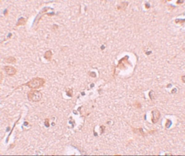
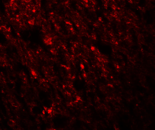
A04259 has IHC data from human brain tissue and IF data from human brain cells (catalog image captions); its listed reactivity is human, mouse and rat (catalog applications/reactivity).

Real IHC data Immunohistochemistry of STAU1 in human brain tissue with STAU1 antibody at 10 μg/mL.
Anti-STAU1 Antibody
Cat # A04259

The sole rendered card, A04259, shows STAU1 staining in human brain tissue by IHC at 10 μg/mL (A04259 IHC image caption). It also shows IF staining in human brain cells at 20 μg/mL (A04259 IF image caption).

Which to pick: For tissue IHC, choose A04259: it lists IHC-P and shows staining in human brain tissue; the caption does not report the fixative (A04259 applications; A04259 IHC image caption). For IF/ICC, A04259 has an IF image in human brain cells, while M04259 and PB9895 list ICC/IF without image evidence in this payload (A04259 IF image caption; M04259 and PB9895 applications/image captions). For work across species, A04259 lists human, mouse and rat reactivity, but its IHC image documents human tissue only (A04259 reactivity; A04259 IHC image caption).

Each figure is that product's own IHC / IF validation image from its datasheet.

References

  1. UniProt Consortium. UniProt entry O95793 (STAU1_HUMAN, Double-stranded RNA-binding protein Staufen homolog 1).
  2. Human Protein Atlas. STAU1 tissue IHC expression (reliability: Approved).
  3. Human Protein Atlas. STAU1 subcellular location (ICC-IF): Localized to the cytosol..
  4. Human Protein Atlas. STAU1 antibody validation summary (3 antibodies).
  5. A human Staufen1 BAC transgenic mouse exhibits abnormal autophagy and neurodegeneration across the central nervous system. Cell death & disease 2026 — PMC13342298.
  6. STAU1 exhibits oncogenic characteristics and modulates alternative splicing and gene expression in lung adenocarcinoma cells. Scientific reports 2025 — PMC12084350.
  7. Machine-Learning Algorithm-Based Prediction of Diagnostic Gene Biomarkers Related to Immune Infiltration in Patients With Chronic Obstructive Pulmonary Disease. Frontiers in immunology 2022 — PMC8957805.
  8. Genome-wide enhancer-gene regulatory maps link causal variants to target genes underlying human cancer risk. Nature communications 2023 — PMC10520073.
  9. PubMed PMID:10022909 — UniProt-cited evidence.
  10. PubMed PMID:10325410 — UniProt-cited evidence.
  11. PubMed PMID:11230166 — UniProt-cited evidence.