This website uses cookies to ensure you get the best experience on our website.


- Table of Contents
Plan paraffin-section STC2 IHC around cytoplasmic or membranous staining in most tissues (HPA tissue IHC). Compare high staining in breast glandular cells with undetected staining in adipocytes (HPA tissue IHC), and account for secretion when interpreting the signal (UniProt).

Expected localisation, validated protocols, controls and antibodies — the at-a-glance facts below, then the full design guide.
| Expected localisation | Cytoplasmic or membranous tissue staining (HPA tissue IHC) | |
| Staining pattern | Cytoplasmic or membranous signal in most tissues (HPA tissue IHC) | |
| Antigen retrieval | EDTA pH 8.0 HIER, heat-mediated (datasheet A05017) | |
| Positive control | Adrenal gland+4 more · see all | |
| Negative control | Adipose tissue+4 more · see all |
| Fixation | Keep fixation conditions consistent across sections. (standard IHC practice; not target-specific) | |
| Caveat | Secreted STC2 may stain beyond producing cells (UniProt) | |
| Regulation | Expression regulation is not established (UniProt) | |
| Isoform / epitope | No annotated isoforms; signal peptide 1–24 is removed (UniProt) |
Compare the catalog antibody’s IHC-P protocol (datasheet: A05017) with four published STC2 IHC protocols (PMC8794563; PMC4186577; PMC12170999; PMC11245224).
| Sample | Paraffin-embedded human breast cancer tissue; fixative not specified (datasheet A05017) |
| Fixation | Image fixative and duration unreported (datasheet A05017); verify before use. |
| Sectioning | 4–5 µm sections on charged slides (standard) |
| Deparaffinisation | Xylene, graded ethanol series to water (standard) |
| Antigen retrieval | Heat retrieval: EDTA pH 8.0 (datasheet A05017); 20 min, 95–100 °C (standard) |
| Peroxidase block | 3% H2O2, 10 min, room temperature (standard) |
| Blocking | 10% goat serum (datasheet A05017) |
| Primary antibody | Rabbit anti-STC2, 2-5 μg/ml (datasheet A05017) |
| Primary incubation | Overnight at 4 °C (datasheet A05017) |
| Detection | HRP-conjugated secondary, DAB chromogen (datasheet A05017) |
| Counterstain | Hematoxylin, blue, dehydrate and mount (standard) |
| Expected result | STC2-positive staining in glandular cells of adrenal gland (HPA tissue IHC: High). HPA tissue profile: Cytoplasmic or membranous expression in most tissues. No signal in the no-primary control. |
STC2 is secreted and has no transmembrane segment (UniProt O76061 topology). In tissue IHC, expect cytoplasmic or membranous staining in most tissues, with high staining in selected glandular, glial, glomerular, immune and macrophage populations (HPA tissue IHC). The tissue profile is Approved, but HPA notes that secreted proteins can show complex RNA–protein relationships and that external verification is pending (HPA tissue IHC reliability).
| Cytoplasmic or membranous staining appears in breast glandular cells or lung macrophages. | This matches the reported compartment and high-staining cell populations (HPA tissue IHC: breast glandular cells High; lung macrophages High). Judge the named cells separately from neighboring cells; secretion can complicate attribution of nearby signal (UniProt O76061: Secreted; HPA tissue IHC reliability). |
| The dominant signal is sharply nuclear, with little cytoplasmic or membranous staining. | A nuclear-only pattern does not match the reported tissue profile or approved ER location in ICC-IF (HPA tissue IHC profile; HPA subcellular). Check morphology, counterstain and detection controls before assigning that signal to STC2 (general IHC practice). |
| Strong staining appears in adipocytes or cardiomyocytes, while expected positive cells are weak. | Those cell types were reported as Not detected (HPA tissue IHC: adipocytes; cardiomyocytes). Consider cross-reactivity or endogenous detection activity, then compare with reagent controls and a known-positive section (general IHC practice). |
| Broad, hazy color covers tissue and background, obscuring cell boundaries. | This is difficult to score against HPA's cell-resolved cytoplasmic or membranous profile (HPA tissue IHC profile). Review blocking, washes, chromogen development and the detection-only control for nonspecific background (general IHC practice). |
| No staining is visible in an expected high-staining population. | Check that the relevant cells are present: breast glandular cells, lung macrophages and kidney glomerular cells are reported High (HPA tissue IHC). If present, review antibody dilution, retrieval and detection performance with a positive control (general IHC practice). |
| Tissue and cell selection | Use a reported High population, such as breast glandular cells, lung macrophages or kidney glomerular cells, to assess sensitivity (HPA tissue IHC). Adipocytes and cardiomyocytes offer reported Not detected comparisons (HPA tissue IHC); interpret each cell type within its own tissue context. |
| Secreted-protein interpretation | STC2 has a signal peptide at residues 1–24, a mature chain at 25–302 and no transmembrane segment (UniProt O76061 topology/processing). HPA cautions that RNA location and protein staining may differ for secreted variants (HPA tissue IHC reliability). |
| Evidence strength | The tissue profile and the listed antibody's IHC status are Approved (HPA tissue IHC reliability; HPA antibody HPA045372). HPA also flags pending external verification (HPA tissue IHC reliability); the supplied record does not establish an Enhanced IHC result (HPA antibody HPA045372). |
| IF/ICC Q&A | Where should STC2 appear in IF/ICC? HPA reports an approved endoplasmic reticulum location and ICC approval for HPA045372 (HPA subcellular; HPA antibody HPA045372). This cell-image finding helps interpret localization; tissue IHC has its own cytoplasmic or membranous profile (HPA tissue IHC profile). |
| Situation | Likely cause | Next action |
|---|---|---|
| A breast section appears negative. | The assessed area may lack the relevant glandular cells, or the staining run may have failed (HPA tissue IHC: breast glandular cells High; general IHC practice). | Confirm glandular cells on the counterstained section, then inspect a known-positive control and the staining run before interpreting absence in the specimen (general IHC practice). |
| A heart muscle section stains strongly throughout cardiomyocytes. | HPA reports cardiomyocytes as Not detected, despite UniProt listing expression in heart among various tissues (HPA tissue IHC: heart muscle; UniProt O76061 tissue specificity). | Verify cell identity and compare reagent controls; do not equate tissue-level expression with cardiomyocyte protein staining (general IHC practice; HPA tissue IHC: cardiomyocytes Not detected). |
| Nuclei dominate the signal. | The compartment conflicts with HPA's cytoplasmic or membranous tissue pattern and approved ER location in ICC-IF (HPA tissue IHC profile; HPA subcellular). | Check counterstain overlap and detection-only controls; reassess localization in a reported High cell population (general IHC practice; HPA tissue IHC levels). |
| Background masks macrophages in lung. | Heavy background prevents assessment of a reported High cell population (HPA tissue IHC: lung macrophages High). | Review blocking, washing and chromogen development; compare a detection-only control, then score only identifiable macrophages above local background (general IHC practice). |
| Signal differs between neighboring cell types. | HPA reports cell-specific levels: kidney glomerular cells are High, while adipocytes are Not detected in adipose tissue (HPA tissue IHC). STC2 secretion can also complicate protein attribution (UniProt O76061: Secreted; HPA reliability). | Score identified cell populations separately, using morphology and an appropriate negative comparison; investigate signal that obscures cell boundaries (general IHC practice). |
| Positive control stains weakly after a protocol change. | Target-specific fixation effects are not established by the supplied assay evidence. Verify with a matched IHC source before attributing a result to fixation. | Check reagent preparation, dilution, retrieval settings and detection controls against the last working run before drawing a biological conclusion (general IHC practice). |
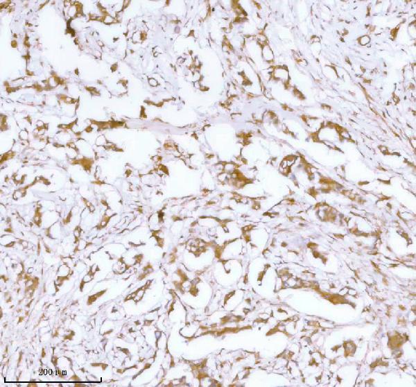
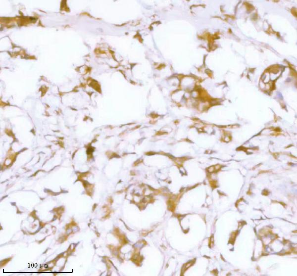
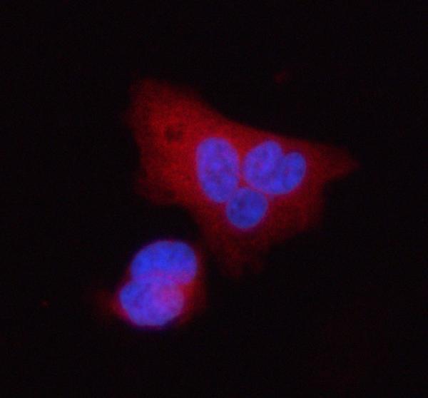

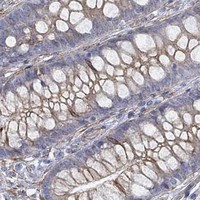


Comprehensive Human Protein Atlas IHC scoring per tissue (reliability: Approved — At least one protein variant secreted, tissue location of RNA and protein might differ and correlation is complex. Pending external verification.). Rows are taken directly from the HPA tissue chart — click any row's HPA link to view the source.
| Tissue | Cell type | Level | Evidence | Source |
|---|---|---|---|---|
| Adrenal gland | Glandular cells | High | Protein (IHC) | HPA → |
| Breast | Glandular cells | High | Protein (IHC) | HPA → |
| Caudate | Glial cells | High | Protein (IHC) | HPA → |
| Cerebral cortex | Glial cells | High | Protein (IHC) | HPA → |
| Gallbladder | Glandular cells | High | Protein (IHC) | HPA → |
| Tissue | Cell type | Level | Evidence | Source |
|---|---|---|---|---|
| Adipose tissue | Adipocytes | Not detected | Protein (IHC) | HPA → |
| Heart muscle | Cardiomyocytes | Not detected | Protein (IHC) | HPA → |
| Parathyroid gland | Glandular cells | Not detected | Protein (IHC) | HPA → |
| Skeletal muscle | Myocytes | Not detected | Protein (IHC) | HPA → |
| Soft tissue | Fibroblasts | Not detected | Protein (IHC) | HPA → |
Use the catalog antibody’s paraffin-section protocol as the IHC starting point, then assess staining against STC2’s secreted biology and reported tissue distribution.
A05017 has IHC data from paraffin-embedded human breast and lung squamous cell carcinomas, plus IF data from PC-3 cells and human cancer sections (A05017 image captions).
A05017 will render with its own IHC figure from a paraffin-embedded human breast cancer section (A05017 IHC image caption). Its IF captions document PC-3 cells and paraffin-embedded human breast and lung cancer sections (A05017 IF image captions).
Which to pick: For tissue IHC, choose A05017: its IHC captions document paraffin-embedded human breast cancer and lung squamous cell carcinoma with EDTA pH 8.0 retrieval; the fixative is unreported (A05017 IHC image captions). For IF/ICC, A05017 has IF data from PC-3 cells and human cancer sections (A05017 IF image captions). For mouse or rat work, A05017 lists both species as reactive, but the supplied IHC and IF images show human samples; clonality is unreported (catalog reactivity; A05017 image captions; catalog clone field).