This website uses cookies to ensure you get the best experience on our website.


- Table of Contents
Plan chromogenic STEAP1 IHC on paraffin sections using the catalog antibody at 2.5 μg/mL (datasheet). Compare the observed cytoplasmic pattern with the reported tissue profile, while treating HPA staining as uncertain because presumed off-target binding awaits external verification (HPA tissue IHC).

Expected localisation, validated protocols, controls and antibodies — the at-a-glance facts below, then the full design guide.
| Expected localisation | Cytoplasmic in tissue (HPA tissue IHC); cell membrane and endosome membrane (UniProt) | |
| Staining pattern | Cytoplasmic in brain cells, pneumocytes and some prostate glandular cells (HPA tissue IHC) | |
| Antigen retrieval | Citrate pH 6.0 HIER, 95–98 °C, 20 min (rule: cytoplasmic / membrane antigen) | |
| Positive control | Cerebellum+4 more · see all | |
| Negative control | Adipose tissue+4 more · see all |
| Fixation | Keep fixation consistent across sections (standard IHC practice; not target-specific) | |
| Caveat | Presumed off-target binding; external verification pending (HPA tissue IHC) | |
| Regulation | Highly expressed in prostate tumors (UniProt) | |
| Isoform / epitope | No annotated isoforms; extracellular vs cytoplasmic epitope affects access (UniProt) |
The catalog antibody’s IHC-P protocol is accompanied by four published STEAP1 tissue IHC protocols (PMC4849046; PMC12710653; PMC9968870; PMC9643485).
| Sample | Tissue sections; selected-image fixative not specified (standard IHC workflow) |
| Fixation | Image fixative and duration unreported (datasheet A07506); verify before use. |
| Sectioning | 4–5 µm sections on charged slides (standard) |
| Deparaffinisation | Xylene, graded ethanol series to water (standard) |
| Antigen retrieval | Heat-induced epitope retrieval in citrate buffer, pH 6.0, 20 min at 95–98 °C (standard rule: cytoplasmic / membrane antigen) |
| Peroxidase block | 3% H2O2, 10 min, room temperature (standard) |
| Blocking | 10% normal serum of the secondary host, 30 min, room temperature (standard) |
| Primary antibody | Rabbit anti-STEAP1, 2.5 μg/mL (datasheet A07506) |
| Primary incubation | Overnight at 4 °C (standard) |
| Detection | HRP-polymer secondary, DAB chromogen 5–10 min (standard) |
| Counterstain | Hematoxylin, blue, dehydrate and mount (standard) |
| Expected result | STEAP1-positive staining in cells in granular layer of cerebellum (HPA tissue IHC: High). HPA tissue profile: Cytoplasmic expression in brain, pneumocytes and subset of prostate glandular cells. No signal in the no-primary control. |
STEAP1 is a six-pass protein assigned to the cell membrane and endosome membrane (UniProt Q9UHE8 topology and subcellular location). In paraffin-section IHC, HPA describes cytoplasmic staining in brain cells, pneumocytes and a subset of prostate glandular cells (HPA tissue IHC). Treat that distribution as provisional: HPA rates its tissue staining Uncertain because presumed off-target binding was observed and disregarded, with external verification pending (HPA tissue IHC).
| A subset of prostate glandular cells shows cytoplasmic staining of moderate intensity. | This matches the reported prostate pattern (HPA tissue IHC: Medium in glandular cells). A membrane-associated or punctate component is biologically plausible from STEAP1's cell membrane and endosome locations, but HPA's observed IHC profile is cytoplasmic and Uncertain (UniProt Q9UHE8 subcellular location; HPA tissue IHC). |
| Alveolar cells or the listed neuronal populations show stronger cytoplasmic staining. | High staining is reported in lung alveolar cells, hippocampal neurons and cerebellar granular-layer cells (HPA tissue IHC). These are provisional comparison patterns, not independently verified positive controls, because the tissue IHC reliability is Uncertain (HPA tissue IHC). |
| Strong nuclear staining dominates, with little staining in the expected cytoplasmic or membrane-associated pattern. | Nuclear dominance does not fit the reported cell membrane and endosome locations or HPA's cytoplasmic IHC profile (UniProt Q9UHE8 subcellular location; HPA tissue IHC). Examine the negative control and staining conditions before interpreting it as STEAP1. |
| Prominent staining appears in bronchial respiratory epithelial cells or appendix glandular cells. | HPA reports STEAP1 as Not detected in those specific cell populations (HPA tissue IHC). Consider nonspecific antibody binding or endogenous detection activity; HPA's Uncertain reliability means an unexpected signal needs independent confirmation, not an automatic exclusion (HPA tissue IHC; general IHC practice). |
| Diffuse color covers tissue and spaces between cells, obscuring cell boundaries. | A widespread deposit cannot be assigned confidently to HPA's reported cell populations (HPA tissue IHC). Review the negative control, detection background, washes and blocking before scoring; diffuse background alone does not establish STEAP1 expression (general IHC practice). |
| Membrane topology and compartment | STEAP1 has 6 transmembrane segments and is assigned to cell and endosome membranes (UniProt Q9UHE8 topology and subcellular location). HPA describes the tissue IHC appearance as cytoplasmic; do not require a sharp surface-only outline to recognize its reported pattern (HPA tissue IHC). |
| Tissue and cell selection | HPA reports Medium staining in prostate glandular cells and High staining in lung alveolar cells, hippocampal neurons and cerebellar granular-layer cells (HPA tissue IHC). Compare the named cell populations rather than whole-section color; all these IHC observations carry HPA's Uncertain reliability (HPA tissue IHC). |
| Antibody-validation limit | The listed antibody, HPA030985, has an Uncertain IHC status, and the tissue profile is pending external verification (HPA antibodies; HPA tissue IHC). Reproducible location and cell-type agreement strengthen an interpretation, but these records alone do not establish IHC specificity. |
| Protein processing and target form | The supplied UniProt record lists one chain spanning residues 1–339, with no signal peptide, propeptide, glycosylation sites or isoforms (UniProt Q9UHE8 processing, glycosylation and isoforms). These annotations give no basis for assigning a distinct staining pattern to a shed or alternative form. |
| IF/ICC: what pattern can be expected? | A membrane-associated signal is consistent with UniProt's cell membrane and endosome assignments, and HPA summarizes its subcellular location as Membrane (UniProt Q9UHE8 subcellular location; HPA subcellular). HPA supplies no ICC-IF image cell lines or main-location detail here, so this record cannot establish an IF cell-type pattern (HPA subcellular). |
| Situation | Likely cause | Next action |
|---|---|---|
| No staining in prostate glandular cells. | The reported prostate signal is only Medium and present in a subset of glandular cells; absence in one field may reflect cell selection or assay sensitivity (HPA tissue IHC). | Check that the relevant glands are present, review a negative control, and optimize the catalog antibody's documented IHC-P conditions if available; record any departure from those conditions (general IHC practice). |
| No staining in a proposed high-signal comparison tissue. | HPA reports High staining in specific alveolar or neuronal cell populations, but rates the tissue pattern Uncertain (HPA tissue IHC). A negative result alone cannot identify the failed step. | Confirm the named cells are present and inspect the detection control. Reassess antigen retrieval and primary-antibody conditions against the antibody's IHC-P instructions, without assuming a STEAP1-specific fixation effect (general IHC practice). |
| Nuclear staining dominates. | The compartment conflicts with the reported cytoplasmic IHC profile and UniProt's membrane assignments (HPA tissue IHC; UniProt Q9UHE8 subcellular location). | Compare with a no-primary control and review antibody dilution and detection background. Score nuclear-only signal separately until an independent method supports its identity (general IHC practice). |
| Many unexpected cell types stain. | Broad staining may reflect cross-reactivity or endogenous detection activity (general IHC practice). HPA also reports presumed off-target binding and Uncertain tissue reliability (HPA tissue IHC). | Check controls and the cell-specific HPA comparisons, including Not detected bronchial respiratory epithelial and appendix glandular cells (HPA tissue IHC). Confirm unexpected staining with an independent approach before assigning it to STEAP1. |
| Diffuse brown background obscures cellular staining. | Nonspecific binding, insufficient washing or endogenous enzyme activity can produce diffuse chromogenic background (general IHC practice). The appearance cannot be matched reliably to HPA's named positive cells. | Inspect no-primary and detection controls, then review blocking, washes and endogenous-activity blocking appropriate to the chromogen system (general IHC practice). Re-score only cell-associated signal after background is controlled. |
| Results vary between sections or staining runs. | Differences in retrieval, antibody concentration, detection or cell composition can change IHC appearance (general IHC practice); the supplied records do not establish STEAP1-specific fixation sensitivity. | Run comparable sections and controls together, document retrieval and antibody conditions, and score the same named cell population each time (general IHC practice; HPA tissue IHC). Keep the HPA Uncertain designation in the interpretation (HPA tissue IHC). |
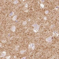
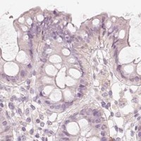
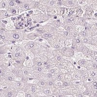
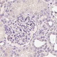
Comprehensive Human Protein Atlas IHC scoring per tissue (reliability: Uncertain — Presumed off target binding observed and disregarded. Pending external verification.). Rows are taken directly from the HPA tissue chart — click any row's HPA link to view the source.
| Tissue | Cell type | Level | Evidence | Source |
|---|---|---|---|---|
| Cerebellum | Cells in granular layer | High | Protein (IHC) | HPA → |
| Hippocampus | Neuronal cells | High | Protein (IHC) | HPA → |
| Lung | Alveolar cells | High | Protein (IHC) | HPA → |
| Cerebral cortex | Neuronal cells | Medium | Protein (IHC) | HPA → |
| Prostate | Glandular cells | Medium | Protein (IHC) | HPA → |
| Tissue | Cell type | Level | Evidence | Source |
|---|---|---|---|---|
| Adipose tissue | Adipocytes | Not detected | Protein (IHC) | HPA → |
| Appendix | Glandular cells | Not detected | Protein (IHC) | HPA → |
| Bone marrow | Hematopoietic cells | Not detected | Protein (IHC) | HPA → |
| Breast | Adipocytes | Not detected | Protein (IHC) | HPA → |
| Bronchus | Respiratory epithelial cells | Not detected | Protein (IHC) | HPA → |
Troubleshoot STEAP1 staining in paraffin sections using membrane and endosome localisation, the selected antibody image, and cautious interpretation of tissue patterns.
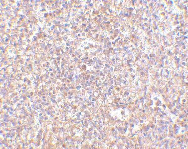
One anti-STEAP1 antibody has a human spleen IHC figure; its catalog lists human, mouse and rat reactivity, with no IF/ICC data (A07506 image caption; catalog: applications/reactivity).
A07506 is listed for IHC-P and for human, mouse and rat reactivity (catalog: A07506 applications/reactivity). Its own figure shows human spleen IHC at 2.5 μg/mL; no IF/ICC figure is provided (A07506 image caption; catalog: IF images).
Which to pick: Choose A07506 for paraffin-section tissue IHC: IHC-P is listed, and its own figure shows human spleen staining at 2.5 μg/mL (catalog: A07506 applications; A07506 image caption). The figure does not report a fixative, and no clone is specified (A07506 image caption; catalog: clone). For IF/ICC, no validated SKU is listed; for cross-species work, A07506 lists human, mouse and rat reactivity, but its reported IHC validation is in human samples only (catalog: A07506 applications/reactivity; datasheet: human IHC validation).