This website uses cookies to ensure you get the best experience on our website.


- Table of Contents
Plan chromogenic STING1 IHC in paraffin sections around cytoplasmic tissue staining (HPA tissue IHC) and an ER/perinuclear molecular expectation (UniProt). The guide highlights strong staining in lung macrophages (HPA tissue IHC) and the catalog antibody’s 2–5 μg/ml IHC range (datasheet A01871-3).

Expected localisation, validated protocols, controls and antibodies — the at-a-glance facts below, then the full design guide.
| Expected localisation | Cytoplasmic tissue staining (HPA tissue IHC); ER/perinuclear location (UniProt) | |
| Staining pattern | Cytoplasmic staining in respiratory, fallopian and lymphoid cells (HPA tissue IHC) | |
| Antigen retrieval | EDTA pH 8.0 HIER, heat-mediated (datasheet A01871-3) | |
| Positive control | Bronchus+4 more · see all | |
| Negative control | Adrenal gland+4 more · see all |
| Fixation | Keep fixation conditions consistent across sections. (standard IHC practice; not target-specific) | |
| Caveat | Lung macrophages may dominate mixed-cell staining (HPA tissue IHC) | |
| Regulation | cGAMP triggers ER-to-ERGIC trafficking (UniProt) | |
| Isoform / epitope | No annotated isoforms; residues 135–379 face cytoplasm (UniProt) |
The catalog antibody’s IHC-P protocol (datasheet A01871-3) is followed by published STING1 IHC methods for cholangiocarcinoma (PMC10468663) and papillary renal neoplasms (PMC11573472).
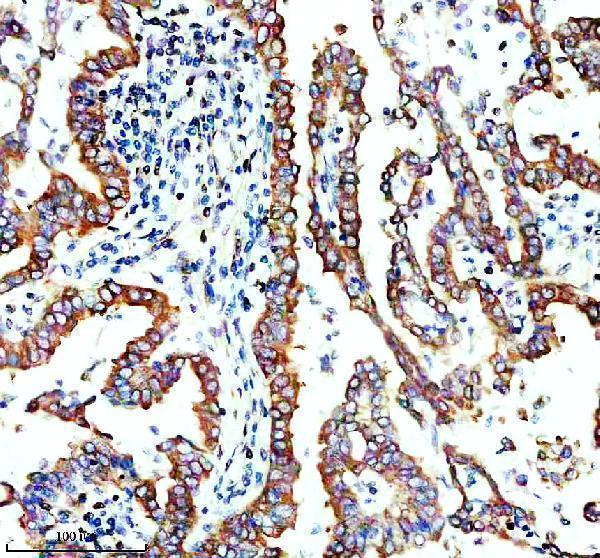
| Sample | Paraffin-embedded human lung cancer tissue; fixative not specified (datasheet A01871-3) |
| Fixation | Image fixative and duration unreported (datasheet A01871-3); verify before use. |
| Sectioning | 4–5 µm sections on charged slides (standard) |
| Deparaffinisation | Xylene, graded ethanol series to water (standard) |
| Antigen retrieval | Heat retrieval: EDTA pH 8.0 (datasheet A01871-3); 20 min, 95–100 °C (standard) |
| Peroxidase block | 3% H2O2, 10 min, room temperature (standard) |
| Blocking | 10% goat serum (datasheet A01871-3) |
| Primary antibody | Rabbit anti-STING1, 2-5 μg/ml (datasheet A01871-3) |
| Primary incubation | Overnight at 4 °C (datasheet A01871-3) |
| Detection | HRP-conjugated secondary, DAB chromogen (datasheet A01871-3) |
| Counterstain | Hematoxylin, blue, dehydrate and mount (standard) |
| Expected result | STING1-positive staining in basal cells of bronchus (HPA tissue IHC: High). HPA tissue profile: Cytoplasmic expression in several tissues including cells in respiratory tract, fallopian tube and cells in lymphoid tissues. No signal in the no-primary control. |
STING1 is a four-pass membrane protein with a large cytoplasmic C-terminal region; inactive STING1 resides at the ER and can move to the ER–Golgi intermediate compartment after activation (UniProt Q86WV6 topology and subcellular location). In tissue IHC, expect cytoplasmic staining in selected cells, especially lung macrophages and bronchial basal cells (HPA: High). HPA rates the tissue pattern Enhanced, with medium consistency between staining and RNA expression (HPA: tissue IHC).
| Cytoplasmic staining is strong in lung macrophages or bronchial basal cells, while neighboring cells differ in intensity (HPA: High in these cell types). | This fits the reported cell-specific tissue pattern. Judge signal in the named cells rather than treating every cell in a positive tissue as a positive control (HPA: tissue IHC). The cytoplasmic appearance also fits STING1 membrane topology and ER localization (UniProt Q86WV6). |
| The dominant signal is nuclear-only, with little cytoplasmic staining in otherwise recognizable positive cells. | This is unexpected for tissue IHC, which HPA describes as cytoplasmic (HPA: tissue IHC). It warrants an artefact check, but location alone cannot prove artefact: HPA reports nucleoplasmic ICC-IF staining with uncertain support (HPA: subcellular). |
| Strong staining appears in a cell population reported as not detected, such as skeletal-muscle myocytes or breast adipocytes (HPA: Not detected). | Assess possible cross-reactivity or endogenous detection activity before calling this STING1. These HPA observations apply to the specified cells, not every cell in those tissues (HPA: tissue IHC). Compare the suspicious signal with a known-positive cell population and a control lacking primary antibody (general IHC practice). |
| Broad, diffuse color covers tissue and spaces between cells, obscuring cellular boundaries. | That distribution does not resolve the cytoplasmic, cell-specific pattern reported by HPA (HPA: tissue IHC). Background from detection reagents or incomplete blocking is a general IHC possibility; the image alone does not identify its source. Review a control lacking primary antibody before scoring weak cells (general IHC practice). |
| No cytoplasmic signal is visible in lung macrophages or bronchial basal cells (HPA: High). | A negative result in these reported high-staining cells makes the run difficult to interpret (HPA: tissue IHC). Check that the relevant cells are present, then review antibody application and detection controls (general IHC practice). HPA tissue staining does not establish a STING1-specific fixation or retrieval failure. |
| Topology and compartment | STING1 has 4 transmembrane segments and a cytoplasmic region spanning residues 135–379 (UniProt Q86WV6 topology). Its ER and related membrane locations make a cytoplasmic tissue pattern plausible (UniProt Q86WV6 subcellular location; HPA: tissue IHC). The supplied record does not identify the catalog antibody's epitope. |
| Activation state | Inactive STING1 localizes to the ER; after cyclic-dinucleotide activation it moves toward the ER–Golgi intermediate compartment (UniProt Q86WV6 subcellular location). A shift in intracellular distribution can therefore be biologically plausible. Routine tissue staining alone does not establish activation state or prove that trafficking occurred. |
| Tissue and antibody evidence | HPA reports High staining in lung macrophages, bronchial and nasopharyngeal basal cells, fallopian-tube ciliated cells, and tonsil squamous epithelial cells (HPA: tissue IHC). Its tissue reliability is Enhanced, with medium staining–RNA consistency (HPA: tissue IHC). HPA038116 and HPA038534 have Enhanced IHC validation (HPA: antibodies); this does not validate every antibody. |
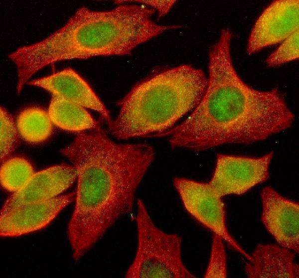
| IF/ICC: what localization is expected? | HPA reports mainly cytosolic ICC-IF localization and additionally supported vesicle staining (HPA: subcellular). It labels nucleoplasm, primary cilium and basal body locations uncertain (HPA: subcellular). Use those observations to interpret IF images; the IHC tissue pattern remains the reference for this paraffin-section guide. |
| Situation | Likely cause | Next action |
|---|---|---|
| Known-positive lung macrophages are unstained, and other expected positive cells are also blank (HPA: High in lung macrophages). | The cells may be absent from the examined field, or the primary-antibody or detection step may have failed (general IHC practice). The supplied sources do not identify a STING1-specific fixation effect. | Confirm cell identity in the section, inspect a positive control, and verify that the primary antibody and chromogenic detection steps were applied (general IHC practice). |
| A positive tissue shows weak staining, making cell boundaries and localization hard to judge. | Low signal or an unsuitable staining run can prevent a clear comparison with HPA's cytoplasmic pattern; HPA intensity is cell-specific (HPA: tissue IHC). The payload gives no catalog dilution or retrieval condition. | Compare the named positive cells with a run control; review the antibody's documented IHC conditions, including retrieval and dilution, before changing them (general IHC practice). Do not infer a STING1-specific retrieval requirement from HPA. |
| Color is widespread even where the primary antibody was omitted. | A control lacking primary antibody can reveal signal from detection chemistry or tissue-associated endogenous activity (general IHC practice). That signal cannot be attributed to STING1 from appearance alone. | Check the detection-system controls and apply the appropriate endogenous-activity blocking step for that system (general IHC practice). Reassess cellular localization after background is controlled. |
| Strong nuclear-only staining dominates an IHC section. | The pattern conflicts with HPA's reported cytoplasmic tissue staining (HPA: tissue IHC). HPA's uncertain nucleoplasmic ICC-IF observation does not by itself establish nuclear-only paraffin IHC as the expected result (HPA: subcellular). | Review the control lacking primary antibody and compare cytoplasmic staining in named positive cells; seek confirmation with an independently validated IHC antibody if the interpretation matters (general IHC practice; HPA: antibodies). |
| Skeletal-muscle myocytes or breast adipocytes stain as strongly as the selected positive cells (HPA: Not detected in those cell types). | Cross-reactivity or nonspecific detection is possible (general IHC practice). HPA's negative calls concern the listed cell types, so staining elsewhere in the same tissue is a separate observation (HPA: tissue IHC). | Verify cell identity and compare a control lacking primary antibody, then review antibody specificity before scoring the unexpected cells as STING1-positive (general IHC practice). |
| A section mixes high, medium and absent staining across distinct cell populations. | HPA reports cell-level differences: lung macrophages are High, appendix glandular cells Medium, and adrenal gland glandular cells Not detected (HPA: tissue IHC). Such variation alone does not mean the run failed. | Record intensity and cytoplasmic localization by cell type. Compare each cell population with its own HPA reference and interpret borderline staining alongside controls (HPA: tissue IHC; general IHC practice). |


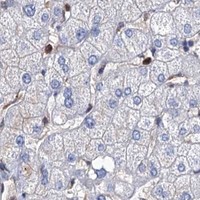
![HPA ICC-IF BJ [Human fibroblast] — STING1 (HPA038534)](https://images.proteinatlas.org/38534/1658_C1_2_blue_red_green.jpg)
![HPA ICC-IF BJ [Human fibroblast] — STING1 (HPA038534)](https://images.proteinatlas.org/38534/1658_C1_3_blue_red_green.jpg)
Comprehensive Human Protein Atlas IHC scoring per tissue (reliability: Enhanced — Medium consistency between antibody staining and RNA expression data.). Rows are taken directly from the HPA tissue chart — click any row's HPA link to view the source.
| Tissue | Cell type | Level | Evidence | Source |
|---|---|---|---|---|
| Bronchus | Basal cells | High | Protein (IHC) | HPA → |
| Fallopian tube | Ciliated cells (cell body) | High | Protein (IHC) | HPA → |
| Lung | Macrophages | High | Protein (IHC) | HPA → |
| Nasopharynx | Basal cells | High | Protein (IHC) | HPA → |
| Tonsil | Squamous epithelial cells | High | Protein (IHC) | HPA → |
| Tissue | Cell type | Level | Evidence | Source |
|---|---|---|---|---|
| Adrenal gland | Glandular cells | Not detected | Protein (IHC) | HPA → |
| Breast | Adipocytes | Not detected | Protein (IHC) | HPA → |
| Cerebellum | Cells in granular layer | Not detected | Protein (IHC) | HPA → |
| Hippocampus | Glial cells | Not detected | Protein (IHC) | HPA → |
| Liver | Cholangiocytes | Not detected | Protein (IHC) | HPA → |
Troubleshoot STING1 staining in paraffin sections by checking retrieval, cell type, subcellular pattern and controls before interpreting chromogenic signal.
A01871-3 has IHC data from human lung cancer paraffin sections and IF/ICC data from SIHA cells; the catalog lists human, mouse and rat reactivity (A01871-3 image captions; catalog reactivity).
A01871-3 was demonstrated by IHC on a human lung cancer paraffin section (A01871-3 IHC caption). The same SKU was demonstrated by IF/ICC in SIHA cells; its listed applications include IHC, IF and ICC (A01871-3 IF caption; catalog applications).
Which to pick: Choose A01871-3 for tissue IHC because its own caption documents staining of a paraffin section with 2 μg/ml primary antibody after EDTA retrieval (A01871-3 IHC caption). Choose the same SKU for IF/ICC because its IF caption documents SIHA cell staining at 5 μg/ml (A01871-3 IF caption). For mouse or rat samples, the catalog lists reactivity, but the supplied IHC and IF examples are human; the tissue fixative is unreported (catalog reactivity; A01871-3 image captions).