STK24 / Serine/threonine-protein kinase 24 · IHC design guide

Design Immunohistochemistry for STK24

Plan STK24 IHC-P around widespread cytoplasmic tissue staining and strong signal in glandular cells (HPA tissue IHC). This guide covers fixation consistency, the catalog antibody’s documented human IHC-P use, and how isoforms may affect epitope interpretation (datasheet A04112-3; UniProt).

Evidence assembled Oct 2026 · For research use; verify linked source records and product datasheet before use
Immunohistochemistry protocol sheet for STK24 (IHC for STK24): expected localisation Cytoplasmic staining in tissue (HPA tissue IHC), antibody A04112-3, validated IHC image, and IHC protocol steps
Printable STK24 IHC protocol sheet — expected localisation Cytoplasmic staining in tissue (HPA tissue IHC), antibody A04112-3, controls and protocol steps. Open the full STK24 IHC guide →

STK24 Immunohistochemistry Experimental Design Guide

Expected localisation, validated protocols, controls and antibodies — the at-a-glance facts below, then the full design guide.

Must know before staining
Expected localisation Cytoplasmic staining in tissue (HPA tissue IHC)
Staining pattern Widespread cytoplasmic signal, high in glandular cells (HPA tissue IHC)
Antigen retrieval EDTA pH 8.0 HIER, heat-mediated (datasheet A04112-3)
Positive control ⓘ Appendix+4 more · see all
Negative control ⓘ None in HPA (detected in all 45 tissues); use no-primary + isotype controls
Important caveats
Reasons your staining may differ from the expected pattern.
Fixation Matched tissue-IHC evidence does not establish the fixation claim. Validate the specimen-specific method before use. (selected-SKU IHC image A04112-3)
Caveat Caudate glial cells show low signal in brain tissue (HPA tissue IHC)
Regulation Isoform B is brain-enriched (UniProt)
Isoform / epitope Two isoforms and 1–325/326–443 chains; epitope location matters (UniProt)
Section 1

Recommended STK24 IHC & IF Protocols

The catalog antibody uses EDTA pH 8.0 heat retrieval (datasheet A04112-3). One published mouse kidney IHC protocol provides additional conditions (PMC7863938).

Recommended immunohistochemistry (IHC-P) protocol parameters
SampleParaffin-embedded mouse brain tissue; fixative not specified (datasheet A04112-3)
FixationImage fixative and duration unreported (datasheet A04112-3); verify before use.
Sectioning4–5 µm sections on charged slides (standard)
DeparaffinisationXylene, graded ethanol series to water (standard)
Antigen retrievalHeat retrieval: EDTA pH 8.0 (datasheet A04112-3); 20 min, 95–100 °C (standard)
Peroxidase block3% H2O2, 10 min, room temperature (standard)
Blocking10% goat serum (datasheet A04112-3)
Primary antibodyRabbit anti-STK24, 2-5 μg/ml (datasheet A04112-3)
Primary incubationOvernight at 4 °C (datasheet A04112-3)
DetectionHRP-conjugated secondary, DAB chromogen (datasheet A04112-3)
CounterstainHematoxylin, blue, dehydrate and mount (standard)
Expected resultSTK24-positive staining in glandular cells of appendix (HPA tissue IHC: High). HPA tissue profile: Ubiquitous cytoplasmic expression. No signal in the no-primary control.
💡Decision noteStart with EDTA pH 8.0 heat retrieval (datasheet A04112-3). The published mouse kidney protocol used Tris in PBS at pH 8.2 (PMC7863938).
Section 2

What Is the Expected STK24 Staining Pattern?

In paraffin-section IHC, expect predominantly cytoplasmic STK24 staining across many tissues (HPA: ubiquitous cytoplasmic expression; Enhanced reliability). Glandular cells in colon, duodenum and appendix are useful high-staining examples (HPA: High in glandular cells). STK24 has no transmembrane segment; UniProt also lists nuclear and membrane localisation, with nuclear translocation of a truncated form (UniProt Q9Y6E0: topology and subcellular location).

What am I looking at on my slide?
Clear cytoplasmic stain in glandular cells, with tissue structure preserved.This fits the reported IHC pattern (HPA: ubiquitous cytoplasmic expression; High in colon, duodenum and appendix glandular cells). Compare signal within the same section before assigning intensity: cell types can differ (HPA: tissue IHC levels).
An isolated membrane rim or extracellular deposit dominates, with little cytoplasmic signal.Treat this as a pattern mismatch requiring review: STK24 lacks a transmembrane segment (UniProt Q9Y6E0: topology), while HPA describes cytoplasmic tissue IHC. Membrane localisation is listed by UniProt, so a membrane component alone does not establish artefact.
Stromal or muscle staining dominates while nearby glandular cells are weak.Consider cross-reactivity or endogenous chromogen activity and inspect controls (general IHC practice). HPA reports High glandular staining in several tissues but Low staining in ovarian stroma, fibroblasts and muscle cells; Low does not mean absent (HPA: tissue IHC).
A uniform haze covers cells and surrounding tissue.The haze obscures the reported cellular pattern (HPA: ubiquitous cytoplasmic expression). Review blocking, antibody concentration, washes and detection controls as general IHC practice; the supplied HPA and UniProt records do not identify its cause in a given slide.
No cellular stain appears in a colon glandular-cell positive control.An assay failure is plausible because colon glandular cells are reported High (HPA: colon tissue IHC). Check section quality, retrieval, primary-antibody application and chromogen development as general IHC practice before interpreting another sample as STK24 negative.
💡Expected STK24 appearanceCall a slide positive when identifiable glandular cells show clear, predominantly cytoplasmic staining consistent with a High HPA reference; isolated extracellular deposits or uniform haze are suspect, and Low-staining cell types are not negative controls (HPA: tissue IHC; general IHC interpretation).
How each factor affects the staining
IHC evidenceHPA rates the tissue profile Enhanced, citing high consistency between antibody staining and RNA expression; HPA026435 and HPA026502 each have Enhanced IHC validation (HPA: tissue profile and antibody records). This supports the reference pattern, not every individual slide.
Tissue and cell selectionAppendix, colon, duodenum, endometrium, fallopian tube and gallbladder glandular cells are reported High (HPA: tissue IHC). Caudate glial cells, cardiomyocytes, myocytes and fibroblasts are reported Low, not negative (HPA: tissue IHC).
CompartmentUse cytoplasmic staining as the principal paraffin IHC comparison (HPA: tissue IHC). UniProt also lists nucleus and membrane and specifies nuclear translocation of truncated MST3/N (UniProt Q9Y6E0: subcellular location); nuclear signal needs context.
IsoformsIsoform A is ubiquitous; isoform B is expressed in brain, particularly hippocampus and cerebral cortex (UniProt Q9Y6E0: tissue specificity). This does not establish which isoform an IHC antibody detects or predict its staining intensity there.
Protein processing and antibody epitopeUniProt lists full-length residues 1–443 and processed chains 1–325 and 326–443 (UniProt Q9Y6E0: processing). Without an epitope location in the supplied records, staining cannot be assigned to a particular chain or processing state.
Detection backgroundEndogenous enzyme activity can produce chromogenic signal, depending on the detection chemistry (general IHC practice). Check a primary-omission control when background is suspected; the supplied STK24 records do not identify a tissue-specific endogenous-activity problem.
Why is my staining missing, weak or wrong?
SituationLikely causeNext action
A known High glandular control has no stain.The assay may have missed an expected signal (HPA: High in colon glandular cells); the supplied records do not identify which workflow step failed.Confirm tissue identity and assay controls, then review retrieval, primary-antibody application and chromogen development (general IHC practice).
All tissue compartments are diffusely brown.Background may reflect nonspecific binding or detection activity (general IHC practice); diffuse haze does not match HPA's cellular cytoplasmic profile (HPA: tissue IHC).Compare a primary-omission control; review blocking, wash steps, antibody concentration and detection chemistry (general IHC practice).
Only a sharp surface rim or extracellular material stains.This differs from the reported cytoplasmic IHC pattern (HPA: tissue IHC). STK24 has no transmembrane segment, although UniProt lists membrane localisation (UniProt Q9Y6E0).Check morphology and control staining, then compare another section before assigning the signal to STK24 (general IHC practice).
Low-reference stromal or muscle cells stain more strongly than expected.Cross-reactivity or endogenous detection activity is possible (general IHC practice); HPA calls these cell types Low, not negative (HPA: tissue IHC).Compare adjacent cell types and the primary-omission control; assess whether cellular staining persists with the IHC-validated antibody (HPA: antibody IHC validation; general IHC practice).
Nuclear signal accompanies cytoplasmic staining in IHC.UniProt lists nuclear localisation and nuclear translocation of truncated MST3/N (UniProt Q9Y6E0: subcellular location); the HPA tissue IHC summary remains cytoplasmic.Record each compartment separately and compare with the cytoplasmic reference; do not infer cleavage from nuclear staining alone (HPA: tissue IHC; UniProt Q9Y6E0: processing).
IF/ICC shows nucleoli and cytosol—is that expected?Yes. HPA reports supported nucleolar and cytosolic localisation in ICC-IF (HPA: subcellular ICC-IF); that evidence comes from a different application than paraffin IHC.Interpret the IF/ICC result against its own supported compartment pattern; use the cytoplasmic tissue profile for the IHC call (HPA: subcellular ICC-IF and tissue IHC).

Sample controls for STK24 IHC & IF

🧪Run colon first and score staining in glandular cells (HPA: High in colon glandular cells). HPA detects STK24 in all 45 scored tissues, so use no-primary and isotype controls for the negative comparison; weakly stained neighboring cells can provide internal contrast but are not a validated negative cell population (HPA: no negative tissue rows).
Positive control tissue: Appendix (Glandular cells, HPA High)
Negative control tissue: None in HPA: STK24 is detected in all 45 scored tissues. Use a no-primary (secondary-only) and an isotype control instead.
ICC-IF cell lines (HPA subcellular resource): HPA ICC-IF images show STK24 in A-431, U-251MG, U2OS, RPTEC/TERT1, hTERT-RPE1 (serum starved), with annotated localisation: Nucleoli (supported), Cytosol (supported) (HPA subcellular).
Technical controls: Include a secondary-only slide, a rabbit IgG isotype control matched to the primary antibody, and STK24-knockout material as a biological negative (caption: rabbit primary antibody). For chromogenic detection in colon, quench endogenous peroxidase and check background before interpreting DAB staining (HPA: colon glandular cells High; caption: peroxidase secondary and DAB).
⚠️Feasibility: Paraffin-section IHC has a demonstrated starting condition of heat retrieval in EDTA at pH 8.0, but the selected A04112-3 caption does not report the fixative, and no target-specific fixation window or fixation effect is reported (selected-SKU tissue-IHC caption). ICC-IF images support examining nucleolar and cytosolic localization, but the supplied evidence does not establish whether IF or frozen sections are easier than paraffin IHC (HPA: supported nucleoli and cytosol; HPA: ICC-IF images). In colon, glandular morphology helps locate the expected positive cells, while endogenous peroxidase can confound chromogenic signal unless controlled (HPA: colon glandular cells High; caption: peroxidase secondary and DAB).

HPA tissue IHC evidence for STK24

Comprehensive Human Protein Atlas IHC scoring per tissue (reliability: Enhanced — High consistency between antibody staining and RNA expression data.). Rows are taken directly from the HPA tissue chart — click any row's HPA link to view the source.

Positive expression · recommended positive controls

TissueCell typeLevelEvidenceSource
Appendix Glandular cells High Protein (IHC) HPA →
Colon Glandular cells High Protein (IHC) HPA →
Duodenum Glandular cells High Protein (IHC) HPA →
Endometrium Glandular cells High Protein (IHC) HPA →
Esophagus Squamous epithelial cells High Protein (IHC) HPA →

Undetected expression · recommended negative controls

TissueCell typeLevelEvidenceSource
None in HPA: STK24 is detected in all 45 scored tissues. Use a no-primary (secondary-only) and an isotype control instead.
Section 3

Advanced STK24 IHC Tips

Troubleshoot STK24 staining in paraffin sections by checking retrieval, compartment, background and scoring against the reported tissue evidence.

Which retrieval conditions should I use when STK24 staining is weak?
Start with heat mediated retrieval in EDTA at pH 8.0 for paraffin sections (datasheet A04112-3). The reported mouse brain staining used that buffer before incubation with 2 μg/ml primary antibody overnight at 4°C (datasheet A04112-3). If staining remains weak, check retrieval heating and section adhesion, then compare a carefully controlled alternative buffer on adjacent sections as a fallback (standard IHC practice). Keep antibody concentration, detection and development consistent across that comparison, and judge improvement by cellular staining alongside background (standard IHC practice). The caption does not specify a retrieval duration, so establish and record one locally (datasheet A04112-3; standard IHC practice).
How should I assess whether fixation is reducing STK24 staining?
The selected paraffin section caption does not state a fixative, so target specific STK24 fixation sensitivity is unknown (datasheet A04112-3). Do not infer a preferred fixative or fixation duration from the tissue expression profile or STK24 modifications (HPA tissue IHC; UniProt Q9Y6E0). For troubleshooting, compare sections with documented fixation histories while holding EDTA retrieval at pH 8.0, antibody concentration and detection constant (datasheet A04112-3; standard IHC practice). Include a consistently staining reference section in each run and assess cellular signal against morphology and background (standard IHC practice). Record fixation and processing differences before attributing a weak result to the antigen (standard IHC practice).
Should nuclear staining count as STK24 positive in IHC?
Assess cytoplasmic staining first, because tissue IHC reports ubiquitous cytoplasmic expression with enhanced reliability (HPA tissue IHC). Nuclear signal can be biologically plausible: STK24 is annotated in the cytoplasm and nucleus, and its truncated MST3/N form translocates to the nucleus (UniProt Q9Y6E0). Supported nucleolar and cytosolic localisation comes from ICC/IF evidence, so use it to guide interpretation without assuming identical patterns in paraffin sections (HPA subcellular; standard IHC practice). Evaluate nuclear and cytoplasmic staining separately in intact cells, using consistent thresholds and a counterstain to define compartments (standard IHC practice). Diffuse staining across damaged areas needs independent scrutiny before scoring (standard IHC practice).
Could isoforms or cleavage explain different STK24 staining patterns?
STK24 has isoforms A and B; A is ubiquitous, whereas B is reported in brain with high expression in hippocampus and cerebral cortex (UniProt Q9Y6E0). The protein also has annotated 1–325 and 326–443 processed chains, and the truncated MST3/N form can enter the nucleus (UniProt Q9Y6E0). Check the catalog antibody’s stated immunogen or epitope before interpreting an altered compartment or tissue pattern as an isoform difference (standard IHC practice). If that information is unavailable, report the observed staining as STK24 immunoreactivity without assigning a specific isoform or processed form (standard IHC practice). Compare matched sections and cellular morphology before drawing that conclusion (standard IHC practice).
How can I check an STK24 IHC pattern with multiplex IF?
For the separate IF/ICC workflow, pair STK24 with a marker identifying the cell population being examined; glandular cells in several tissues show high tissue IHC staining (HPA tissue IHC; standard IF practice). Choose spectrally separated fluorophores and place the weaker signal in a channel with less tissue autofluorescence, using single stain controls to assess bleed through (standard IF practice). STK24 has no transmembrane segment and is annotated in cytoplasm and nucleus, with supported cytosolic and nucleolar ICC/IF localisation (UniProt Q9Y6E0; HPA subcellular). Permeabilise sufficiently for access to intracellular epitopes, adjusting conditions to the antibody’s epitope and preserving morphology (standard IF practice). Interpret colocalisation at the cell and compartment level (standard IF practice).
What should I change when DAB background obscures STK24?
The reported paraffin section method used 10% goat serum block, 2 μg/ml primary antibody overnight at 4°C, and a peroxidase conjugated secondary for 30 minutes at 37°C (datasheet A04112-3). It developed signal with DAB, so check whether diffuse brown color persists in a primary omitted control (datasheet A04112-3; standard IHC practice). Include an endogenous peroxidase block and review washing, secondary reagent binding and DAB development as general chromogenic troubleshooting steps (standard IHC practice). Compare background and cellular signal on the same run before changing primary antibody conditions (standard IHC practice). Avoid scoring pigment or tissue deposits as cellular staining (standard IHC practice).
How should I quantify STK24 staining across sections? ⚠ ANSWER MARKED FOR VERIFICATION
Define the cell population and compartment before scoring, because tissue IHC describes widespread cytoplasmic STK24 while other evidence also supports nuclear localisation (HPA tissue IHC; UniProt Q9Y6E0). Use a consistent H-score for intensity and positive fraction, or report % positive cells when intensity grading is unreliable (standard IHC practice). For spatial questions, count positive cells per mm² of viable tissue and state the compartment scored (standard IHC practice). Normalise to the relevant total cell count or viable tissue area, and keep thresholds, imaging settings and DAB development comparable across slides (standard IHC practice). Exclude folds, edges and necrotic areas by a predefined rule (standard IHC practice).
How can I distinguish true STK24 signal from staining artefact?
A convincing result is cellular and reproducible in intact tissue, with cytoplasmic staining consistent with the tissue IHC profile (HPA tissue IHC; standard IHC practice). High staining in glandular cells of the appendix, colon and duodenum provides context, while glial cells in caudate are listed as low (HPA tissue IHC). Nuclear signal requires compartment aware assessment because truncated MST3/N can translocate to the nucleus; a pattern alone does not identify that form (UniProt Q9Y6E0; standard IHC practice). Treat section edges, necrosis and signal in a primary omitted control as possible artefacts, including endogenous enzyme activity in chromogenic detection (standard IHC practice). Confirm disputed patterns on adjacent sections with morphology preserved (standard IHC practice).
Boster reagents

Best STK24 / Serine/threonine-protein kinase 24 IHC Antibodies

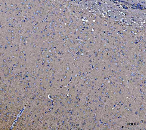
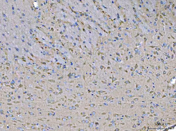
A04112-3 has IHC images of paraffin-embedded human, mouse and rat brain sections (catalog image captions). No IF/ICC images or application are listed (catalog IF images; catalog applications).

Real IHC data IHC analysis of MST3/STK24 using anti-MST3/STK24 antibody (A04112-3). MST3/STK24 was detected in a paraffin-embedded section of mouse brain tissue. Heat mediated antigen retrieval was performed in EDTA buffer (pH 8.0, epitope retrieval solution). The tissue section was blocked with 10% goat serum. The tissue section was then incubated with 2 μg/ml rabbit anti-MST3/STK24 Antibody (A04112-3) overnight at 4°C. Peroxidase Conjugated Goat Anti-rabbit IgG was used as secondary antibody and incubated for 30 minutes at 37°C. The tissue section was developed using HRP Conjugated Rabbit IgG Super Vision Assay Kit (Catalog # SV0002) with DAB as the chromogen.
Anti-MST3/STK24 Antibody ®
Cat # A04112-3

A04112-3 is the sole card; it lists IHC and human, mouse and rat reactivity (catalog applications; catalog reactivity). Its IHC captions document paraffin-embedded brain sections from all three species using 2 μg/ml primary antibody (A04112-3 image captions).

Which to pick: Choose A04112-3 for chromogenic tissue IHC: its captions document EDTA retrieval at pH 8.0, goat-serum blocking and HRP/DAB detection in paraffin sections (A04112-3 image captions). It is also the cross-species choice, with IHC brain images from human, mouse and rat (A04112-3 image captions). No IF/ICC application or clone identity is reported, so an IF/ICC recommendation is unsupported; the IHC captions do not report a fixative (catalog applications; catalog clone; A04112-3 image captions).

Each figure is that product's own IHC / IF validation image from its datasheet.

References

  1. UniProt Consortium. UniProt entry Q9Y6E0 (STK24_HUMAN, Serine/threonine-protein kinase 24).
  2. Human Protein Atlas. STK24 tissue IHC expression (reliability: Enhanced).
  3. Human Protein Atlas. STK24 subcellular location (ICC-IF): Localized to the nucleoli and cytosol..
  4. Human Protein Atlas. STK24 antibody validation summary (2 antibodies).
  5. Knockdown of serine/threonine-protein kinase 24 promotes tumorigenesis and myeloid-derived suppressor cell expansion in an orthotopic immunocompetent gastric cancer animal model. Journal of Cancer 2020 — PMC6930401.
  6. Stk24 deficiency causes disrupted hippocampal neurogenesis and anxiety-like behavior in mice. Communications biology 2025 — PMC12032016.
  7. MST3 Involvement in Na(+) and K(+) Homeostasis with Increasing Dietary Potassium Intake. International journal of molecular sciences 2021 — PMC7863938.
  8. PubMed PMID:9353338 — UniProt-cited evidence.
  9. PubMed PMID:10644707 — UniProt-cited evidence.
  10. PubMed PMID:15057823 — UniProt-cited evidence.