This website uses cookies to ensure you get the best experience on our website.


- Table of Contents
Plan STK26 paraffin IHC around cytoplasmic staining in selected tissues (HPA tissue IHC). Use the IHC-validated antibody’s dilution and compare staining with controls because tissue staining has low consistency with RNA expression (HPA tissue IHC).

Expected localisation, validated protocols, controls and antibodies — the at-a-glance facts below, then the full design guide.
| Expected localisation | Cytoplasmic tissue staining (HPA tissue IHC); Golgi location (UniProt) | |
| Staining pattern | Cytoplasmic staining in glandular cells and myocytes (HPA tissue IHC) | |
| Antigen retrieval | EDTA pH 8.0 HIER, heat-mediated (datasheet A09552-2) | |
| Positive control | Gallbladder+4 more · see all | |
| Negative control | Adipose tissue+4 more · see all |
| Fixation | Keep fixation consistent across sections. (standard IHC practice; not target-specific) | |
| Caveat | Staining has low consistency with RNA expression (HPA tissue IHC) | |
| Regulation | Serum stimulation shifts Golgi signal to puncta (UniProt) | |
| Isoform / epitope | 3 isoforms; epitope coverage needs checking (UniProt) |
The catalog antibody uses EDTA pH 8.0 heat retrieval (datasheet A09552-2). One published choriocarcinoma IHC protocol provides a comparison (PMC7683511).
| Sample | Paraffin-embedded human ovarican cancer tissue; fixative not specified (datasheet A09552-2) |
| Fixation | Image fixative and duration unreported (datasheet A09552-2); verify before use. |
| Sectioning | 4–5 µm sections on charged slides (standard) |
| Deparaffinisation | Xylene, graded ethanol series to water (standard) |
| Antigen retrieval | Heat retrieval: EDTA pH 8.0 (datasheet A09552-2); 20 min, 95–100 °C (standard) |
| Peroxidase block | 3% H2O2, 10 min, room temperature (standard) |
| Blocking | 10% goat serum (datasheet A09552-2) |
| Primary antibody | Rabbit anti-STK26, 2-5 μg/ml (datasheet A09552-2) |
| Primary incubation | Overnight at 4 °C (datasheet A09552-2) |
| Detection | HRP-conjugated secondary, DAB chromogen (datasheet A09552-2) |
| Counterstain | Hematoxylin, blue, dehydrate and mount (standard) |
| Expected result | STK26-positive staining in glandular cells of gallbladder (HPA tissue IHC: High). HPA tissue profile: Cytoplasmic expression in a few tissues. No signal in the no-primary control. |
STK26 is a cytoplasmic, Golgi-associated kinase without a transmembrane segment (UniProt Q9P289 topology and subcellular location). In paraffin-section IHC, expect cytoplasmic staining in gallbladder and small-intestinal glandular cells and skeletal-muscle myocytes (HPA: High). Treat these as reference patterns, not universal positives: HPA rates the tissue IHC antibody Approved but reports low agreement between staining and RNA expression, pending external verification (HPA: tissue IHC reliability).
| Cytoplasmic staining in gallbladder or small-intestinal glandular cells, or skeletal-muscle myocytes. | This matches the reported High cell-specific IHC pattern (HPA: tissue IHC). Assess the named cells against neighboring structures and background on the same section. The observed compartment also agrees with the cytoplasmic location (UniProt Q9P289 subcellular location). |
| Strong nuclear staining dominates the IHC slide, with little cytoplasmic signal. | This departs from the reported tissue IHC profile of cytoplasmic expression (HPA: tissue IHC). Check morphology and staining controls before calling it STK26. HPA ICC-IF lists nucleoplasm as an uncertain main location, so that separate observation does not by itself validate a nuclear IHC pattern (HPA: subcellular ICC-IF). |
| Prominent staining appears in cells reported as undetected, such as bone-marrow hematopoietic cells or adipocytes. | Consider off-target binding or endogenous detection activity, then compare controls and cell identity; one unexpected section cannot establish cross-reactivity. HPA reports these cells as Not detected by IHC, yet lists bone marrow among RNA-enhanced tissues, a reminder that the protein and RNA observations disagree (HPA: tissue IHC and RNA specificity). |
| Diffuse color covers multiple compartments or obscures cell boundaries. | Treat an indistinct, section-wide signal as background until controls and morphology support a cellular pattern. The useful reference is cytoplasmic staining in a few tissues (HPA: tissue IHC), with cytoplasm and Golgi localization described independently (UniProt Q9P289 subcellular location). General IHC practice is to inspect detection-only controls and background before scoring. |
| No interpretable signal appears in gallbladder glandular cells or skeletal-muscle myocytes. | A blank known-positive comparator suggests a failed or insensitive run, but the HPA reference is an observed High pattern rather than a guarantee for every specimen (HPA: tissue IHC). Review tissue preservation, retrieval, antibody application, and detection as general IHC checks; avoid scoring other samples as truly negative until the comparator works. |
| Cell type and reference tissue | Choose the named High IHC cell populations when judging whether a run can reveal STK26; adrenal and colon glandular cells, cerebellar granular-layer cells, and lung macrophages are reported Medium (HPA: tissue IHC). A negative result in a different cell population carries less weight. |
| Cellular compartment and cell state | Cytoplasm and Golgi are annotated locations (UniProt Q9P289 subcellular location). UniProt describes Golgi colocalization with RIPOR1 in serum-starved cells and relocation to cytoplasmic puncta after serum stimulation (UniProt Q9P289, PubMed:27807006). This cell-state observation is not an IHC tissue scoring rule. |
| Antibody evidence and RNA discordance | The listed HPA antibody, HPA059921, is IHC Approved, while the tissue profile has low agreement with RNA and awaits external verification (HPA: antibody validation and tissue IHC reliability). Bone marrow illustrates the limit: RNA is tissue enhanced, but hematopoietic-cell IHC is Not detected (HPA: tissue IHC and RNA specificity). |
| IF/ICC Q&A: should nuclear fluorescence be expected? | HPA reports nucleoplasm as an uncertain main ICC-IF location, with supported Golgi and cytosol locations; its listed antibody is ICC Uncertain (HPA: subcellular ICC-IF and antibody validation). Interpret nuclear fluorescence with that uncertainty and assess localization on the separate IF/ICC guide page. |
| Situation | Likely cause | Next action |
|---|---|---|
| Known-positive comparator is blank. | General IHC possibilities include insufficient retrieval, missed primary antibody, or failed detection; none is a demonstrated STK26-specific fixation effect. | Repeat with a named High cell population and working assay controls (HPA: tissue IHC); review retrieval and detection records as general IHC checks before interpreting study negatives. |
| Nuclear signal exceeds cytoplasmic signal. | Dominant nuclear IHC conflicts with the reported cytoplasmic tissue pattern (HPA: tissue IHC), although HPA separately marks nucleoplasmic ICC-IF localization uncertain (HPA: subcellular ICC-IF). | Verify cell boundaries and counterstain; compare an appropriate negative control and a positive tissue before assigning nuclear STK26. Keep the IHC and ICC-IF observations distinct. |
| A reported negative cell population stains strongly. | Possible nonspecific antibody binding or endogenous detection activity; HPA lists adipocytes and bone-marrow hematopoietic cells as Not detected by IHC (HPA: tissue IHC). | Confirm the cell type, inspect primary-omission and detection controls, and compare a named positive population. Treat a disagreement as unresolved until the signal is independently supported. |
| Diffuse chromogen makes localization unreadable. | General IHC causes include excess detection background or inadequate blocking; a diffuse field cannot be matched confidently to HPA's cytoplasmic profile (HPA: tissue IHC). | Inspect a detection-only control, review blocking and reagent exposure, and score only cells with distinguishable morphology and compartmental signal. |
| Only weak signal appears in a candidate tissue. | Signal may reflect the sampled cell population: HPA reports Medium in lung macrophages and Not detected in heart-muscle cardiomyocytes (HPA: tissue IHC). | Identify the stained cells before comparing intensity; include a reported High comparator and use the same scoring criteria across sections (HPA: tissue IHC). |
| RNA data suggest expression where IHC is negative. | HPA reports bone marrow and epididymis as RNA-enhanced tissues, yet the specified hematopoietic and epididymal glandular cells are Not detected by IHC; overall RNA–staining agreement is low (HPA: tissue IHC and RNA specificity). | Report the protein-level observation and the discordance separately. Check an IHC-positive comparator and seek independent validation before calling the negative result a technical failure. |
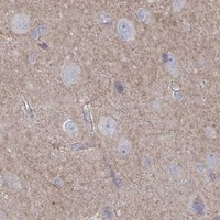
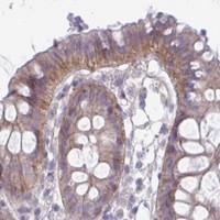
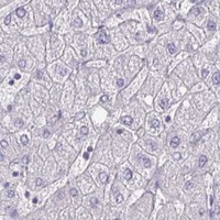
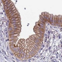


Comprehensive Human Protein Atlas IHC scoring per tissue (reliability: Approved — Low consistency between antibody staining and RNA expression data. Pending external verification.). Rows are taken directly from the HPA tissue chart — click any row's HPA link to view the source.
| Tissue | Cell type | Level | Evidence | Source |
|---|---|---|---|---|
| Gallbladder | Glandular cells | High | Protein (IHC) | HPA → |
| Skeletal muscle | Myocytes | High | Protein (IHC) | HPA → |
| Small intestine | Glandular cells | High | Protein (IHC) | HPA → |
| Adrenal gland | Glandular cells | Medium | Protein (IHC) | HPA → |
| Cerebellum | Cells in granular layer | Medium | Protein (IHC) | HPA → |
| Tissue | Cell type | Level | Evidence | Source |
|---|---|---|---|---|
| Adipose tissue | Adipocytes | Not detected | Protein (IHC) | HPA → |
| Bone marrow | Hematopoietic cells | Not detected | Protein (IHC) | HPA → |
| Breast | Adipocytes | Not detected | Protein (IHC) | HPA → |
| Caudate | Glial cells | Not detected | Protein (IHC) | HPA → |
| Endometrium | Cells in endometrial stroma | Not detected | Protein (IHC) | HPA → |
Troubleshoot STK26 staining in paraffin sections by checking retrieval, cell type, compartment and controls before comparing staining intensity (datasheet A09552-2; HPA tissue IHC; UniProt Q9P289).
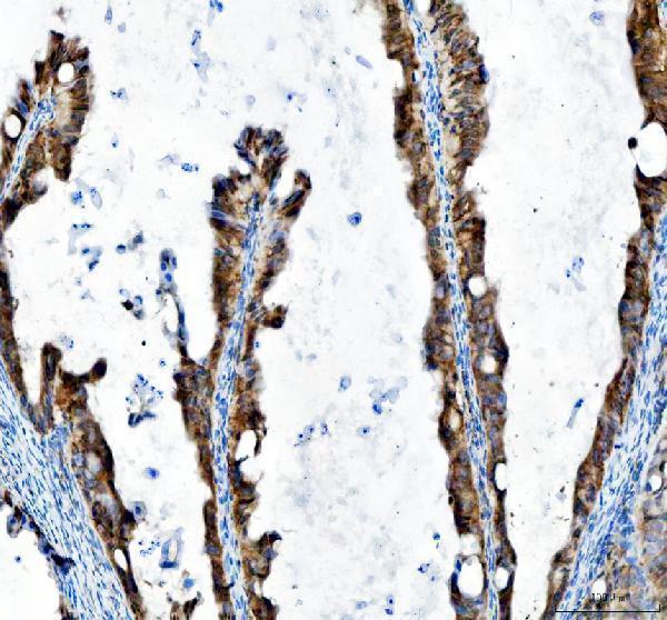
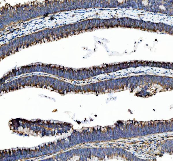
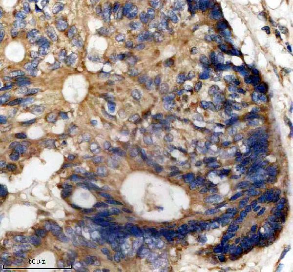
Two anti-STK26 antibodies have IHC images from human paraffin sections (A09552-2 and M09552 image captions). Catalog reactivity covers human and rat for A09552-2, and human, mouse, and rat for M09552 (catalog reactivity).
A09552-2 has IHC images from human paraffin-embedded ovarian and colon cancer tissue (A09552-2 image captions). M09552 has an IHC image from human paraffin-embedded colon cancer tissue (M09552 image caption).
Which to pick: For tissue IHC, choose A09552-2 for the ovarian or colon examples, or monoclonal M09552 for the colon example (catalog clone; each SKU’s image captions). Neither SKU lists IF/ICC validation or an IF image, so there is no evidence-based IF/ICC pick (catalog applications and image lists). For mouse tissue, M09552 is the listed option; both list human and rat reactivity, but their IHC captions show human paraffin sections only, and neither caption reports the fixative (catalog reactivity; each SKU’s image captions).