This website uses cookies to ensure you get the best experience on our website.


- Table of Contents
Plan STK36 staining in paraffin sections around the widespread cytoplasmic pattern reported in tissue IHC (HPA tissue IHC). This guide covers controls, staining interpretation and the uncertainty raised by low agreement between antibody staining and RNA expression (HPA tissue IHC).

Expected localisation, validated protocols, controls and antibodies — the at-a-glance facts below, then the full design guide.
| Expected localisation | Cytoplasmic tissue staining (HPA tissue IHC) | |
| Staining pattern | Widespread cytoplasmic staining across cell types (HPA tissue IHC) | |
| Antigen retrieval | Tris-EDTA pH 9.0 HIER, 95–98 °C, 20 min (rule: nuclear antigen) | |
| Positive control | Adrenal gland+4 more · see all | |
| Negative control | None in HPA (detected in all 45 tissues); use no-primary + isotype controls |
| Fixation | Keep fixation consistent across sections. (standard IHC practice; not target-specific) | |
| Caveat | Staining has low consistency with RNA data (HPA tissue IHC) | |
| Regulation | Expression regulation unreported (UniProt) | |
| Isoform / epitope | 2 isoforms; epitope coverage is unknown (UniProt) |
The catalog antibody’s IHC-P protocol is accompanied by one published prostate tissue microarray protocol (PMC10771505).
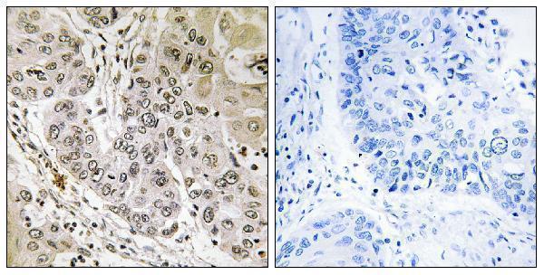
| Sample | Paraffin-embedded human lung carcinoma tissue; fixative not specified (datasheet A09526-1) |
| Fixation | Image fixative and duration unreported (datasheet A09526-1); verify before use. |
| Sectioning | 4–5 µm sections on charged slides (standard) |
| Deparaffinisation | Xylene, graded ethanol series to water (standard) |
| Antigen retrieval | Heat-induced epitope retrieval in Tris-EDTA buffer, pH 9.0, 20 min at 95–98 °C (standard rule: nuclear antigen) |
| Peroxidase block | 3% H2O2, 10 min, room temperature (standard) |
| Blocking | 10% normal serum of the secondary host, 30 min, room temperature (standard) |
| Primary antibody | Rabbit anti-STK36, 1:100 - 1:300 (datasheet A09526-1) |
| Primary incubation | Overnight at 4 °C (standard) |
| Detection | HRP-polymer secondary, DAB chromogen 5–10 min (standard) |
| Counterstain | Hematoxylin, blue, dehydrate and mount (standard) |
| Expected result | STK36-positive staining in glandular cells of adrenal gland (HPA tissue IHC: High). HPA tissue profile: Ubiquitous cytoplasmic expression. No signal in the no-primary control. |
STK36 is an intracellular protein associated with cytoplasm, cilia and, at low levels, nuclei (UniProt Q9NRP7 localization; UniProt Q9NRP7 topology). In paraffin-section IHC, expect predominantly cytoplasmic cell-body staining, including bronchial ciliated cells and several glandular cell populations (HPA: ubiquitous cytoplasmic expression; HPA: High in bronchial ciliated cells and glandular cells). HPA rates its tissue IHC “Approved” but reports low agreement between staining and RNA expression, pending external verification (HPA: reliability description).
| Cytoplasmic staining in bronchial ciliated cell bodies, with little emphasis on nuclei. | This fits the reported bronchial pattern and predominant cytoplasmic distribution (HPA: High in bronchial ciliated cell bodies; HPA: ubiquitous cytoplasmic expression). STK36 can also localize to cilia, so an apical signal may be plausible, but the HPA tissue record specifically scores the cell body (UniProt Q9NRP7 localization; HPA: bronchus). |
| Strong nuclear-only or crisp surface staining dominates an otherwise weak section. | Treat this as discordant with the expected dominant cytoplasmic pattern (HPA: ubiquitous cytoplasmic expression). Low nuclear levels are reported, so occasional nuclear signal alone is not proof of artefact (UniProt Q9NRP7 localization). A membrane-rim pattern needs scrutiny because STK36 has no transmembrane segment (UniProt Q9NRP7 topology). |
| The strongest color is outside expected cell populations, or appears in tissue structures without convincing intracellular detail. | Compare cell identity before calling it cross-reactivity: HPA also reports high signal in colon endothelial cells and cerebellar Purkinje cells (HPA: colon and cerebellum). Staining unrelated to recognizable cells may reflect nonspecific antibody binding or endogenous detection activity (general IHC practice). |
| Color spreads across tissue or appears similarly in positive and low-signal regions. | Diffuse background prevents a reliable compartment call (general IHC practice). Weak glial staining in hippocampus and caudate can serve as a relative comparator, but neither population is documented as negative (HPA: Low in hippocampal and caudate glial cells; HPA: negative list empty). |
| No visible staining in bronchial ciliated cells or another HPA high-signal population. | First consider an assay failure, including detection, retrieval or antibody conditions (general IHC practice). The HPA high-signal designation makes the sample useful for investigation, but its tissue IHC has low staining-to-RNA consistency and awaits external verification (HPA: bronchus; HPA: reliability description). |
| Cell population and compartment | HPA reports broad cytoplasmic expression, with high levels in bronchial ciliated cell bodies, several glandular populations, colon endothelium and Purkinje cells (HPA: tissue IHC). Choose the comparator by cell type within the section; glial cells in hippocampus and caudate are reported Low, not absent (HPA: Low glial cells). |
| Ciliary and nuclear localization | Cilium, axoneme and cytoplasm are documented locations; low nuclear levels are also reported (UniProt Q9NRP7 localization). STK36 and ULK4 depend on each other for localization to the ciliary tip (UniProt Q9NRP7 localization). These facts allow a focused ciliary interpretation without making a visible ciliary tip a required IHC result. |
| Topology and processing | STK36 has no transmembrane segment, signal sequence or propeptide, and its listed chain spans residues 1–1315 (UniProt Q9NRP7 topology and processing). The record therefore supports an intracellular expectation; it supplies no evidence for a shed extracellular staining pattern or a target-specific fixation effect. |
| Antibody validation and isoforms | Three listed antibodies have IHC “Approved” status; that designation coexists with low agreement between staining and RNA expression (HPA: HPA027409, HPA027453, HPA030058 IHC; HPA: reliability description). Two isoforms are listed, but no epitope information here establishes which an individual IHC antibody detects (UniProt Q9NRP7 isoforms). |
| Situation | Likely cause | Next action |
|---|---|---|
| A high-signal comparator is blank. | The staining run may have failed, or the selected section may lack the relevant cells (general IHC practice). | Confirm the cell population is present, then inspect the run control and the documented retrieval, antibody dilution and detection steps (general IHC practice). Use bronchial ciliated cell bodies or another HPA high-signal population as a comparator, while retaining the HPA reliability caveat (HPA: tissue IHC; HPA: reliability description). |
| Nuclei dominate the stain. | A predominantly nuclear result conflicts with HPA's cytoplasmic profile, although low nuclear STK36 is possible (HPA: ubiquitous cytoplasmic expression; UniProt Q9NRP7 localization). | Check a recognizable high-signal cell population and compare its cytoplasm with adjacent nuclei; review a negative reagent control for nonspecific nuclear color (HPA: tissue IHC; general IHC practice). Do not reject every weak nuclear trace solely on compartment grounds (UniProt Q9NRP7 localization). |
| A sharp membrane rim or extracellular deposit is called positive. | The pattern lacks support from STK36 topology, which lists no transmembrane segment or signal sequence (UniProt Q9NRP7 topology and processing). | Reassess cellular boundaries and counterstain, and compare with a control section for edge deposits or detection artefact (general IHC practice). Require convincing intracellular staining before interpreting the result as STK36 (HPA: cytoplasmic profile; UniProt Q9NRP7 localization). |
| Diffuse color obscures cell boundaries. | Background from nonspecific binding or endogenous detection activity can defeat compartment scoring (general IHC practice). | Check negative reagent controls and the detection system; adjust blocking or endogenous-enzyme suppression when appropriate to that system (general IHC practice). Re-score only when cytoplasm can be distinguished from surrounding tissue (HPA: ubiquitous cytoplasmic expression). |
| Unexpected cells stain more strongly than the chosen comparator. | The comparator may be inappropriate: HPA reports high STK36 staining across several cell types, while its IHC pattern has limited RNA agreement (HPA: tissue IHC; HPA: reliability description). | Identify the stained cells before judging specificity; compare them with the exact HPA cell-level entries and a negative reagent control (HPA: tissue IHC; general IHC practice). Treat a discrepancy as unresolved rather than declaring an unlisted cell type definitively negative. |
| ICC/IF shows cytosolic signal but IHC is difficult to interpret. | HPA supports cytosolic localization by ICC/IF, but that observation alone does not validate a particular paraffin-section pattern (HPA: subcellular ICC/IF; HPA: tissue IHC reliability description). | Use the ICC/IF result as a compartment cross-check, then judge the IHC slide against its own cell-type observations and controls (HPA: cytosol supported; HPA: tissue IHC; general IHC practice). |


Comprehensive Human Protein Atlas IHC scoring per tissue (reliability: Approved — Low consistency between antibody staining and RNA expression data. Pending external verification.). Rows are taken directly from the HPA tissue chart — click any row's HPA link to view the source.
| Tissue | Cell type | Level | Evidence | Source |
|---|---|---|---|---|
| Adrenal gland | Glandular cells | High | Protein (IHC) | HPA → |
| Appendix | Glandular cells | High | Protein (IHC) | HPA → |
| Breast | Glandular cells | High | Protein (IHC) | HPA → |
| Bronchus | Ciliated cells (cell body) | High | Protein (IHC) | HPA → |
| Cerebellum | Purkinje cells | High | Protein (IHC) | HPA → |
| Tissue | Cell type | Level | Evidence | Source |
|---|---|---|---|---|
| None in HPA: STK36 is detected in all 45 scored tissues. Use a no-primary (secondary-only) and an isotype control instead. | ||||
Use the catalog antibody’s paraffin-section image as a reference, and assess staining against STK36 localisation and cell-type evidence.
A09526-1 has a real IHC image from paraffin-embedded human lung carcinoma (IHC image caption); IF is listed without an IF image (catalog); human and mouse reactivity is listed (catalog).
A09526-1 will render with an IHC image of paraffin-embedded human lung carcinoma, including a peptide-blocked comparison (IHC image caption). IHC and IF/ICC are listed applications, with human and mouse reactivity listed; no IF image is supplied (catalog).
Which to pick: For tissue IHC, choose A09526-1: its own image shows paraffin-embedded human lung carcinoma, but the fixative is unreported (IHC image caption); the listed IHC dilution is 1:100–1:300 (datasheet). For IF/ICC, A09526-1 lists those applications and an IF dilution of 1:50, without an IF image in the payload (catalog). For human or mouse samples, A09526-1 is the listed polyclonal option with reactivity to both species; its pictured IHC sample is human (catalog; IHC image caption).