This website uses cookies to ensure you get the best experience on our website.


- Table of Contents
This guide maps cytoplasmic STMN1 staining in paraffin sections to positive cell populations (HPA tissue IHC). It covers the catalog antibody’s IHC conditions and practical fixation and cell specific scoring (datasheet M01194; standard IHC practice).

Expected localisation, validated protocols, controls and antibodies — the at-a-glance facts below, then the full design guide.
| Expected localisation | Cytoplasmic staining in most tissues (HPA tissue IHC) | |
| Staining pattern | Cytoplasmic in most tissues; germinal center cells stain strongly (HPA tissue IHC) | |
| Antigen retrieval | EDTA pH 8.0 HIER, heat-mediated (datasheet M01194) | |
| Positive control | Bone marrow+4 more · see all | |
| Negative control | Adipose tissue+4 more · see all |
| Fixation | Keep paraffin section fixation consistent across samples (standard IHC practice; not target-specific) | |
| Caveat | Colon endocrine cells can stain strongly (HPA tissue IHC) | |
| Regulation | Brain enriched expression (UniProt) | |
| Isoform / epitope | 2 isoforms; isoform epitope coverage is unspecified (UniProt; datasheet M01194) |
The catalog antibody IHC-P protocol (datasheet: M01194) is accompanied by four published STMN1 IHC protocols (PMC9944248; PMC5564402; PMC4317896; PMC6129035).
| Sample | Paraffin-embedded human colon tissue; fixative not specified (datasheet M01194) |
| Fixation | Image fixative and duration unreported (datasheet M01194); verify before use. |
| Sectioning | 4–5 µm sections on charged slides (standard) |
| Deparaffinisation | Xylene, graded ethanol series to water (standard) |
| Antigen retrieval | Heat retrieval: EDTA pH 8.0 (datasheet M01194); 20 min, 95–100 °C (standard) |
| Peroxidase block | 3% H2O2, 10 min, room temperature (standard) |
| Blocking | 10% goat serum (datasheet M01194) |
| Primary antibody | Rabbit monoclonal (clone DDA-19) anti-STMN1, 1:50 (datasheet M01194) |
| Primary incubation | Overnight at 4 °C (datasheet M01194) |
| Detection | HRP-conjugated secondary, DAB chromogen (datasheet M01194) |
| Counterstain | Hematoxylin, blue, dehydrate and mount (standard) |
| Expected result | STMN1-positive staining in hematopoietic cells of bone marrow (HPA tissue IHC: High). HPA tissue profile: Cytoplasmic expression in most tissues, including brain and lymphoid tissues. No signal in the no-primary control. |
STMN1 should appear mainly in the cytoplasm, consistent with its cytoplasmic cytoskeleton annotation and lack of a transmembrane segment (UniProt P16949). In paraffin tissue sections, expect staining in selected cell populations, especially bone marrow hematopoietic cells, lymph node germinal center cells, and intestinal endocrine cells (HPA tissue IHC: High). HPA rates the tissue staining evidence Enhanced, with medium consistency between staining and RNA data (HPA tissue IHC).
| Cytoplasmic staining in bone marrow hematopoietic cells or lymph node germinal center cells, with weaker or absent staining in other cells. | This fits the expected compartment and the High staining recorded for those cell populations (UniProt P16949; HPA tissue IHC). Judge cell types individually: HPA reports cytoplasmic expression in most tissues, but its High calls apply to specified cells, not every cell in a section (HPA tissue IHC). |
| Strong staining confined to nuclei, with little or no cytoplasmic signal. | Treat this as a localisation mismatch because UniProt places STMN1 in the cytoplasm and HPA reports cytoplasmic tissue expression (UniProt P16949; HPA tissue IHC). Check morphology, counterstain and detection controls before attributing the nuclear signal to STMN1 (general IHC practice). |
| Prominent signal in a cell type HPA lists as Not detected, such as adipocytes or cardiomyocytes. | This conflicts with the named HPA cell type, though it does not establish that the entire tissue must be negative (HPA tissue IHC). Cross-reactivity or endogenous detection activity are possibilities; compare an established positive cell population and appropriate detection controls before interpreting the signal (general IHC practice). |
| Uniform colour across cells, stroma or tissue edges, obscuring cell boundaries. | Diffuse background cannot establish the cytoplasmic, cell-selective pattern reported for STMN1 (HPA tissue IHC). Excess detection signal or inadequate blocking are general IHC possibilities; evaluate a section processed without primary antibody and review reagent conditions (general IHC practice). |
| No visible signal in bone marrow hematopoietic cells or lymph node germinal center cells. | These are High staining populations in HPA, so a blank result warrants a run-level check before concluding STMN1 is absent (HPA tissue IHC). Confirm that the positive control, primary antibody and chromogenic detection steps performed as intended (general IHC practice). |
| Where should IHC signal localise? | STMN1 is annotated to the cytoplasm and cytoskeleton, with no transmembrane segment; HPA describes cytoplasmic staining in most tissues (UniProt P16949; HPA tissue IHC). Assess signal against the cell boundary and nuclear counterstain (general IHC practice). |
| Which cells make useful tissue references? | HPA records High staining in bone marrow hematopoietic cells, lymph node germinal center cells and several intestinal endocrine cell populations; it records adipocytes and cardiomyocytes as Not detected (HPA tissue IHC). These are cell-specific references, not whole-tissue guarantees. |
| How strong is the tissue-pattern evidence? | HPA labels tissue IHC reliability Enhanced and describes medium consistency between antibody staining and RNA expression (HPA tissue IHC). Its listed antibodies HPA068438 and CAB010107 each have Enhanced IHC status (HPA antibodies). Interpret an individual section alongside its controls (general IHC practice). |
| What do sequence features tell us? | UniProt lists two STMN1 isoforms and multiple modified residues, including phosphoserines (UniProt P16949). Without an epitope or modification-specific validation record, these facts do not predict which forms a particular antibody detects or how strongly a section will stain. |
| IF/ICC Q&A: where is the signal expected? | HPA's ICC-IF summary places STMN1 in the cytosol, with Cytosol approved as its main location (HPA subcellular). That supports a cytosolic localisation check in the separate IF/ICC guide; tissue IHC intensity and cell-type expectations come from HPA tissue IHC. |
| Does this evidence predict a fixation or retrieval effect? | Target-specific fixation effects are not established by the supplied assay evidence. Verify with a matched IHC source before attributing a result to fixation. |
| Situation | Likely cause | Next action |
|---|---|---|
| Known High cell populations are blank. | A failed staining run or insufficient assay signal is possible; HPA reports High staining in bone marrow hematopoietic cells and lymph node germinal center cells (HPA tissue IHC). | Check the positive control and confirm primary-antibody, retrieval and detection steps; optimise the assay using its validated instructions (general IHC practice). Do not assign a target-specific retrieval requirement from these sources. |
| Positive cells stain, but the signal is too faint to score. | The assay may have insufficient contrast; HPA's High category refers to specified cell populations and does not set a numeric intensity threshold for this run (HPA tissue IHC). | Compare the positive control, counterstain and detection timing, then adjust conditions within the antibody's validated IHC workflow (general IHC practice). Record the observed cell type and compartment. |
| Diffuse colour covers tissue and obscures cells. | Nonspecific background or excessive chromogen development is possible (general IHC practice); this appearance does not resolve HPA's cell-specific cytoplasmic pattern (HPA tissue IHC). | Inspect the section without primary antibody, review blocking and washes, and reduce detection intensity if controls support that change (general IHC practice). |
| Signal appears predominantly nuclear. | The compartment conflicts with UniProt's cytoplasmic annotation and HPA's cytoplasmic tissue profile (UniProt P16949; HPA tissue IHC). | Check the counterstain and cell boundaries, then compare a known positive section and detection control before calling the nuclear signal specific (general IHC practice). |
| Strong colour appears in adipocytes or cardiomyocytes. | HPA lists those particular cells as Not detected; cross-reactivity or endogenous detection activity is possible (HPA tissue IHC; general IHC practice). | Use a no-primary control to assess detection background, and compare morphology and staining with a High cell population in a control section (general IHC practice; HPA tissue IHC). |
| Chromogenic signal persists in the no-primary control. | Background from the detection system, including endogenous enzyme activity where enzyme-based detection is used, is possible (general IHC practice). | Review the relevant endogenous-activity block, secondary reagent and detection conditions before interpreting sample staining (general IHC practice). |




Comprehensive Human Protein Atlas IHC scoring per tissue (reliability: Enhanced — Medium consistency between antibody staining and RNA expression data.). Rows are taken directly from the HPA tissue chart — click any row's HPA link to view the source.
| Tissue | Cell type | Level | Evidence | Source |
|---|---|---|---|---|
| Bone marrow | Hematopoietic cells | High | Protein (IHC) | HPA → |
| Colon | Endocrine cells | High | Protein (IHC) | HPA → |
| Duodenum | Endocrine cells | High | Protein (IHC) | HPA → |
| Endometrium | Glandular cells | High | Protein (IHC) | HPA → |
| Lymph node | Germinal center cells | High | Protein (IHC) | HPA → |
| Tissue | Cell type | Level | Evidence | Source |
|---|---|---|---|---|
| Adipose tissue | Adipocytes | Not detected | Protein (IHC) | HPA → |
| Adrenal gland | Glandular cells | Not detected | Protein (IHC) | HPA → |
| Bronchus | Respiratory epithelial cells | Not detected | Protein (IHC) | HPA → |
| Epididymis | Glandular cells | Not detected | Protein (IHC) | HPA → |
| Fallopian tube | Glandular cells | Not detected | Protein (IHC) | HPA → |
Troubleshoot STMN1 staining in paraffin sections by checking retrieval, cell type, cytoplasmic localisation and controls before comparing staining intensity.
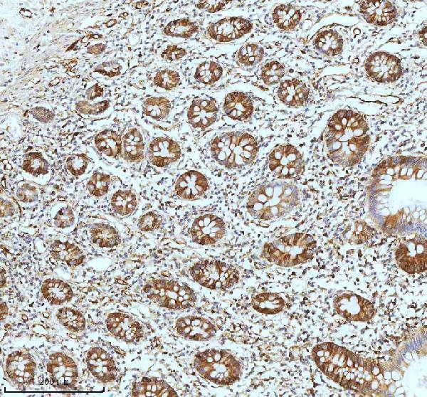
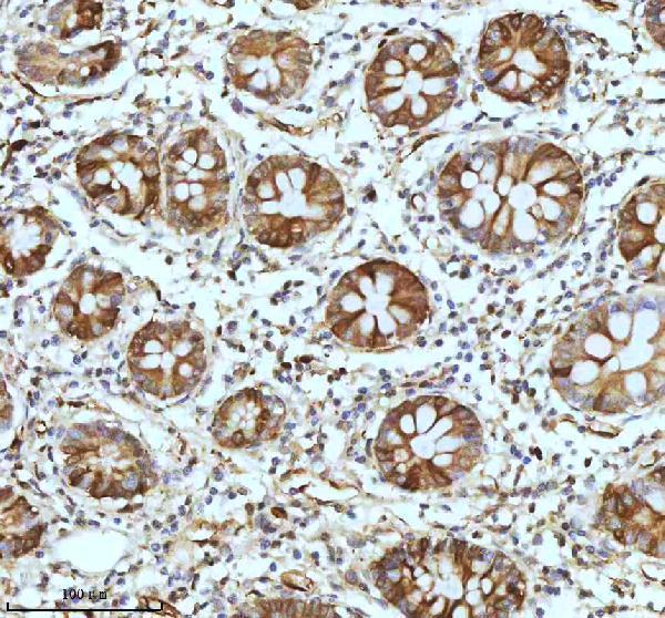
Anti-STMN1 antibodies have IHC images from human colon, placenta, breast carcinoma, and mouse and rat brain; PB9560 also has an IF image in MCF7 cells (catalog image captions).
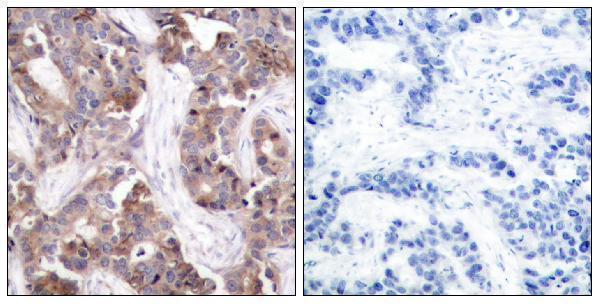
M01194 has paraffin-section IHC images from human colon and colon cancer, while PB9560 has paraffin-section IHC images from human placenta and intestinal cancer and mouse and rat brain, plus an IF image in MCF7 cells (catalog image captions). A01194S25 has a phospho-Ser24 IHC image from paraffin-embedded human breast carcinoma with a phosphopeptide-blocked comparison (A01194S25 IHC caption).
Which to pick: For tissue IHC, choose M01194 for paraffin human colon (1:50; rabbit monoclonal DDA-19; M01194 IHC caption and catalog) or A01194S25 when phospho-Ser24 staining is needed (paraffin human breast carcinoma with phosphopeptide block; A01194S25 IHC caption). For IF/ICC, PB9560 has an MCF7 IF image (PB9560 IF caption; catalog: IF/ICC); for cross-species tissue IHC, it has paraffin-section images from human placenta and mouse and rat brain (PB9560 IHC captions). Fixation is unreported in the IHC captions for all three SKUs (M01194, PB9560 and A01194S25 IHC captions).