This website uses cookies to ensure you get the best experience on our website.


- Table of Contents
Plan chromogenic STOML2 IHC on paraffin sections with the catalog antibody at 2–5 μg/ml (datasheet A04108-1). Use high-staining colon glandular cells and adipocytes reported as not detected to anchor scoring (HPA tissue IHC).

Expected localisation, validated protocols, controls and antibodies — the at-a-glance facts below, then the full design guide.
| Expected localisation | Granular cytoplasm in most tissues (HPA tissue IHC) | |
| Staining pattern | Granular cytoplasm in colon glandular cells (HPA tissue IHC) | |
| Antigen retrieval | EDTA pH 8.0 HIER, heat-mediated (datasheet A04108-1) | |
| Positive control | Adrenal gland+4 more · see all | |
| Negative control | Adipose tissue |
| Fixation | Matched tissue-IHC evidence does not establish the fixation claim. Validate the specimen-specific method before use. (selected-SKU IHC image A04108-1) | |
| Caveat | Hematopoietic cells can contribute strong signal (HPA tissue IHC) | |
| Regulation | No intensity regulation reported (UniProt) | |
| Isoform / epitope | 2 isoforms; chain 29–356; epitope coverage unreported (UniProt) |
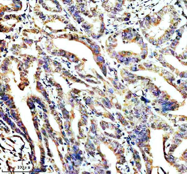
The catalog antibody's IHC-P protocol (datasheet A04108-1) is presented alongside published STOML2 IHC methods for hepatocellular, gastric, and colorectal cancer samples (PMC12695159; PMC6090455; PMC3069341).
| Sample | Paraffin-embedded human colon cancer tissue; fixative not specified (datasheet A04108-1) |
| Fixation | Image fixative and duration unreported (datasheet A04108-1); verify before use. |
| Sectioning | 4–5 µm sections on charged slides (standard) |
| Deparaffinisation | Xylene, graded ethanol series to water (standard) |
| Antigen retrieval | Heat retrieval: EDTA pH 8.0 (datasheet A04108-1); 20 min, 95–100 °C (standard) |
| Peroxidase block | 3% H2O2, 10 min, room temperature (standard) |
| Blocking | 10% goat serum (datasheet A04108-1) |
| Primary antibody | Rabbit anti-STOML2, 2-5 μg/ml (datasheet A04108-1) |
| Primary incubation | Overnight at 4 °C (datasheet A04108-1) |
| Detection | HRP-conjugated secondary, DAB chromogen (datasheet A04108-1) |
| Counterstain | Hematoxylin, blue, dehydrate and mount (standard) |
| Expected result | STOML2-positive staining in glandular cells of adrenal gland (HPA tissue IHC: High). HPA tissue profile: Cytoplasmic expression with a granular pattern in most tissues. No signal in the no-primary control. |
STOML2 is primarily associated with mitochondria, including the inner membrane and intermembrane space; it has no detectable transmembrane segment, and a minor pool can associate with the cell surface in activated T cells (UniProt Q9UJZ1). In paraffin tissue sections, expect granular cytoplasmic staining in many cell types, with intensity varying by tissue and cell type (HPA tissue IHC). HPA rates the tissue pattern Supported, with medium consistency between staining and RNA expression (HPA tissue IHC).
| Discrete granular cytoplasmic signal in glandular cells of colon, breast, appendix, or adrenal gland. | This matches the reported tissue pattern; each listed cell population is rated High (HPA tissue IHC). Assess the cytoplasmic granules within the identified cells, and compare intensity with controls processed in the same run (general IHC practice). |
| Predominantly nuclear staining, or a smooth cell-boundary outline without appreciable cytoplasmic granules. | Neither is the characteristic paraffin-section pattern of granular cytoplasmic expression (HPA tissue IHC). UniProt also records a minor surface-associated pool in activated T cells, so a membrane accent alone is not proof of a false signal; investigate whether the overall pattern and controls agree (UniProt Q9UJZ1; general IHC practice). |
| Strong staining in adipocytes while nearby cellular staining is otherwise unconvincing. | HPA reports STOML2 as Not detected in adipocytes, making this a warning for nonspecific binding or endogenous chromogen-generating activity rather than a supported positive result (HPA tissue IHC; general IHC practice). Confirm cell identity and inspect appropriate detection controls before interpreting it. |
| Uniform haze across cells and surrounding tissue obscures granular detail. | The expected IHC pattern is cellular and granular, so diffuse background prevents a confident localization call (HPA tissue IHC). Review blocking, antibody concentration, washing, and detection controls as general IHC variables; the supplied sources do not identify a STOML2-specific cause (general IHC practice). |
| No cytoplasmic signal in a documented High-staining tissue and cell population. | A blank result in, for example, colon glandular cells or bone marrow hematopoietic cells conflicts with HPA's reported High staining (HPA tissue IHC). Check the run's positive control and detection steps before calling the specimen negative; HPA's Supported rating does not guarantee every preparation will stain (general IHC practice; HPA tissue IHC). |
| Compartment and topology (UniProt Q9UJZ1). | STOML2 is chiefly mitochondrial despite lacking a detectable transmembrane segment; UniProt also lists other cellular locations (UniProt Q9UJZ1). Routine chromogenic IHC resolves the reported granular cytoplasmic pattern, but that appearance alone cannot assign individual granules to a mitochondrial subcompartment (HPA tissue IHC; general IHC practice). |
| Choice of tissue and cell-level comparator (HPA tissue IHC). | HPA rates several glandular, epithelial, neuronal, and hematopoietic populations High, skeletal myocytes and chondrocytes Low, and adipocytes Not detected (HPA tissue IHC). Compare the named cell populations rather than treating an entire section as uniformly positive or negative (general IHC practice). |
| Strength of IHC evidence (HPA tissue IHC; HPA antibodies). | The tissue pattern is Supported with medium staining-to-RNA consistency, and HPA062016 has Supported IHC status (HPA tissue IHC; HPA antibodies). These classifications support use as comparators, while leaving an unexpected result to be checked against morphology and controls in the actual run (general IHC practice). |
| IF/ICC Q: Should plasma-membrane or cytosolic fluorescence be expected? | A: HPA reports supported plasma-membrane and cytosol localization in ICC-IF; UniProt describes a minor plasma-membrane pool in activated T cells (HPA subcellular ICC-IF; UniProt Q9UJZ1). These observations can inform interpretation, but the ICC-IF localization is a separate assay and does not replace the granular cytoplasmic IHC benchmark (HPA tissue IHC). |
| Retrieval and epitope uncertainty (general IHC practice; UniProt Q9UJZ1). | Antigen retrieval is a routine paraffin-IHC variable, and UniProt records a processed STOML2 chain spanning residues 29–356 (general IHC practice; UniProt Q9UJZ1). The supplied sources give no antibody epitope or target-specific fixation sensitivity, so they do not predict which retrieval condition will improve STOML2 staining. |
| Situation | Likely cause | Next action |
|---|---|---|
| A High-staining control population gives little or no signal. | The observed result disagrees with the reported High cell-level pattern; the supplied sources do not establish whether retrieval, antibody concentration, or detection caused the loss (HPA tissue IHC). | Verify the positive control's cell identity, then review retrieval, primary-antibody application, and chromogenic detection records for that run (general IHC practice). Keep the result unresolved until the control performs as expected. |
| The whole section has diffuse color with no clear cytoplasmic granules. | Background may reflect nonspecific binding, inadequate washing, or detection-system activity; these are general IHC possibilities, not documented STOML2-specific effects (general IHC practice). | Inspect a primary-antibody omission control, review blocking and wash steps, and adjust working conditions against a known positive tissue (general IHC practice; HPA tissue IHC). Score only cellular signal distinguishable from background. |
| Nuclei dominate the stain. | Nuclear predominance does not match HPA's granular cytoplasmic tissue pattern (HPA tissue IHC). The supplied sources cannot identify the particular reagent or preparation step responsible. | Check the counterstain and detection controls, then compare the same run with a High-staining reference cell population (general IHC practice; HPA tissue IHC). Withhold a STOML2 localization call if the nuclear signal persists without cytoplasmic granules. |
| Adipocytes stain strongly. | HPA rates adipocytes Not detected; apparent positivity could arise from nonspecific staining or endogenous detection activity (HPA tissue IHC; general IHC practice). | Confirm that the colored structures are adipocytes, examine omission and detection controls, and compare a documented positive cell population on the same run (general IHC practice; HPA tissue IHC). Do not use adipocyte staining alone as a positive control. |
| A Low-staining population appears weak or blank. | Skeletal myocytes and chondrocytes are rated Low, so absence of an obvious signal there is a weak test of assay performance (HPA tissue IHC). | Judge run performance using an HPA High population, such as colon glandular cells or bone marrow hematopoietic cells, under the same staining conditions (HPA tissue IHC; general IHC practice). |
| Cell boundaries stain more strongly than the cytoplasm. | A minor surface-associated STOML2 pool is documented in activated T cells, but the HPA paraffin-tissue summary describes granular cytoplasmic expression in most tissues (UniProt Q9UJZ1; HPA tissue IHC). | Record the cell type and check for convincing cytoplasmic granules and appropriate control behavior before accepting a membrane-dominant IHC result (general IHC practice; HPA tissue IHC). |
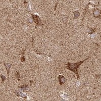


Comprehensive Human Protein Atlas IHC scoring per tissue (reliability: Supported — Medium consistency between antibody staining and RNA expression data.). Rows are taken directly from the HPA tissue chart — click any row's HPA link to view the source.
| Tissue | Cell type | Level | Evidence | Source |
|---|---|---|---|---|
| Adrenal gland | Glandular cells | High | Protein (IHC) | HPA → |
| Appendix | Glandular cells | High | Protein (IHC) | HPA → |
| Bone marrow | Hematopoietic cells | High | Protein (IHC) | HPA → |
| Breast | Glandular cells | High | Protein (IHC) | HPA → |
| Bronchus | Respiratory epithelial cells | High | Protein (IHC) | HPA → |
| Tissue | Cell type | Level | Evidence | Source |
|---|---|---|---|---|
| Adipose tissue | Adipocytes | Not detected | Protein (IHC) | HPA → |
Troubleshoot STOML2 staining in paraffin sections by checking retrieval, compartmental pattern, controls and scoring before interpreting differences between samples.
A04108-1 has human paraffin-section IHC data (image caption); M04108-1 is listed for ICC/IF in human, mouse and rat (catalog: applications and reactivity), with no supplied IF figure.
A04108-1 is the pictured IHC card, showing staining in a paraffin-embedded human colon cancer section (A04108-1 image caption). M04108-1 is listed for ICC/IF and reactivity with human, mouse and rat, but has no supplied IF image (M04108-1 catalog).
Which to pick: Choose A04108-1 for chromogenic tissue IHC: its image shows a human paraffin section with EDTA pH 8.0 retrieval, 2 μg/ml primary antibody and DAB detection; the fixative is unreported (A04108-1 image caption). Choose M04108-1 for IF/ICC because it is a rabbit monoclonal listed for that application, although no IF figure is supplied (M04108-1 catalog). Both list human, mouse and rat reactivity, but the supplied IHC image documents human tissue only (catalog: reactivity; A04108-1 image caption).