STRN / Striatin · IHC design guide

Design Immunohistochemistry for STRN

Plan STRN chromogenic IHC in paraffin sections around the cytoplasmic pattern reported in most tissues (HPA tissue IHC). Start the catalog antibody at 1:100 (datasheet: 1:100), and compare high staining in glandular cells with low staining in pancreatic exocrine cells (HPA tissue IHC).

Evidence assembled Oct 2026 · For research use; verify linked source records and product datasheet before use
Immunohistochemistry protocol sheet for STRN (IHC for STRN): expected localisation Cytoplasmic in most tissues (HPA tissue IHC), antibody A05855, validated IHC image, and IHC protocol steps
Printable STRN IHC protocol sheet — expected localisation Cytoplasmic in most tissues (HPA tissue IHC), antibody A05855, controls and protocol steps. Open the full STRN IHC guide →

STRN Immunohistochemistry Experimental Design Guide

Expected localisation, validated protocols, controls and antibodies — the at-a-glance facts below, then the full design guide.

Must know before staining
Expected localisation Cytoplasmic in most tissues (HPA tissue IHC)
Staining pattern Cytoplasmic staining; high in selected glandular cells (HPA tissue IHC)
Antigen retrieval Citrate pH 6.0 HIER, 95–98 °C, 20 min (rule: cytoplasmic / membrane antigen)
Positive control ⓘ Appendix+4 more · see all
Negative control ⓘ None in HPA (detected in all 45 tissues); use no-primary + isotype controls
Important caveats
Reasons your staining may differ from the expected pattern.
Fixation Keep fixation consistent across paraffin samples. (standard IHC practice; not target-specific)
Caveat Low consistency between staining and RNA expression (HPA tissue IHC)
Regulation Expression regulation not specified (UniProt)
Isoform / epitope 2 isoforms; epitope coverage unspecified (UniProt)
Section 1

Recommended STRN IHC & IF Protocols

Compare the catalog antibody’s IHC-P protocol with two published STRN IHC methods (PMC7125443; PMC8734210).

Recommended immunohistochemistry (IHC-P) protocol parameters
SampleParaffin-embedded Mouse Brain tissue; fixative not specified (datasheet A05855)
FixationImage fixative and duration unreported (datasheet A05855); verify before use.
Sectioning4–5 µm sections on charged slides (standard)
DeparaffinisationXylene, graded ethanol series to water (standard)
Antigen retrievalHeat-induced epitope retrieval in citrate buffer, pH 6.0, 20 min at 95–98 °C (standard rule: cytoplasmic / membrane antigen)
Peroxidase block3% H2O2, 10 min, room temperature (standard)
Blocking10% normal serum of the secondary host, 30 min, room temperature (standard)
Primary antibodyRabbit anti-STRN, 1:100 (datasheet A05855)
Primary incubationOvernight at 4 °C (standard)
DetectionHRP-polymer secondary, DAB chromogen 5–10 min (standard)
CounterstainHematoxylin, blue, dehydrate and mount (standard)
Expected resultSTRN-positive staining in glandular cells of appendix (HPA tissue IHC: High). HPA tissue profile: Cytoplasmic expression in most tissues. No signal in the no-primary control.
💡Decision noteStart with citrate pH 6.0 retrieval at 95–98 °C for 20 min (page antigen retrieval); the published excerpts give no retrieval conditions.
Section 2

What Is the Expected STRN Staining Pattern?

STRN should appear mainly cytoplasmic in tissue IHC (HPA: cytoplasmic expression in most tissues). High staining is reported in several glandular, epithelial, germinal center and trophoblastic cell populations (HPA: tissue IHC). UniProt also places STRN at membranes and dendritic spines, with no transmembrane segment (UniProt O43815: subcellular location and topology). Treat the tissue pattern as provisional: HPA rates the IHC antibody Approved but reports low consistency with RNA data and pending external verification (HPA: tissue IHC reliability).

What am I looking at on my slide?
Clear cytoplasmic staining in appendix or duodenal glandular cells.This matches reported High staining in those cells and the predominant tissue compartment (HPA: tissue IHC). Evaluate the named cell population, rather than assigning one score to the whole section (general IHC practice).
Predominantly nuclear staining in tissue IHC, with little cytoplasmic signal.This departs from the reported tissue IHC pattern and warrants an artifact check (HPA: tissue IHC; general IHC practice). Nuclear signal alone is not definitive evidence of cross-reactivity: HPA also reports nucleoplasm as an additional location in ICC-IF (HPA: subcellular ICC-IF).
Strong staining confined to an unexpected cell population.Check cell identity and compare with reported populations before calling it nonspecific (HPA: tissue IHC). Broad cytoplasmic expression is reported, so an unlisted cell type is not automatically negative; discordance could reflect cross-reactivity or endogenous detection activity (HPA: tissue IHC; general IHC practice).
Diffuse color across cells and extracellular spaces, without a discernible cytoplasmic pattern.This is difficult to score as STRN because the reported tissue pattern is cytoplasmic (HPA: tissue IHC). Compare a no-primary control and review blocking, washing and detection background before interpreting cell-level signal (general IHC practice).
No discernible signal in a reported High-staining cell population.A negative result in, for example, rectal glandular cells conflicts with the HPA observation (HPA: High in rectal glandular cells). Check tissue identity and the IHC run before concluding absence of STRN; HPA reports low antibody–RNA consistency (HPA: tissue IHC reliability; general IHC practice).
💡Expected STRN appearanceCall a result consistent when cytoplasmic staining is clear in a reported High-staining population, such as small-intestinal glandular cells (HPA: tissue IHC); predominantly nuclear-only or diffuse, cell-independent color warrants investigation (HPA: tissue IHC; general IHC practice).
How each factor affects the staining
Compartment and topologyCytoplasm is the main tissue IHC expectation (HPA: tissue IHC). Membrane and dendritic-spine locations are annotated, but STRN has no transmembrane segment (UniProt O43815: location and topology); do not require a continuous cell-surface rim.
Choice of reference tissue and cellsAppendix, duodenum, endometrium and rectum have reported High glandular-cell staining; exocrine pancreatic cells are reported Low (HPA: tissue IHC). Compare named cell populations when choosing a reference slide.
Evidence limitsThe listed HPA017286 antibody is Approved for IHC, while the tissue profile has low consistency with RNA and awaits external verification (HPA: antibody validation and tissue IHC reliability). Agreement with the reported pattern supports interpretation but does not establish specificity by itself (general IHC practice).
Isoforms and antigen preparationUniProt lists 2 isoforms, but the supplied record gives no antibody epitope or isoform-reactivity data (UniProt O43815: isoforms; supplied HPA antibody record). Target-specific fixation sensitivity and antigen-retrieval effects are unreported here; do not predict either from topology or staining levels.
Why is my staining missing, weak or wrong?
SituationLikely causeNext action
Reported High-staining glandular cells are negative.The section or cell population may be misidentified, or the IHC run may have failed (general IHC practice); High is an HPA observation, not a guarantee for every preparation (HPA: tissue IHC).Confirm morphology, then inspect the run’s positive control, detection reagents and the catalog antibody’s IHC-P instructions (general IHC practice). Do not infer STRN-specific fixation sensitivity from this result.
Signal is mostly nuclear in paraffin IHC.The pattern differs from HPA’s cytoplasmic tissue profile; nonspecific detection is possible (HPA: tissue IHC; general IHC practice).Review the no-primary control and compare cytoplasmic signal in a reported High-staining population (general IHC practice; HPA: tissue IHC). Do not use nuclear signal alone to validate the stain.
Unlisted cells stain strongly while the expected population does not.Misidentified cells, cross-reactivity or endogenous detection activity may explain the mismatch (general IHC practice). HPA also reports cytoplasmic expression across most tissues (HPA: tissue IHC).Verify morphology and compare a no-primary control with a reported High-staining population on the same run (general IHC practice; HPA: tissue IHC).
Diffuse chromogen obscures cell boundaries.Detection background can prevent assessment of the reported cytoplasmic pattern (general IHC practice; HPA: tissue IHC).Check the no-primary control, blocking and wash steps; score STRN only where the cellular pattern can be distinguished from background (general IHC practice).
A low-staining reference tissue gives a faint result.Low staining is reported for exocrine pancreatic cells, myocytes, fibroblasts and adipocytes (HPA: tissue IHC). A faint result there gives limited leverage for judging assay failure.Use a reported High-staining cell population to assess whether the IHC run detects the expected pattern (HPA: tissue IHC; general IHC practice).
IF/ICC shows cytosolic signal with some nucleoplasmic signal.That combination matches HPA’s approved main cytosolic and additional nucleoplasmic ICC-IF locations (HPA: subcellular ICC-IF).Interpret it within the separate IF/ICC guide; for this paraffin IHC section, compare the tissue result with HPA’s cytoplasmic IHC profile (HPA: subcellular ICC-IF and tissue IHC).

Sample controls for STRN IHC & IF

🧪Run appendix first and score its glandular cells, where STRN staining is High (HPA: High in appendix glandular cells). HPA detects STRN in all 45 scored tissues, so there is no supported negative tissue; use no-primary and isotype controls, and treat cells with little staining on the positive slide as background references rather than confirmed STRN-negative cells (HPA: no negative tissue rows).
Positive control tissue: Appendix (Glandular cells, HPA High)
Negative control tissue: None in HPA: STRN is detected in all 45 scored tissues. Use a no-primary (secondary-only) and an isotype control instead.
ICC-IF cell lines (HPA subcellular resource): HPA ICC-IF images show STRN in A-431, U-251MG, U2OS, with annotated localisation: Cytosol (approved) (HPA subcellular).
Technical controls: Include a no-primary, secondary-only control and an isotype-matched control appropriate to the primary antibody’s host species and clonality; a STRN knockout specimen or validated peptide block would provide a biological specificity control. For chromogenic appendix IHC, block endogenous peroxidase and check the glandular region for background staining (HPA: High in appendix glandular cells).
⚠️Feasibility: The selected antibody caption shows paraffin-section IHC in mouse brain at 1:100, but reports no fixative; a target-specific fixation window or fixation effect is unreported (selected A05855 tissue-IHC caption). Retrieval conditions and dependence are also unreported, so optimize retrieval empirically rather than inferring it from STRN localization (UniProt O43815: cytoplasm, membrane and dendritic spine). No supplied evidence establishes that frozen sections or IF are easier; ICC-IF images exist for A-431, U-251MG and U2OS, with mainly cytosolic and additional nucleoplasmic localization (HPA subcellular), while appendix-specific staining artefacts are unreported.

HPA tissue IHC evidence for STRN

Comprehensive Human Protein Atlas IHC scoring per tissue (reliability: Approved — Low consistency between antibody staining and RNA expression data. Pending external verification.). Rows are taken directly from the HPA tissue chart — click any row's HPA link to view the source.

Positive expression · recommended positive controls

TissueCell typeLevelEvidenceSource
Appendix Glandular cells High Protein (IHC) HPA →
Duodenum Glandular cells High Protein (IHC) HPA →
Endometrium Glandular cells High Protein (IHC) HPA →
Esophagus Squamous epithelial cells High Protein (IHC) HPA →
Lymph node Germinal center cells High Protein (IHC) HPA →

Undetected expression · recommended negative controls

TissueCell typeLevelEvidenceSource
None in HPA: STRN is detected in all 45 scored tissues. Use a no-primary (secondary-only) and an isotype control instead.
Section 3

Advanced STRN IHC Tips

Troubleshoot STRN staining in paraffin sections by checking retrieval, compartment, cell type and controls before interpreting chromogenic signal.

How should I adjust retrieval when STRN staining is weak in paraffin sections?
Start with citrate buffer at pH 6.0, heated to 95–98 °C for 20 minutes (page retrieval specification). If staining is weak, compare a longer citrate incubation or an alkaline retrieval buffer on matched sections, while keeping antibody dilution and detection conditions constant (standard IHC practice). Score both signal and tissue preservation: stronger staining accompanied by torn sections or widespread background does not establish better retrieval (standard IHC practice). STRN has cytoplasmic and membrane annotations but no transmembrane segment, so assess intracellular staining across the same cell populations rather than treating a crisp membrane outline as the required endpoint (UniProt O43815 topology and subcellular annotation).
Can fixation explain inconsistent STRN staining across paraffin blocks?
STRN-specific sensitivity to fixation is unknown from the supplied evidence; the selected mouse brain caption identifies paraffin embedding but does not state a fixative (catalog antibody A05855 IHC caption). Record each block’s fixative and fixation interval, then compare sections processed with the same retrieval, staining run and detection settings (standard IHC practice). If archived blocks differ, use internal tissue morphology and a no-primary control to distinguish poor preservation from nonspecific chromogen deposition (standard IHC practice). Do not infer STRN’s fixation tolerance from its phosphorylation sites, lack of a transmembrane segment, or tissue staining patterns (UniProt O43815 modified residues and topology; HPA tissue IHC profile).
Which STRN staining pattern should I accept in tissue sections?
Prioritize cytoplasmic signal, because STRN is annotated in the cytoplasm and membrane, while tissue IHC describes cytoplasmic expression in most tissues (UniProt O43815 subcellular annotation; HPA tissue IHC profile). Membrane association may appear as localized peripheral staining, but a continuous membrane rim is not required for a protein without a transmembrane segment (UniProt O43815 topology and subcellular annotation). Nuclear staining deserves separate assessment: approved cell imaging reports cytosol as the main location and nucleoplasm as an additional location (HPA subcellular). Compare compartments in intact cells at matched exposure or chromogen development, and check whether an unexpected pattern also appears in the no-primary control (standard IHC practice).
How can I assess whether this antibody detects both STRN isoforms?
STRN has 2 annotated isoforms, but the supplied evidence does not map the catalog antibody’s epitope or establish isoform coverage (UniProt O43815 isoforms; catalog antibody A05855 IHC caption). Obtain the immunogen sequence and align it to both isoforms before describing staining as shared or isoform specific (standard antibody validation practice). If the epitope remains undisclosed, report staining as STRN immunoreactivity and avoid assigning a particular isoform to individual cells (standard antibody validation practice). Compare independent antibodies with mapped, nonoverlapping epitopes on adjacent sections when available, using the same cell compartments and controls to judge agreement (standard IHC practice).
How should I use IF to check the cell source of STRN IHC signal?
Use the separate IF/ICC workflow to pair STRN with a validated marker of the cell population being examined, then assess colocalization within individual cells (standard IF practice). Select spectrally separated fluorophores and reserve a channel with low tissue autofluorescence for a weak STRN signal; include unstained and single-label controls (standard IF practice). Because STRN is mainly cytosolic and has no transmembrane segment, permeabilize cells to assess intracellular epitopes, then titrate detergent strength to preserve morphology (HPA subcellular; UniProt O43815 topology; standard IF practice). The mouse brain paraffin IHC caption supplies neither an IF fixation condition nor an IF antibody dilution, so establish both in the IF/ICC workflow (catalog antibody A05855 IHC caption).
What controls help distinguish STRN signal from diffuse brown background?
Run a no-primary control through the same peroxidase block, secondary reagent and DAB development to identify detection-related staining (standard chromogenic IHC practice). If pigment persists, inspect unstained tissue and adjust chromogen development before changing the STRN antibody, since tissue pigment can resemble brown reaction product (standard IHC practice). Titrate the primary antibody around the caption’s 1:100 starting dilution on matched paraffin sections, keeping retrieval and development time fixed (catalog antibody A05855 IHC caption; standard IHC practice). Judge improvement by cytoplasmic contrast in intact cells, while treating staining at folds, damaged edges or necrotic regions as suspect (HPA tissue IHC profile; standard IHC practice).
How should I quantify STRN across specimens with different cell mixtures? ⚠ ANSWER MARKED FOR VERIFICATION
Define the cell population and cytoplasmic compartment before scoring, because the tissue profile reports cytoplasmic expression in most tissues and differing levels among cell types (HPA tissue IHC profile). Use an H-score based on staining intensity and the percentage of positive cells, or report the percentage positive within the annotated population (standard IHC scoring practice). For spatial comparisons, report positive-cell density per mm² of viable tissue and normalize to the area or number of eligible cells, rather than total section area alone (standard IHC scoring practice). Keep retrieval, antibody dilution, DAB development and image thresholds consistent across specimens, with blinded review of ambiguous cells (standard IHC practice).
When should apparent STRN positivity be treated as an artefact?
Give most weight to reproducible cytoplasmic staining in intact cells, consistent with the reported tissue profile and STRN’s cytoplasmic annotation (HPA tissue IHC profile; UniProt O43815 subcellular annotation). Unexpected nuclear-only staining needs scrutiny, although nucleoplasm is an additional location in cell imaging, so compartment alone cannot settle specificity (HPA subcellular). Treat signal concentrated at section edges, folds or necrotic areas as suspect and compare it with a no-primary control for residual endogenous peroxidase or detection background (standard IHC practice). Cell identity also matters: high staining is reported in several glandular and epithelial populations, while some muscle and stromal populations are low, so confirm identity from morphology and adjacent markers before assigning STRN expression (HPA tissue IHC profile; standard IHC practice).
Boster reagents

Best STRN / Striatin IHC Antibodies

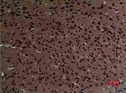
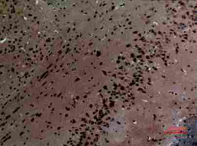
A05855 has an IHC image from paraffin-embedded mouse brain (catalog image caption); IF is listed without an IF image (catalog applications; catalog image records). Human, mouse and rat reactivity is listed (catalog reactivity).

Real IHC data Immunohistochemistry (IHC) analysis of paraffin-embedded Mouse Brain, antibody was diluted at 1:100.
Anti-Striatin STRN Antibody
Cat # A05855

A05855 will render with an IHC figure from paraffin-embedded mouse brain at 1:100 (catalog image caption). A05855 lists IHC and IF applications and human, mouse and rat reactivity; no IF image is provided (catalog applications; catalog reactivity; catalog image records).

Which to pick: For tissue IHC, choose A05855: its own image documents paraffin-embedded mouse brain at 1:100; the fixative is unreported (catalog image caption). For IF/ICC, A05855 lists IF at 1:50, but provides no IF image (catalog applications; catalog IF dilution; catalog image records). For work across human, mouse and rat, A05855 is the listed rabbit polyclonal option; the IHC image demonstrates mouse tissue only (catalog reactivity; catalog host; catalog dilution record; catalog image caption).

Each figure is that product's own IHC / IF validation image from its datasheet.

References

  1. UniProt Consortium. UniProt entry O43815 (STRN_HUMAN, Striatin).
  2. Human Protein Atlas. STRN tissue IHC expression (reliability: Approved).
  3. Human Protein Atlas. STRN subcellular location (ICC-IF): Mainly localized to the cytosol. In addition localized to the nucleoplasm..
  4. Human Protein Atlas. STRN antibody validation summary (1 antibodies).
  5. High STRN Expression Promotes HCC Invasion and Migration but Not Cell Proliferation or Apoptosis through Facilitating Epithelial-Mesenchymal Transition. BioMed research international 2020 — PMC7125443.
  6. Peritoneal Metastasis in Advanced Lung Adenocarcinoma Harboring EGFR Exon 19 Deletion/T790M with STRN::ALK Fusion and EGFR Kinase Domain Duplication: Two Case Reports and a Narrative Literature Summary. Cancer management and research 2026 — PMC13525806.
  7. STK25 enhances hepatocellular carcinoma progression through the STRN/AMPK/ACC1 pathway. Cancer cell international 2022 — PMC8734210.
  8. CircTNPO3 promotes hepatocellular carcinoma progression by sponging miR-199b-5p and regulating STRN expression. The Kaohsiung journal of medical sciences 2023 — PMC11895897.
  9. PubMed PMID:9693043 — UniProt-cited evidence.
  10. PubMed PMID:15815621 — UniProt-cited evidence.
  11. PubMed PMID:15489334 — UniProt-cited evidence.