This website uses cookies to ensure you get the best experience on our website.


- Table of Contents
Plan chromogenic STX18 IHC in paraffin sections around the reported cytoplasmic tissue staining (HPA tissue IHC). The catalog antibody has an IHC dilution range of 2–5 μg/ml (datasheet A11895); interpret staining with care because antibody staining and RNA expression have low consistency (HPA tissue IHC).

Expected localisation, validated protocols, controls and antibodies — the at-a-glance facts below, then the full design guide.
| Expected localisation | General cytoplasmic staining (HPA tissue IHC); ER/Golgi membrane location (UniProt) | |
| Staining pattern | Colon glandular cells: high cytoplasmic staining (HPA tissue IHC) | |
| Antigen retrieval | EDTA pH 8.0 HIER, heat-mediated (datasheet A11895) | |
| Positive control | Colon+4 more · see all | |
| Negative control | Soft tissue+2 more · see all |
| Fixation | Keep fixation consistent across sections (standard IHC practice; not target-specific). Selected-image fixative and duration unreported (datasheet A11895); verify before use. | |
| Caveat | Staining has low consistency with RNA expression (HPA tissue IHC) | |
| Regulation | Expression regulation not specified (UniProt) | |
| Isoform / epitope | No isoforms; cytoplasmic vs luminal epitope matters (UniProt) |
Compare the catalog antibody’s IHC-P protocol (datasheet: A11895) with a published STX18 protocol using human prostate tissue microarrays (PMC4749310: Methods).
| Sample | Paraffin-embedded human appendix adenocarcinoma tissue; fixative not specified (datasheet A11895) |
| Fixation | Image fixative and duration unreported (datasheet A11895); verify before use. |
| Sectioning | 4–5 µm sections on charged slides (standard) |
| Deparaffinisation | Xylene, graded ethanol series to water (standard) |
| Antigen retrieval | Heat retrieval: EDTA pH 8.0 (datasheet A11895); 20 min, 95–100 °C (standard) |
| Peroxidase block | 3% H2O2, 10 min, room temperature (standard) |
| Blocking | 10% goat serum (datasheet A11895) |
| Primary antibody | Rabbit anti-STX18, 2-5 μg/ml (datasheet A11895) |
| Primary incubation | Overnight at 4 °C (datasheet A11895) |
| Detection | HRP-conjugated secondary, DAB chromogen (datasheet A11895) |
| Counterstain | Hematoxylin, blue, dehydrate and mount (standard) |
| Expected result | STX18-positive staining in glandular cells of colon (HPA tissue IHC: High). HPA tissue profile: General cytoplasmic expression. No signal in the no-primary control. |
STX18 is an ER and Golgi membrane protein with a cytoplasmic region spanning residues 1–309 (UniProt Q9P2W9 topology). In paraffin IHC, expect mainly cytoplasmic staining, especially in the epithelial and glandular cell groups reported as high by HPA (HPA tissue IHC). Interpret intensity cautiously: HPA rates the tissue IHC profile Approved but reports low consistency between antibody staining and RNA expression, pending external verification (HPA tissue IHC).
| Glandular cells stain in colon, duodenum, endometrium, gallbladder, rectum or small intestine. | These cell groups are reported as High, and a cytoplasmic pattern fits the tissue profile (HPA tissue IHC). An ER-associated distribution is biologically plausible because STX18 resides on the ER membrane (UniProt Q9P2W9 subcellular location). Judge the relevant cells within the section; a dark field alone does not establish the expected pattern (general IHC practice). |
| Predominantly nuclear staining, or staining confined to an unexpected compartment, dominates the slide. | That pattern conflicts with HPA’s general cytoplasmic tissue profile and UniProt’s ER/Golgi membrane locations (HPA tissue IHC; UniProt Q9P2W9 subcellular location). Treat it as a possible staining artefact and review controls and morphology before assigning STX18 expression (general IHC practice). A membrane component can occur in IF/ICC, so compartment calls should remain application specific (HPA subcellular ICC-IF). |
| A cell group reported as Not detected stains strongly, while expected glandular cells do not. | HPA reports no detection in peripheral nerve in soft tissue, splenic red-pulp cells and vaginal squamous epithelial cells (HPA tissue IHC). Such discordance raises concern for cross-reactivity or endogenous detection activity (general IHC practice). These are comparison groups, not absolute biological negatives: the HPA tissue profile has low staining–RNA consistency and awaits external verification (HPA tissue IHC). |
| Color spreads across extracellular spaces or obscures cell borders throughout the section. | Diffuse background prevents a reliable cytoplasmic call, even where HPA reports High glandular-cell staining (HPA tissue IHC; general IHC practice). Check the detection-only control, blocking, washes and chromogen development before scoring; none of those workflow checks demonstrates a target-specific STX18 effect (general IHC practice). |
| No signal appears in a section selected for HPA High glandular-cell staining. | First confirm that the reported cell group is present and preserved; examples include colon and duodenal glandular cells (HPA tissue IHC; general IHC practice). A negative result may reflect the assay or the specimen and cannot by itself revise the HPA profile. Its Approved rating still carries a low-consistency, pending-verification caveat (HPA tissue IHC). |
| Cell type and reference intensity | HPA reports High staining in several glandular-cell groups, respiratory epithelial cells of nasopharynx, and cells in testicular seminiferous ducts (HPA tissue IHC). It reports Low staining in ovarian stromal cells, smooth muscle cells and adipocytes (HPA tissue IHC). Compare like cell groups when judging a section; the reported levels are observations, not universal thresholds (general IHC practice). |
| Membrane topology and processing | STX18 has one transmembrane segment at residues 310–330, a cytoplasmic region at 1–309, and no listed signal peptide or propeptide (UniProt Q9P2W9 topology/processing). The record lists no isoforms, glycosylation sites or modified residues (UniProt Q9P2W9). These annotations guide compartment interpretation; they do not identify the antibody epitope or establish antigen-retrieval conditions. |
| Evidence and antibody validation | The listed HPA antibody, HPA003019, has Approved IHC status; the tissue profile has low consistency with RNA expression and is pending external verification (HPA antibodies; HPA tissue IHC). Avoid treating a single intense field or a reported Not detected group as definitive proof of specificity or absence (general IHC practice). |
| IF/ICC Q: What localization should appear? | A: HPA supports ER and plasma-membrane localization in ICC-IF and lists images from A-431, U-251MG and U2OS; UniProt also places STX18 at ER and Golgi membranes (HPA subcellular ICC-IF; UniProt Q9P2W9 subcellular location). This localization answer does not specify an IF/ICC protocol. The paraffin IHC tissue description remains general cytoplasmic expression (HPA tissue IHC). |
| Situation | Likely cause | Next action |
|---|---|---|
| A selected HPA High tissue gives no interpretable signal. | The reported positive cell group may be absent, or an assay step may have failed (HPA tissue IHC; general IHC practice). | Check morphology and a suitable positive control, then review the catalog antibody’s IHC-P procedure, detection reagents and development (general IHC practice). Do not infer STX18-specific fixation sensitivity; none is reported here. |
| The whole section has weak, widespread color. | Background from nonspecific binding, incomplete washing or detection chemistry may obscure cells (general IHC practice). | Inspect a detection-only control; review blocking, washes and development, then score only clearly cellular staining (general IHC practice). |
| Nuclei are the strongest stained compartment. | A dominant nuclear pattern disagrees with the cytoplasmic tissue profile and ER/Golgi membrane annotation (HPA tissue IHC; UniProt Q9P2W9 subcellular location). | Check counterstain and detection-only controls, then repeat interpretation against cell morphology and a reported High cell group (general IHC practice; HPA tissue IHC). |
| Splenic red-pulp or vaginal squamous cells stain strongly. | These cell groups are reported Not detected, so cross-reactivity or endogenous detection activity is possible (HPA tissue IHC; general IHC practice). | Check controls for endogenous activity and compare staining with an appropriate HPA High cell group; retain the HPA low-consistency caveat (general IHC practice; HPA tissue IHC). |
| Only tissue edges or damaged areas show intense color. | Edge concentration or damaged morphology can make local color unreliable for cell-level scoring (general IHC practice). | Assess intact interior cells and review section handling and detection controls before calling the expected cytoplasmic pattern (general IHC practice; HPA tissue IHC). |
| Staining is present, but its intensity differs from the HPA example. | HPA’s High and Low labels describe observed cell groups, while its tissue profile has low staining–RNA consistency (HPA tissue IHC). | Compare the same cell type and compartment, document controls and score the specimen on its own merits; avoid treating HPA intensity as a fixed cutoff (general IHC practice; HPA tissue IHC). |


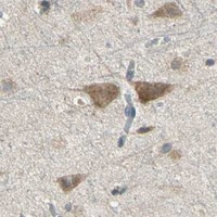


Comprehensive Human Protein Atlas IHC scoring per tissue (reliability: Approved — Low consistency between antibody staining and RNA expression data. Pending external verification.). Rows are taken directly from the HPA tissue chart — click any row's HPA link to view the source.
| Tissue | Cell type | Level | Evidence | Source |
|---|---|---|---|---|
| Colon | Glandular cells | High | Protein (IHC) | HPA → |
| Duodenum | Glandular cells | High | Protein (IHC) | HPA → |
| Endometrium | Glandular cells | High | Protein (IHC) | HPA → |
| Gallbladder | Glandular cells | High | Protein (IHC) | HPA → |
| Nasopharynx | Respiratory epithelial cells | High | Protein (IHC) | HPA → |
Use compartment, cell type, and matched controls together when assessing STX18 staining in paraffin sections (UniProt Q9P2W9 localisation; HPA tissue IHC).
The catalog antibody has IHC and IF images from human paraffin tissue sections (A11895 image captions; catalog reactivity).
A11895 is listed for human IHC and IF (catalog applications and reactivity). Its IHC captions show appendix adenocarcinoma, breast cancer, endometrioid adenocarcinoma and penis squamous cell carcinoma; its IF captions show colon and breast cancer (A11895 image captions).
Which to pick: For tissue IHC, choose A11895 for human paraffin sections; its own IHC caption documents EDTA pH 8.0 retrieval and 2 μg/ml primary antibody (A11895 IHC caption). For IF, the same SKU has human paraffin-section images, while ICC validation is unreported (A11895 IF captions; catalog applications). No cross-species option is documented, the clone is unspecified, and the IHC caption does not report the fixative (catalog reactivity and clone; A11895 IHC caption).