SUB1 / Activated RNA polymerase II transcriptional coactivator p15 · IHC design guide

Design Immunohistochemistry for SUB1

Plan SUB1 IHC on paraffin sections around widespread nuclear staining (HPA tissue IHC) and a catalog antibody starting range of 2–5 μg/ml (datasheet: M02698-3). Use liver hepatocytes as a positive reference (HPA tissue IHC) and a no-primary control to assess background (standard IHC practice).

Evidence assembled Oct 2026 · For research use; verify linked source records and product datasheet before use
Immunohistochemistry protocol sheet for SUB1 (IHC for SUB1): expected localisation Nuclear staining across tissues (HPA tissue IHC), antibody M02698-3, validated IHC image, and IHC protocol steps
Printable SUB1 IHC protocol sheet — expected localisation Nuclear staining across tissues (HPA tissue IHC), antibody M02698-3, controls and protocol steps. Open the full SUB1 IHC guide →

SUB1 Immunohistochemistry Experimental Design Guide

Expected localisation, validated protocols, controls and antibodies — the at-a-glance facts below, then the full design guide.

Must know before staining
Expected localisation Nuclear staining across tissues (HPA tissue IHC)
Staining pattern Widespread nuclear staining across cell types (HPA tissue IHC)
Antigen retrieval EDTA pH 8.0 HIER, heat-mediated (datasheet M02698-3)
Positive control ⓘ Appendix+4 more · see all
Negative control ⓘ None in HPA (detected in all 45 tissues); use no-primary + isotype controls
Important caveats
Reasons your staining may differ from the expected pattern.
Fixation Keep fixation consistent across sections (standard IHC practice; not target-specific)
Caveat Ubiquitous nuclear signal limits negative-cell controls (HPA tissue IHC)
Regulation Low tissue specificity (HPA tissue RNA)
Isoform / epitope No isoforms; mature chain aa 2–127; no TM segment (UniProt)
Section 1

Recommended SUB1 IHC & IF Protocols

The catalog antibody’s IHC-P protocol is accompanied by one published SUB1 IHC protocol using prostate tissues (PMC5140777).

Recommended immunohistochemistry (IHC-P) protocol parameters
SampleParaffin-embedded human colorectal adenocarcinoma tissue; fixative not specified (datasheet M02698-3)
FixationImage fixative and duration unreported (datasheet M02698-3); verify before use.
Sectioning4–5 µm sections on charged slides (standard)
DeparaffinisationXylene, graded ethanol series to water (standard)
Antigen retrievalHeat retrieval: EDTA pH 8.0 (datasheet M02698-3); 20 min, 95–100 °C (standard)
Peroxidase block3% H2O2, 10 min, room temperature (standard)
Blocking10% goat serum (datasheet M02698-3)
Primary antibodyMouse monoclonal (clone 8D9D1) anti-SUB1, 2-5 μg/ml (datasheet M02698-3)
Primary incubationOvernight at 4 °C (datasheet M02698-3)
DetectionHRP-conjugated secondary, DAB chromogen (datasheet M02698-3)
CounterstainHematoxylin, blue, dehydrate and mount (standard)
Expected resultSUB1-positive staining in glandular cells of appendix (HPA tissue IHC: High). HPA tissue profile: Ubiquitous nuclear expression. No signal in the no-primary control.
💡Decision noteStart with heat-mediated EDTA pH 8.0 retrieval for the catalog antibody (datasheet M02698-3); the published prostate protocol does not specify retrieval (PMC5140777).
Section 2

What Is the Expected SUB1 Staining Pattern?

SUB1 should appear chiefly in nuclei across many cell types (UniProt P53999: nucleus; HPA tissue IHC: ubiquitous nuclear expression). In HPA tissue sections, high staining is reported in several glandular populations, hepatocytes and bone marrow hematopoietic cells; some muscle, follicle and fibroblast populations show low staining (HPA tissue IHC). HPA rates the tissue staining evidence Supported, with medium consistency between antibody staining and RNA expression (HPA tissue IHC).

What am I looking at on my slide?
Nuclear staining in glandular cells of colon, breast or appendix, with recognizable tissue structure.This fits the reported high staining in those cell populations and the ubiquitous nuclear profile (HPA tissue IHC). Score the fraction and intensity of stained nuclei within each identified population; do not treat all cells in a section as equally positive (general IHC practice).
Predominantly cytoplasmic or membrane staining, with little nuclear signal.The compartment conflicts with the nuclear tissue pattern and UniProt localization (HPA tissue IHC; UniProt P53999: nucleus). SUB1 has no annotated transmembrane segment or signal sequence (UniProt P53999 topology). Consider nonspecific staining or a detection artefact before calling this SUB1 positive.
Strong staining in cells expected to be low, such as skeletal myocytes or smooth muscle cells.HPA reports low staining in these cells, but supplies no negative tissue population (HPA tissue IHC). Recheck cell identity and nuclear localization. A strong, misplaced signal may reflect cross-reactivity or endogenous detection activity (general IHC practice); intensity alone cannot establish either cause.
Diffuse color across tissue, including spaces between cells, obscures nuclear boundaries.This cannot be scored confidently as the reported nuclear pattern (HPA tissue IHC). Uneven reagent removal, excess primary antibody or detection background are possible general IHC causes (general IHC practice). Compare a control without primary antibody and inspect whether color follows identifiable nuclei.
No convincing nuclear signal in a section chosen for a reported high-staining population.A negative result in colon glandular cells, liver hepatocytes or bone marrow hematopoietic cells conflicts with the reported high staining (HPA tissue IHC). It does not by itself establish absent SUB1: check tissue identity, assay controls and retrieval and detection performance (general IHC practice).
💡Expected SUB1 appearanceCall a result positive when identifiable nuclei stain in the relevant cells, with strong signal plausible in HPA high-staining populations; dominant cytoplasmic, membrane or diffuse tissue color is inconsistent with the reported SUB1 pattern (HPA tissue IHC; UniProt P53999: nucleus).
How each factor affects the staining
Which compartment should drive the IHC score?Nuclear staining: UniProt places SUB1 in the nucleus, and HPA describes ubiquitous nuclear tissue expression (UniProt P53999; HPA tissue IHC). HPA ICC-IF further supports nucleoplasmic and nucleolar localization (HPA subcellular). Use a nuclear counterstain and intact cell morphology to judge compartment (general IHC practice).
Which tissue comparisons are informative?HPA reports high staining in colon glandular cells and liver hepatocytes, and low staining in skeletal myocytes and soft-tissue fibroblasts (HPA tissue IHC). These are relative pattern checks, not absolute positive and negative controls: the HPA record lists no negative populations (HPA tissue IHC).
How strong is the antibody evidence?Two listed rabbit polyclonal antibodies, HPA001311 and CAB015351, each have Supported IHC status (HPA antibodies). Overall tissue staining reliability is Supported, with medium staining–RNA consistency (HPA tissue IHC). These summaries support pattern comparison but do not establish that every staining difference reflects SUB1 abundance.
What does IF/ICC add here?It offers a localization cross-check: HPA reports supported nucleoplasmic and nucleolar staining, with images from A-431, U-251MG and U2OS cells (HPA subcellular). Those ICC-IF observations do not define an IHC paraffin-section protocol or prove that nucleoli must be distinguishable in every chromogenic section.
Is target-specific fixation sensitivity known?No target-specific fixation effect is supplied by UniProt or HPA here (UniProt P53999; HPA tissue IHC). If retrieval conditions are compared, interpret the outcome as an assay optimization result rather than a documented SUB1-specific fixation response (general IHC practice).
Why is my staining missing, weak or wrong?
SituationLikely causeNext action
High-staining reference tissue shows no nuclear signal.Tissue selection, retrieval or detection may have failed; the chosen cell population is reported as high by HPA (HPA tissue IHC; general IHC practice).Confirm the population on the section, check assay controls, then compare validated retrieval and detection settings within the IHC workflow (general IHC practice).
Only cytoplasm or cell outlines stain.That distribution conflicts with the nuclear localization and lack of a transmembrane segment (UniProt P53999; HPA tissue IHC). Nonspecific primary or detection binding is possible (general IHC practice).Inspect a control without primary antibody; reassess primary dilution and blocking, then score only identifiable nuclear signal (general IHC practice).
Every area develops diffuse chromogen.Detection background or incomplete washing can obscure the nuclear pattern (general IHC practice; HPA tissue IHC: ubiquitous nuclear expression).Compare a control without primary antibody, improve washing and titrate the primary or detection reagents as appropriate (general IHC practice).
Low-staining muscle appears as strong as a high-staining reference population.HPA reports low staining in skeletal and smooth muscle cells, so the relative pattern warrants review (HPA tissue IHC). Cell misidentification or assay background is possible (general IHC practice).Identify cells on the counterstained section, compare nuclear intensity across matched runs and examine a control without primary antibody (general IHC practice).
Only some nuclei stain within an expected positive region.HPA reports population-level staining, not a required positive fraction for every section or nucleus (HPA tissue IHC). Section quality and cell identity can affect interpretation (general IHC practice).Record intensity and positive fraction by cell population; inspect morphology and compare a reported high-staining tissue in the same run (HPA tissue IHC; general IHC practice).
A candidate pattern differs between antibody preparations.The two HPA-listed antibodies are Supported for IHC, while overall staining–RNA consistency is medium (HPA antibodies; HPA tissue IHC). Discordance needs assessment rather than automatic acceptance of either pattern.Compare nuclear localization, tissue distribution and controls for each preparation; treat unsupported compartment staining cautiously (UniProt P53999; HPA tissue IHC; general IHC practice).

Sample controls for SUB1 IHC & IF

🧪Run appendix first and score its glandular cells for SUB1 staining (HPA: High in appendix glandular cells). HPA detects SUB1 in all 45 scored tissues and lists no negative tissue; no-primary and isotype controls carry the negative comparison, while unstained cells on the positive slide provide a local background reference rather than a verified SUB1-negative population (HPA: no negative rows; SUB1 detected in all 45 scored tissues).
Positive control tissue: Appendix (Glandular cells, HPA High)
Negative control tissue: None in HPA: SUB1 is detected in all 45 scored tissues. Use a no-primary (secondary-only) and an isotype control instead.
ICC-IF cell lines (HPA subcellular resource): HPA ICC-IF images show SUB1 in A-431, U-251MG, U2OS, with annotated localisation: Nucleoplasm (supported), Nucleoli (supported) (HPA subcellular).
Technical controls: Include a no-primary, secondary-only section and a mouse IgG isotype control matched to the primary antibody; use SUB1 knockout material as a biological negative if available (caption: mouse primary and goat anti-mouse secondary; standard IHC practice). Block endogenous peroxidase and check inflammatory cells for DAB background in appendix sections (HPA: appendix; caption: HRP/DAB detection; standard IHC practice).
⚠️Feasibility: No matched source reports a target-specific fixation window or fixation effect, and the selected SKU caption does not state the fixative (caption: M02698-3, fixative unreported). The caption documents heat retrieval in EDTA at pH 8.0 before chromogenic detection, but does not establish whether retrieval is required for every specimen (caption: M02698-3). Frozen-section IHC and IF cannot be ranked as easier from the supplied evidence; HPA ICC-IF images support nucleoplasmic and nucleolar localisation, while appendix inflammatory cells warrant attention to peroxidase background in chromogenic IHC (HPA: supported nucleoplasm and nucleoli; HPA: appendix; standard IHC practice).

HPA tissue IHC evidence for SUB1

Comprehensive Human Protein Atlas IHC scoring per tissue (reliability: Supported — Medium consistency between antibody staining and RNA expression data.). Rows are taken directly from the HPA tissue chart — click any row's HPA link to view the source.

Positive expression · recommended positive controls

TissueCell typeLevelEvidenceSource
Appendix Glandular cells High Protein (IHC) HPA →
Bone marrow Hematopoietic cells High Protein (IHC) HPA →
Breast Glandular cells High Protein (IHC) HPA →
Cerebellum Cells in molecular layer High Protein (IHC) HPA →
Cervix Glandular cells High Protein (IHC) HPA →

Undetected expression · recommended negative controls

TissueCell typeLevelEvidenceSource
None in HPA: SUB1 is detected in all 45 scored tissues. Use a no-primary (secondary-only) and an isotype control instead.
Section 3

Advanced SUB1 IHC Tips

Use nuclear staining, matched controls and the documented retrieval conditions to troubleshoot SUB1 in chromogenic paraffin-section IHC (UniProt P53999: nucleus; datasheet M02698-3).

How should I adjust retrieval when SUB1 nuclear staining is weak?
Start with heat-mediated antigen retrieval in EDTA at pH 8.0 for paraffin sections (datasheet M02698-3). The selected image used this retrieval before 10% goat-serum blocking and 2 μg/ml primary antibody overnight at 4°C (datasheet M02698-3). If nuclear signal remains weak, compare shorter and longer heat exposures on adjacent sections while holding detection constant (standard IHC practice). Include a positive colon glandular-cell control, because these cells show high staining (HPA: High in colon glandular cells). Check morphology and background before increasing retrieval intensity further, since excessive heating can damage tissue structure (standard IHC practice).
What fixation variables matter when comparing SUB1 staining across paraffin sections?
The selected paraffin-section caption does not state its fixative, so SUB1-specific fixation sensitivity is unknown (datasheet M02698-3). For a comparative series, keep fixation type, duration, section thickness and processing consistent across samples (standard IHC practice). Record those conditions and use adjacent positive-control sections when assessing nuclear staining intensity (standard IHC practice; UniProt P53999: nucleus). Apply the documented EDTA retrieval at pH 8.0 before changing processing conditions (datasheet M02698-3). If nuclei remain faint or morphology is compromised, compare controlled processing conditions and treat staining differences as method effects until independently validated (standard IHC practice).
Which compartment should count as convincing SUB1 staining in IHC?
Expect predominantly nuclear chromogenic signal, consistent with the annotated location and tissue profile (UniProt P53999: nucleus; HPA: ubiquitous nuclear expression). At higher resolution, nucleoplasm and nucleoli are supported locations, although DAB may not resolve these subnuclear patterns reliably (HPA: nucleoplasm and nucleoli; standard IHC practice). Score nuclei separately from cytoplasm and record mixed staining as a possible technical flag (standard IHC practice). Use colon glandular cells as a positive reference because their staining is high (HPA: High in colon glandular cells). Compare diffuse cytoplasmic deposits with primary-omission controls before interpreting them as SUB1 (standard IHC practice).
Could variable SUB1 staining reflect an isoform or modified epitope?
UniProt lists 0 annotated SUB1 isoforms, so an isoform-specific IHC explanation is unsupported here (UniProt P53999: isoforms). SUB1 lacks an annotated transmembrane segment and has phosphorylated N-terminal serines, including residues 4 and 9 (UniProt P53999: topology and modified residues). The selected caption does not identify the antibody epitope, so its sensitivity to those modifications cannot be predicted (datasheet M02698-3). If staining varies, compare matched retrieval and processing conditions, then seek epitope information or an independent antibody (standard IHC practice). Assign a phosphorylation-dependent staining change only after independent validation (standard IHC practice).
How can IF help investigate uncertain SUB1 IHC localisation?
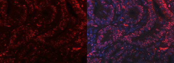
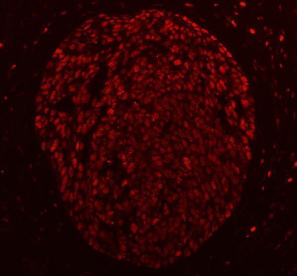
Treat IF as a separate validation experiment: the selected paraffin-section image documents chromogenic IHC (datasheet M02698-3). In colon, multiplex SUB1 with a validated glandular-cell marker, since glandular cells show high IHC staining (HPA: High in colon glandular cells; standard IF practice). Choose a far-red fluorophore and examine an unstained section to assess tissue autofluorescence (standard IF practice). SUB1 is nuclear and lacks a transmembrane segment, so optimize gentle permeabilisation for nuclear epitope access (UniProt P53999: nucleus and topology; standard IF practice). Compare any nucleoplasmic or nucleolar signal with no-primary controls before relating it to the IHC pattern (HPA: nucleoplasm and nucleoli; standard IF practice).
How do I reduce diffuse DAB background without losing nuclear SUB1 signal?
First compare stained sections with a primary-omission control to separate antibody-dependent signal from detection background (standard IHC practice). The selected image used 10% goat serum, 2 μg/ml primary antibody overnight at 4°C, and a peroxidase-conjugated secondary (datasheet M02698-3). Keep washes consistent, then titrate the primary concentration or incubation if background persists (standard IHC practice). Include a peroxidase-blocking step and control DAB development time, as these are general chromogenic IHC measures (standard IHC practice). Preserve readable nuclear staining in positive colon glandular cells while judging each adjustment (HPA: High in colon glandular cells; standard IHC practice).
How should I quantify SUB1 across sections with different cellular composition? ⚠ ANSWER MARKED FOR VERIFICATION
Define a nuclear positivity threshold using matched controls before scoring, because SUB1 has a nuclear tissue profile (HPA: ubiquitous nuclear expression; standard IHC practice). Report the percentage of positive nuclei and an H-score from 0–300 when intensity categories are reproducible (standard IHC practice). For spatial comparisons, report positive nuclei per mm² of viable, annotated tissue (standard IHC practice). Normalize each result to the nuclei of the specified cell population, and report that denominator alongside the score (standard IHC practice). Keep retrieval, staining batch, imaging and threshold settings consistent across sections, with colon glandular cells as a reference where available (datasheet M02698-3; HPA: High in colon glandular cells; standard IHC practice).
How can I distinguish true SUB1 positivity from tissue artefacts?
Convincing signal should be nuclear and occur in intact cells, matching SUB1 localisation and its ubiquitous nuclear tissue profile (UniProt P53999: nucleus; HPA: ubiquitous nuclear expression). Colon glandular cells provide a positive reference, whereas smooth-muscle cells have reported low staining (HPA: High in colon glandular cells; HPA: Low in smooth-muscle cells). Treat isolated cytoplasmic deposits or signal concentrated at section edges as suspect until controls support them (standard IHC practice). Exclude necrotic areas from scoring and compare suspicious DAB deposits with primary-omission and peroxidase controls (standard IHC practice). HPA rates tissue staining as Supported with medium RNA-to-protein consistency, so corroborate an unexpected cellular pattern independently (HPA: Supported; medium consistency).
Boster reagents

Best SUB1 / Activated RNA polymerase II transcriptional coactivator p15 IHC Antibodies

Four anti-SUB1 antibodies have human paraffin-section IHC images (catalog IHC captions); three also have human paraffin-section IF images (catalog IF captions). All list human, mouse, and rat reactivity (catalog reactivity).

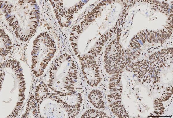
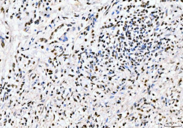
Real IHC data IHC analysis of PC4/SUB1 using anti-PC4/SUB1 antibody (M02698-3). PC4/SUB1 was detected in a paraffin-embedded section of human colorectal adenocarcinoma tissue. Heat mediated antigen retrieval was performed in EDTA buffer (pH 8.0, epitope retrieval solution). The tissue section was blocked with 10% goat serum. The tissue section was then incubated with 2 μg/ml mouse anti-PC4/SUB1 Antibody (M02698-3) overnight at 4°C. Peroxidase Conjugated Goat Anti-mouse IgG was used as secondary antibody and incubated for 30 minutes at 37°C. The tissue section was developed using HRP Conjugated Mouse IgG Super Vision Assay Kit (Catalog # SV0001) with DAB as the chromogen.
Anti-PC4/SUB1 Antibody ® (monoclonal, 8D9D1)
Cat # M02698-3
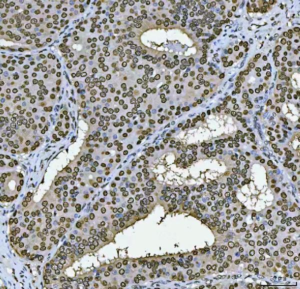
Real IHC data IHC analysis of PC4/SUB1 using anti-PC4/SUB1 antibody (M02698-2). PC4/SUB1 was detected in a paraffin-embedded section of human colonic adenoma tissue. Heat mediated antigen retrieval was performed in EDTA buffer (pH 8.0, epitope retrieval solution). The tissue section was blocked with 10% goat serum. The tissue section was then incubated with 2 μg/ml mouse anti-PC4/SUB1 Antibody (M02698-2) overnight at 4°C. Peroxidase Conjugated Goat Anti-mouse IgG was used as secondary antibody and incubated for 30 minutes at 37°C. The tissue section was developed using HRP Conjugated Mouse IgG Super Vision Assay Kit (Catalog # SV0001) with DAB as the chromogen.
Anti-PC4/SUB1 Antibody ® (monoclonal, 6B5B10)
Cat # M02698-2
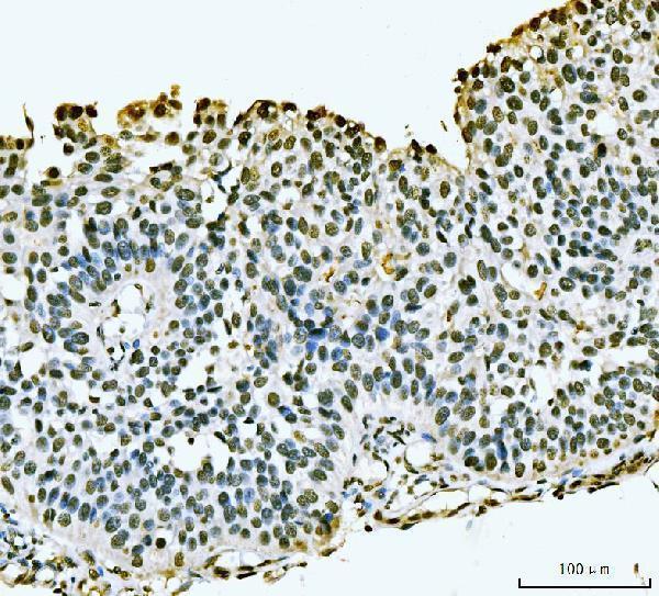
Real IHC data IHC analysis of PC4/SUB1 using anti-PC4/SUB1 antibody (A02698-1). PC4/SUB1 was detected in a paraffin-embedded section of human bladder epithelial carcinoma tissue. Heat mediated antigen retrieval was performed in EDTA buffer (pH 8.0, epitope retrieval solution). The tissue section was blocked with 10% goat serum. The tissue section was then incubated with 2 μg/ml rabbit anti-PC4/SUB1 Antibody (A02698-1) overnight at 4°C. Biotinylated goat anti-rabbit IgG was used as secondary antibody and incubated for 30 minutes at 37°C. The tissue section was developed using Strepavidin-Biotin-Complex (SABC) (Catalog # SA1022) with DAB as the chromogen.
Anti-PC4/SUB1 Antibody ®
Cat # A02698-1
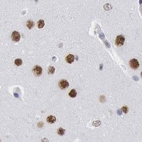
Real IHC data IHC analysis of PC4/SUB1 using anti-PC4/SUB1 antibody (PB10098). PC4/SUB1 was detected in a paraffin-embedded section of human esophageal squamous carcinoma tissue. Heat mediated antigen retrieval was performed in EDTA buffer (pH 8.0, epitope retrieval solution). The tissue section was blocked with 10% goat serum. The tissue section was then incubated with 2 μg/ml rabbit anti-PC4/SUB1 Antibody (PB10098) overnight at 4°C. Peroxidase Conjugated Goat Anti-rabbit IgG was used as secondary antibody and incubated for 30 minutes at 37°C. The tissue section was developed using HRP Conjugated Rabbit IgG Super Vision Assay Kit (Catalog # SV0002) with DAB as the chromogen.
Anti-PC4/SUB1 Antibody ®
Cat # PB10098

M02698-3 was imaged by IHC in human colorectal adenocarcinoma, and M02698-2 in human colonic adenoma (respective catalog IHC captions). A02698-1 was imaged by IHC in human bladder epithelial carcinoma, and PB10098 in human esophageal squamous carcinoma (respective catalog IHC captions).

Which to pick: For tissue IHC, choose monoclonal M02698-2 for its human paraffin-section example using EDTA retrieval at pH 8.0 and 2 μg/ml primary antibody; the fixative is unreported (M02698-2 catalog IHC caption; catalog clone). For IF/ICC planning, M02698-3 lists IF and has a human paraffin-section IF image at 5 μg/ml; ICC validation is unreported (M02698-3 catalog applications and IF caption). For cross-species IHC, M02698-3 lists IHC testing in human, mouse, and rat at 2–5 μg/ml, while its pictured paraffin-section IHC example is human and does not report the fixative (M02698-3 catalog dilution data and IHC caption).

Each figure is that product's own IHC / IF validation image from its datasheet.

References

  1. UniProt Consortium. UniProt entry P53999 (TCP4_HUMAN, Activated RNA polymerase II transcriptional coactivator p15).
  2. Human Protein Atlas. SUB1 tissue IHC expression (reliability: Supported).
  3. Human Protein Atlas. SUB1 subcellular location (ICC-IF): Localized to the nucleoplasm and nucleoli..
  4. Human Protein Atlas. SUB1 antibody validation summary (2 antibodies).
  5. MicroRNA-101 regulated transcriptional modulator SUB1 plays a role in prostate cancer. Oncogene 2016 — PMC5140777.
  6. Proteogenomic characterization of molecular and cellular targets for treatment-resistant subtypes in locally advanced cervical cancers. Molecular cancer 2025 — PMC11908047.
  7. PubMed PMID:8062392 — UniProt-cited evidence.
  8. PubMed PMID:8062391 — UniProt-cited evidence.
  9. PubMed PMID:15489334 — UniProt-cited evidence.