SUCLG2 / Succinate--CoA ligase [GDP-forming] subunit beta, mitochondrial · IHC design guide

Design Immunohistochemistry for SUCLG2

Plan SUCLG2 paraffin IHC around cytoplasmic tissue staining (HPA tissue IHC) and its mitochondrial location (UniProt). Start the IHC-validated antibody at 2–5 μg/ml (datasheet A08268-1), using glandular or endocrine cells as positive references and adipocytes as a low-signal comparison (HPA tissue IHC).

Evidence assembled Oct 2026 · For research use; verify linked source records and product datasheet before use
Immunohistochemistry protocol sheet for SUCLG2 (IHC for SUCLG2): expected localisation Cytoplasmic staining (HPA tissue IHC); mitochondrial protein (UniProt), antibody A08268-1, validated IHC image, and IHC protocol steps
Printable SUCLG2 IHC protocol sheet — expected localisation Cytoplasmic staining (HPA tissue IHC); mitochondrial protein (UniProt), antibody A08268-1, controls and protocol steps. Open the full SUCLG2 IHC guide →

SUCLG2 Immunohistochemistry Experimental Design Guide

Expected localisation, validated protocols, controls and antibodies — the at-a-glance facts below, then the full design guide.

Must know before staining
Expected localisation Cytoplasmic staining (HPA tissue IHC); mitochondrial protein (UniProt)
Staining pattern Glandular and endocrine cells show cytoplasmic staining (HPA tissue IHC)
Antigen retrieval EDTA pH 8.0 HIER, heat-mediated (datasheet A08268-1)
Positive control ⓘ Adrenal gland+4 more · see all
Negative control ⓘ Adipose tissue+3 more · see all
Important caveats
Reasons your staining may differ from the expected pattern.
Fixation Keep fixation consistent across sections. (standard IHC practice; not target-specific)
Caveat Staining intensity may diverge from RNA levels (HPA tissue IHC)
Regulation Mainly expressed in liver, kidney and heart (UniProt)
Isoform / epitope 2 isoforms; mature chain 38–432; epitope unknown (UniProt)
Section 1

Recommended SUCLG2 IHC & IF Protocols

The catalog antibody’s IHC-P protocol (datasheet: A08268-1) is accompanied by 3 published paraffin-section IHC protocols (PMC9096168; PMC7218928; PMC12623996).

Recommended immunohistochemistry (IHC-P) protocol parameters
SampleParaffin-embedded human breast cancer tissue; fixative not specified (datasheet A08268-1)
FixationImage fixative and duration unreported (datasheet A08268-1); verify before use.
Sectioning4–5 µm sections on charged slides (standard)
DeparaffinisationXylene, graded ethanol series to water (standard)
Antigen retrievalHeat retrieval: EDTA pH 8.0 (datasheet A08268-1); 20 min, 95–100 °C (standard)
Peroxidase block3% H2O2, 10 min, room temperature (standard)
Blocking10% goat serum (datasheet A08268-1)
Primary antibodyRabbit anti-SUCLG2, 2-5 μg/ml (datasheet A08268-1)
Primary incubationOvernight at 4 °C (datasheet A08268-1)
DetectionHRP-conjugated secondary, DAB chromogen (datasheet A08268-1)
CounterstainHematoxylin, blue, dehydrate and mount (standard)
Expected resultSUCLG2-positive staining in glandular cells of adrenal gland (HPA tissue IHC: High). HPA tissue profile: Ubiquitous cytoplasmic expression. No signal in the no-primary control.
💡Decision noteStart with heat-mediated EDTA retrieval at pH 8.0 for the catalog antibody (datasheet: A08268-1); pH 9.0 EDTA is a published alternative (PMC9096168).
Section 2

What Is the Expected SUCLG2 Staining Pattern?

SUCLG2 is mitochondrial and has no transmembrane segment (UniProt Q96I99). In paraffin-section IHC, expect predominantly cytoplasmic staining, with strong signal in selected glandular, endocrine, and respiratory epithelial cells (HPA: ubiquitous cytoplasmic expression; High in the listed cell populations). HPA rates the tissue staining profile Enhanced, but reports only medium agreement with RNA data and says external verification is pending (HPA: tissue IHC reliability).

What am I looking at on my slide?
Strong cytoplasmic staining in adrenal gland or breast glandular cells, bronchial respiratory epithelial cells, or colon endocrine cells (HPA: High in these cell populations).This matches the reported IHC pattern in those specific cells (HPA: ubiquitous cytoplasmic expression; High in the listed cell populations). Score the cell population and compartment together; a positive tissue section need not stain every cell equally (general IHC practice).
Predominantly nuclear staining, with little cytoplasmic signal.A dominant nuclear pattern conflicts with the reported cytoplasmic IHC profile and mitochondrial location (HPA: tissue IHC profile; UniProt Q96I99). Suspect nonspecific staining or a detection artefact, and check controls before calling it SUCLG2 (general IHC practice).
Strong staining in adipocytes or in glial cells of the caudate or hippocampus.Those specified cell populations were reported as Not detected (HPA: tissue IHC). Review morphology and controls for cross-reactivity or endogenous detection activity (general IHC practice). An unexpected positive result alone does not identify its cause, and HPA's tissue profile is pending external verification (HPA: reliability description).
Uniform color across tissue, blank areas, or many unrelated cell populations.This obscures the reported cell-associated cytoplasmic pattern (HPA: tissue IHC profile). Consider excess antibody, incomplete blocking or washing, and chromogenic detection background; compare a no-primary control under the same detection conditions (general IHC practice).
No staining in an expected positive population, such as adrenal glandular cells or appendix endocrine cells (HPA: High in these populations).A negative result there conflicts with the reported IHC observations (HPA: tissue IHC). First check section quality, retrieval, antibody and detection performance against a same-run positive control (general IHC practice). HPA does not establish a SUCLG2-specific fixation sensitivity (HPA: supplied tissue IHC evidence).
💡Expected SUCLG2 appearanceCall a section positive when selected reported High cell populations show clear cytoplasmic staining (HPA: tissue IHC); dominant nuclear staining or color spread across blank areas warrants artefact review (UniProt Q96I99; general IHC practice).
How each factor affects the staining
Which cells offer a useful positive reference?Adrenal and breast glandular cells, bronchial respiratory epithelial cells, and appendix or colon endocrine cells are reported High (HPA: tissue IHC). Select the relevant cell population within the section, rather than treating the whole organ as uniformly positive (general IHC practice).
Which cells offer a low-signal comparison?Adipocytes and specified glial populations are reported Not detected; ovarian stroma and smooth muscle cells are Low (HPA: tissue IHC). These are comparative observations, not proof that every such specimen must be negative (HPA: medium RNA–staining consistency; external verification pending).
How should compartment be judged?UniProt places SUCLG2 in mitochondria and reports no transmembrane segment (UniProt Q96I99). HPA describes its tissue IHC profile as cytoplasmic (HPA: tissue IHC). At light-microscope resolution, use cytoplasmic distribution for the IHC call; do not require visible individual mitochondria (general IHC practice).
What do isoforms and processing establish?UniProt lists two isoforms and a processed chain spanning residues 38–432 (UniProt Q96I99). The supplied record gives no antibody epitope, so it cannot establish whether either isoform or the removed precursor region changes staining (UniProt Q96I99; supplied HPA antibody summary).
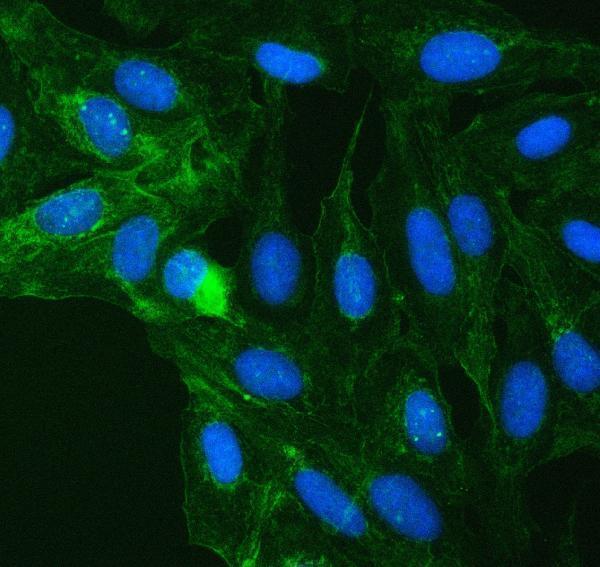
IF/ICC: what pattern should I expect?HPA reports mainly mitochondrial localization and an additional plasma-membrane location in ICC-IF (HPA: subcellular localization). Those observations help interpret IF images; they do not establish that membrane-only chromogenic IHC is the expected tissue pattern (HPA: cytoplasmic tissue IHC profile).
How much confidence does antibody validation add?Both listed rabbit polyclonal antibodies, HPA046705 and HPA051998, have Enhanced IHC status (HPA: antibody validation). HPA defines that status through agreement between independent antibodies or orthogonal data; its tissue profile still has medium RNA–staining consistency and awaits external verification (HPA: reliability description).
Why is my staining missing, weak or wrong?
SituationLikely causeNext action
Expected positive cells are blank.The staining run may have underperformed; the supplied sources identify expected positive cells but no target-specific retrieval condition (HPA: tissue IHC; supplied evidence).Check a same-run positive section and the antibody and detection controls; then optimize retrieval and dilution using the antibody's IHC instructions (general IHC practice).
Every cell and blank area shows chromogen.Broad background can result from antibody concentration, inadequate washing or blocking, or detection-reagent activity (general IHC practice).Compare a no-primary control; improve washing and blocking, and adjust antibody concentration or development time according to the assay controls (general IHC practice).
Signal is mainly nuclear.This conflicts with the mitochondrial assignment and cytoplasmic tissue profile (UniProt Q96I99; HPA: tissue IHC).Inspect nuclear counterstain and no-primary controls, then repeat with an independently validated IHC antibody if available (general IHC practice; HPA: two Enhanced IHC antibodies listed).
Unexpected adipocyte or glial staining is strong.The specified adipocytes and glial populations are reported Not detected, although HPA flags pending external verification (HPA: tissue IHC; reliability description).Confirm the cell identity, compare nearby expected-positive cells, and test a no-primary control before interpreting cross-reactivity or endogenous activity (general IHC practice).
A purported negative area stains weakly.Some populations are Low rather than Not detected, including ovarian stroma and smooth muscle cells (HPA: tissue IHC).Record intensity and exact cell type; compare with a reported High population stained in the same run instead of forcing a binary call (HPA: tissue IHC; general IHC practice).
IF shows membrane signal alongside mitochondrial signal.HPA reports an additional plasma-membrane location in ICC-IF (HPA: subcellular localization).Interpret that signal against IF controls and cell morphology (general IF practice). Use the separate IF/ICC guide for imaging workflow; the tissue IHC expectation remains cytoplasmic (HPA: tissue IHC profile).

Sample controls for SUCLG2 IHC & IF

🧪Run breast tissue first: glandular cells should stain strongly (HPA: High in breast glandular cells; selected-SKU IHC caption: breast cancer paraffin section). Use adipose tissue as a negative comparator, where adipocytes are reported as not detected (HPA: Not detected in adipocytes). On the positive slide, compare glandular staining with adjacent nonglandular cells as an internal background reference; their SUCLG2 status is not established by the supplied HPA row.
Positive control tissue: Adrenal gland (Glandular cells, HPA High)
Negative control tissue: Adipose tissue (HPA Not detected)
ICC-IF cell lines (HPA subcellular resource): HPA ICC-IF images show SUCLG2 in A-431, Hep-G2, U2OS, NIH 3T3, with annotated localisation: Mitochondria (approved) (HPA subcellular).
Technical controls: Include a no-primary, secondary-only control; a concentration-matched rabbit IgG isotype control (selected-SKU IHC caption: rabbit primary antibody); and a biological specificity control using SUCLG2 knockout material or peptide blocking if an appropriate immunizing peptide is available. Quench endogenous peroxidase and assess background DAB staining in the breast section (selected-SKU IHC caption: peroxidase detection with DAB).
⚠️Feasibility: Paraffin-section IHC is documented with heat retrieval in EDTA at pH 8.0, but the selected-SKU caption does not report a fixative, and no target-specific fixation window or fixation effect is supplied (selected-SKU IHC caption: retrieval conditions; fixative unreported). Retrieval was used in that example; its necessity relative to other conditions and whether frozen sections are easier are unreported. ICC-IF images support testing fluorescent localization, but do not establish that IF is easier than IHC (HPA subcellular: ICC-IF images and approved mitochondrial localization); assess endogenous peroxidase background in breast DAB sections (selected-SKU IHC caption: peroxidase detection with DAB).

HPA tissue IHC evidence for SUCLG2

Comprehensive Human Protein Atlas IHC scoring per tissue (reliability: Enhanced — Medium consistency between antibody staining and RNA expression data. Pending external verification.). Rows are taken directly from the HPA tissue chart — click any row's HPA link to view the source.

Positive expression · recommended positive controls

TissueCell typeLevelEvidenceSource
Adrenal gland Glandular cells High Protein (IHC) HPA →
Appendix Endocrine cells High Protein (IHC) HPA →
Breast Glandular cells High Protein (IHC) HPA →
Bronchus Respiratory epithelial cells High Protein (IHC) HPA →
Cervix Glandular cells High Protein (IHC) HPA →

Undetected expression · recommended negative controls

TissueCell typeLevelEvidenceSource
Adipose tissue Adipocytes Not detected Protein (IHC) HPA →
Caudate Glial cells Not detected Protein (IHC) HPA →
Cerebellum Cells in granular layer Not detected Protein (IHC) HPA →
Hippocampus Glial cells Not detected Protein (IHC) HPA →
Section 3

Advanced SUCLG2 IHC Tips

Troubleshoot SUCLG2 staining in paraffin sections using the catalog antibody’s tissue IHC conditions and compartment-specific controls.

What retrieval should I try first when SUCLG2 staining is weak in paraffin sections?
Start with heat-mediated retrieval in EDTA pH 8.0 for paraffin sections (datasheet A08268-1). The selected tissue IHC image used this retrieval before incubation with 2 μg/ml antibody overnight at 4°C, so keep those conditions consistent while assessing retrieval (datasheet A08268-1). Compare adjacent sections processed with and without retrieval, and inspect tissue preservation alongside staining intensity (standard IHC practice). If staining remains weak, test an alternative retrieval condition on matched sections as a fallback, recording the buffer, pH, heating and cooling conditions for each comparison (standard IHC practice).
How should I troubleshoot variable SUCLG2 staining across differently fixed paraffin samples?
Treat SUCLG2-specific fixation sensitivity as unknown: the selected paraffin tissue caption does not state a fixative (datasheet A08268-1). Record the fixative, fixation duration, tissue thickness and processing history for each sample before attributing differences to SUCLG2 abundance (standard IHC practice). Run matched sections together with the same EDTA pH 8.0 retrieval and antibody conditions to reduce staining-run variation (datasheet A08268-1; standard IHC practice). Check morphology and an appropriate tissue control, then report fixation differences as a limitation if the samples cannot be matched (standard IHC practice).
Which staining pattern supports mitochondrial SUCLG2 in a chromogenic section?
Look for cytoplasmic staining consistent with a mitochondrial protein, while recognizing that DAB does not resolve individual mitochondria reliably in every section (UniProt Q96I99 subcellular location; standard IHC practice). HPA describes ubiquitous cytoplasmic tissue staining and identifies mitochondria as the main subcellular location (HPA tissue IHC; HPA subcellular). Evaluate the signal within intact cells and compare adjacent tissue compartments under the same retrieval and detection conditions (standard IHC practice). HPA also reports an additional plasma membrane location, so membrane-associated signal merits review rather than automatic rejection, particularly when cytoplasmic staining is present (HPA subcellular).
Can this IHC stain distinguish SUCLG2 isoforms or processing-dependent epitopes?
Do not assign staining to a particular isoform without an antibody epitope map and isoform-specific validation; the record lists 2 isoforms (UniProt Q96I99 isoforms; standard IHC practice). The annotated mature chain spans residues 38–432, but the supplied tissue caption gives no epitope position (UniProt Q96I99 processing; datasheet A08268-1). SUCLG2 has an ATP-grasp domain at residues 46–274 and annotated modified residues, including phosphoserine 161 (UniProt Q96I99 domains and modified residues). If staining changes between samples, compare retrieval and tissue quality first; an epitope or modification explanation requires separate evidence (standard IHC practice).
How can IF help assess whether a disputed SUCLG2 IHC pattern is specific?
Use IF/ICC as a separate localisation check, since the supplied catalog image documents paraffin tissue IHC rather than IF conditions (datasheet A08268-1). Multiplex SUCLG2 with a validated marker for the cell population under study and, where useful, a mitochondrial marker to assess within-cell distribution (UniProt Q96I99 subcellular location; standard IF practice). Choose fluorophores after examining unstained tissue autofluorescence and include single-stain controls when interpreting overlap (standard IF practice). For an intracellular mitochondrial epitope, assess permeabilisation against signal and morphology; the antibody epitope and its membrane-facing side are not specified here (UniProt Q96I99 subcellular location; datasheet A08268-1; standard IF practice).
How do I reduce diffuse DAB background without losing SUCLG2 signal?
Check a no-primary control and inspect whether DAB appears in cells, damaged areas or across the section before changing antibody concentration (standard IHC practice). The selected image used 10% goat serum block and 2 μg/ml primary antibody, providing documented starting conditions for this reagent (datasheet A08268-1). Include a peroxidase block and review wash steps, secondary-only staining and DAB development time when background is widespread (standard chromogenic IHC practice). Titrate detection conditions on adjacent sections while retaining a tissue control, because overdevelopment can obscure the cytoplasmic pattern reported for SUCLG2 (HPA tissue IHC; standard IHC practice).
What should I measure when comparing SUCLG2 IHC across sections? ⚠ ANSWER MARKED FOR VERIFICATION
Define the cell population and viable tissue area before scoring, then apply the same rules to every section (standard IHC practice). For cellular staining, report percent positive cells and an intensity-weighted H-score on its conventional 0–300 scale; density per mm² can be useful when positive cells are sparse (standard IHC practice). Normalize counts to the relevant cell population or viable tissue area, and compare only sections stained and imaged under matched conditions (standard IHC practice). Record cytoplasmic staining separately from any membrane-associated signal, given HPA’s main mitochondrial and additional plasma membrane locations (HPA subcellular).
When should I question an apparently positive SUCLG2 IHC result?
Question staining confined to nuclei or extracellular material, because SUCLG2 is annotated as mitochondrial and HPA reports mainly cytoplasmic tissue expression (UniProt Q96I99 subcellular location; HPA tissue IHC). Compare the stained cell population with an appropriate control: HPA reports high glandular-cell staining in breast and no detected adipocyte staining in adipose tissue (HPA tissue IHC). Review section edges and necrotic areas for processing artefacts, and use a no-primary control to assess detection-related signal (standard IHC practice). A peroxidase-sensitive DAB signal or inconsistent compartment pattern calls for repeat staining and independent validation before a biological conclusion (standard chromogenic IHC practice).
Boster reagents

Best SUCLG2 / Succinate--CoA ligase [GDP-forming] subunit beta, mitochondrial IHC Antibodies

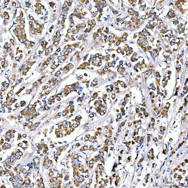
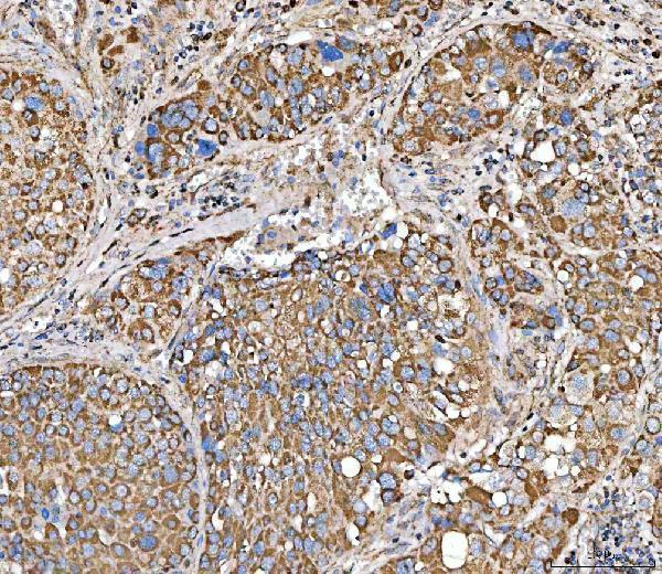
A08268-1 has documented IHC images from human paraffin sections and IF images from human tissue and cells (catalog image captions); the catalog lists human, mouse and rat reactivity (catalog: reactivity).

Real IHC data IHC analysis of SUCLG2 using anti-SUCLG2 antibody (A08268-1). SUCLG2 was detected in a paraffin-embedded section of human breast cancer tissue. Heat mediated antigen retrieval was performed in EDTA buffer (pH 8.0, epitope retrieval solution). The tissue section was blocked with 10% goat serum. The tissue section was then incubated with 2 μg/ml rabbit anti-SUCLG2 Antibody (A08268-1) overnight at 4°C. Peroxidase Conjugated Goat Anti-rabbit IgG was used as secondary antibody and incubated for 30 minutes at 37°C. The tissue section was developed using HRP Conjugated Rabbit IgG Super Vision Assay Kit (Catalog # SV0002) with DAB as the chromogen.
Anti-SUCLG2 Antibody ®
Cat # A08268-1

A08268-1 has IHC images from paraffin sections of human breast cancer, liver cancer, ovarian serous adenocarcinoma and prostate adenocarcinoma (catalog: IHC image captions). Its IF images show human ovarian cancer tissue and U20S cells (catalog: IF image captions).

Which to pick: For tissue IHC, choose A08268-1: its documented procedure used EDTA retrieval at pH 8.0 and 2 μg/ml primary antibody on paraffin sections (catalog: IHC image captions); the fixative is unreported (catalog: IHC image captions). For IF/ICC, A08268-1 is also listed for both applications, with a recommended concentration of 5 μg/ml (catalog: applications and dilution); follow the separate IF/ICC guide. For mouse or rat samples, A08268-1 lists reactivity with both species (catalog: reactivity), although the supplied IHC and IF images show human samples (catalog: image captions).

Each figure is that product's own IHC / IF validation image from its datasheet.

References

  1. UniProt Consortium. UniProt entry Q96I99 (SUCB2_HUMAN, Succinate--CoA ligase [GDP-forming] subunit beta, mitochondrial).
  2. Human Protein Atlas. SUCLG2 tissue IHC expression (reliability: Enhanced).
  3. Human Protein Atlas. SUCLG2 subcellular location (ICC-IF): Mainly localized to the mitochondria. In addition localized to the plasma membrane..
  4. Human Protein Atlas. SUCLG2 antibody validation summary (2 antibodies).
  5. Exploring the prognostic significance and therapeutic potential of SUCLG2 in prostate cancer. Frontiers in genetics 2025 — PMC12310446.
  6. Value of Immunohistochemical Expression of Apelin, Succinate Dehydrogenase B, Chromogranin B, Human Epidermal Growth Factor Receptor-2, Contactin 4, and Succinyl-CoA Synthetase Subunit Beta in Differentiating Metastatic From Non-Metastatic Pheochromocytoma and Paraganglioma. Frontiers in endocrinology 2022 — PMC9096168.
  7. Mesothelial Cell HIF1α Expression Is Metabolically Downregulated by Metformin to Prevent Oncogenic Tumor-Stromal Crosstalk. Cell reports 2019 — PMC7218928.
  8. Knockdown of SUCLG2 inhibits glioblastoma proliferation and promotes apoptosis through LMNA acetylation and the mediation of H4K16la lactylation. Cell death discovery 2025 — PMC12623996.
  9. PubMed PMID:14702039 — UniProt-cited evidence.
  10. PubMed PMID:16641997 — UniProt-cited evidence.
  11. PubMed PMID:15489334 — UniProt-cited evidence.