SULT2B1 / Sulfotransferase 2B1 · IHC design guide

Design Immunohistochemistry for SULT2B1

Plan SULT2B1 chromogenic IHC in paraffin sections using cytoplasmic squamous epithelial staining as the tissue reference (HPA tissue IHC). The guide covers fixation, staining controls and interpretation of possible nuclear signal associated with Ser-348 phosphorylation (UniProt).

Evidence assembled Oct 2026 · For research use; verify linked source records and product datasheet before use
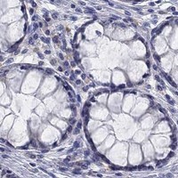
Immunohistochemistry protocol sheet for SULT2B1 (IHC for SULT2B1): expected localisation Predominantly cytoplasmic in squamous epithelium (HPA tissue IHC), antibody PB9822, validated IHC image, and IHC protocol steps
Printable SULT2B1 IHC protocol sheet — expected localisation Predominantly cytoplasmic in squamous epithelium (HPA tissue IHC), antibody PB9822, controls and protocol steps. Open the full SULT2B1 IHC guide →

SULT2B1 Immunohistochemistry Experimental Design Guide

Expected localisation, validated protocols, controls and antibodies — the at-a-glance facts below, then the full design guide.

Must know before staining
Expected localisation Predominantly cytoplasmic in squamous epithelium (HPA tissue IHC)
Staining pattern Cytoplasmic staining in squamous epithelial cells (HPA tissue IHC)
Antigen retrieval EDTA pH 8.0 HIER, heat-mediated (datasheet PB9822)
Positive control ⓘ Cervix+4 more · see all
Negative control ⓘ Adipose tissue+4 more · see all
Important caveats
Reasons your staining may differ from the expected pattern.
Fixation Matched tissue-IHC evidence does not establish the fixation claim. Validate the specimen-specific method before use. (selected-SKU IHC image PB9822)
Caveat Ser-348 phosphorylation may shift staining toward nuclei (UniProt)
Regulation Expression regulation is unreported (UniProt)
Isoform / epitope 2 isoforms; epitope coverage is unspecified (UniProt; datasheet PB9822)
Section 1

Recommended SULT2B1 IHC & IF Protocols

The catalog antibody’s IHC-P protocol (datasheet: PB9822) is accompanied by published SULT2B1 staining protocols for liver, colon and gastric tissue (PMC13485696; PMC8683511; PMC6874824).

Recommended immunohistochemistry (IHC-P) protocol parameters
SampleParaffin-embedded rat intestine tissue; fixative not specified (datasheet PB9822)
FixationImage fixative and duration unreported (datasheet PB9822); verify before use.
Sectioning4–5 µm sections on charged slides (standard)
DeparaffinisationXylene, graded ethanol series to water (standard)
Antigen retrievalHeat retrieval: EDTA pH 8.0 (datasheet PB9822); 20 min, 95–100 °C (standard)
Peroxidase block3% H2O2, 10 min, room temperature (standard)
Blocking10% goat serum (datasheet PB9822)
Primary antibodyRabbit anti-SULT2B1, 0.5-1μg/ml (datasheet PB9822)
Primary incubationOvernight at 4 °C (datasheet PB9822)
DetectionStreptavidin-biotin complex (SABC), DAB chromogen (datasheet PB9822)
CounterstainHematoxylin, blue, dehydrate and mount (standard)
Expected resultSULT2B1-positive staining in squamous epithelial cells of cervix (HPA tissue IHC: High). HPA tissue profile: Cytoplasmic expression mainly in squamous epithelial cells, including placenta and intestines. No signal in the no-primary control.
💡Decision noteStart with heat-mediated EDTA retrieval at pH 8.0 (datasheet: PB9822); potassium citrate microwave retrieval is a published alternative (PMC8683511).
Section 2

What Is the Expected SULT2B1 Staining Pattern?

SULT2B1 should stain mainly the cytoplasm of selected epithelial cells in paraffin sections, especially squamous epithelial cells and skin granular-layer cells (HPA tissue IHC: Enhanced reliability). Cytosolic and microsomal localization is consistent with a protein lacking a transmembrane segment; nuclear localization is also reported and depends on Ser-348 phosphorylation (UniProt O00204).

What am I looking at on my slide?
Strong cytoplasmic signal in cervical or esophageal squamous epithelial cells, or skin granular-layer cells.This matches the cell-specific High staining reported by HPA tissue IHC. Judge the stained cells within their tissue context; an unstained neighboring cell population does not negate a positive result (HPA tissue IHC).
Predominantly nuclear staining, with little cytoplasmic signal, in an expected positive epithelium.Reassess the pattern and controls before calling it specific. HPA tissue IHC describes mainly cytoplasmic staining. UniProt O00204 also reports nuclear localization dependent on Ser-348 phosphorylation, so nuclear signal alone cannot establish an artefact.
Strong signal in adipocytes or adrenal glandular cells.These cell populations are Not detected by HPA tissue IHC. Consider cross-reactivity or endogenous chromogenic detection activity; confirm with an expected positive tissue and detection controls before assigning SULT2B1 expression.
Uniform haze across epithelium, stroma, and empty regions of the section.A distribution that ignores cell boundaries is background rather than the cell-specific epithelial pattern reported by HPA tissue IHC. Check blocking, antibody concentration, washes, and chromogen development as general IHC practice.
No staining in an intact cervical squamous epithelium or skin granular layer.Both are High cell populations in HPA tissue IHC. First confirm that the expected cells are present; then review the IHC-validated antibody, antigen retrieval, detection reagents, and a positive control using general IHC practice.
💡Expected SULT2B1 appearanceCall a section positive when selected epithelial cells show clear, mainly cytoplasmic staining, potentially High in the HPA-listed squamous epithelia or skin granular layer; widespread compartment-blind haze or strong staining confined to HPA Not detected cell populations warrants investigation (HPA tissue IHC).
How each factor affects the staining
Positive and negative reference cellsHPA tissue IHC reports High staining in cervical, esophageal, oral, tonsillar, and vaginal squamous cells and skin granular-layer cells; adipocytes and adrenal glandular cells are Not detected. Compare matching cell populations, not whole organs.
Subcellular interpretationUniProt O00204 lists cytosol, microsomes, and nucleus, with Ser-348 phosphorylation required for nuclear translocation. HPA tissue IHC describes mainly cytoplasmic expression. A nuclear component needs context; its absence does not invalidate epithelial cytoplasmic staining.
Antibody validationHPA041724, HPA043539, and HPA076510 each have Enhanced IHC status (HPA antibodies). That supports the reported tissue pattern but does not establish that a different catalog antibody has identical specificity or staining intensity.
Isoforms and epitope coverageUniProt O00204 lists two isoforms but supplies no antibody epitope map here. If antibodies disagree, check their documented immunogens and isoform coverage; do not infer isoform-specific IHC staining from this record.
IF/ICC question: should vesicles be visible?HPA subcellular ICC-IF reports vesicles as the main location and cytosol as an additional location. This is an IF/ICC observation, not an IHC-P protocol or a requirement that chromogenic paraffin sections resolve vesicles.
Endogenous detection activityIn chromogenic IHC, endogenous enzyme activity can mimic antibody signal as a general IHC concern. A suitable detection control helps assess it; HPA tissue IHC does not identify a SULT2B1-specific endogenous activity problem.
Why is my staining missing, weak or wrong?
SituationLikely causeNext action
Expected positive epithelium has no visible signal.The relevant cells may be absent, or the staining workflow may have failed; HPA tissue IHC reports High staining only for specified cell populations.Verify the cell population on the counterstain, then check an IHC positive control, antibody dilution, retrieval, and detection reagents using general IHC practice.
Signal appears only in nuclei.This differs from HPA tissue IHC's mainly cytoplasmic pattern, although nuclear localization is possible after Ser-348 phosphorylation (UniProt O00204).Compare cytoplasmic signal and controls, repeat with an independently validated IHC antibody if available, and avoid calling nuclear-only staining specific from localization alone.
Strong staining appears in adipocytes or adrenal glandular cells.Those populations are Not detected in HPA tissue IHC; cross-reactivity or endogenous detection activity is possible.Run a detection control, review blocking and chromogen conditions, and compare a High epithelial reference tissue. Treat the unexpected result as unresolved until corroborated.
Diffuse brown background obscures cell boundaries.Excess nonspecific staining or chromogen deposition is possible under general IHC practice; HPA tissue IHC describes a cell-specific cytoplasmic pattern.Review antibody dilution, blocking, wash steps, and development time; use a detection control to locate the background source.
An epithelial section looks weak while a different tissue stains strongly.Expression is cell and tissue dependent: HPA tissue IHC lists some glandular or neuronal populations as Low and selected epithelial populations as High.Score the specified cell population and compare sections with matched detection conditions. Do not use organ-wide color intensity as a substitute for cell-specific interpretation.
ICC-IF shows puncta, but IHC-P looks broadly cytoplasmic.HPA subcellular ICC-IF places SULT2B1 mainly in vesicles, while HPA tissue IHC summarizes mainly cytoplasmic staining; the methods report different visual detail.Interpret each assay against its own HPA reference pattern. Use the separate IF/ICC guide for that assay; do not impose a punctate criterion on chromogenic IHC-P.

Sample controls for SULT2B1 IHC & IF

🧪Run cervix first and require staining in squamous epithelial cells (HPA: High in cervical squamous epithelial cells). Run adipose tissue as a negative, expecting adipocytes to lack specific staining (HPA: Not detected in adipocytes); on the cervical slide, use adjacent nonepithelial cells as candidate internal background controls only after confirming their identity and staining pattern.
Positive control tissue: Cervix (Squamous epithelial cells, HPA High)
Negative control tissue: Adipose tissue (HPA Not detected)
ICC-IF cell lines (HPA subcellular resource): HPA ICC-IF images show SULT2B1 in A-431, MCF-7, U2OS, OE19, with annotated localisation: Vesicles (enhanced) (HPA subcellular).
Technical controls: Include a no-primary, secondary-only control and a concentration-matched rabbit isotype control alongside the rabbit catalog antibody (PB9822 caption: rabbit primary). Use SULT2B1 knockout material as a biological negative if available; for chromogenic detection, block endogenous peroxidase and check for endogenous biotin background because the reported detection uses a biotinylated secondary and avidin–biotin complex (PB9822 caption).
⚠️Feasibility: A target-specific fixation window or fixation effect is unreported in the supplied evidence, and the fixative for the selected paraffin-section example is unreported (PB9822 caption). Heat retrieval with EDTA at pH 8.0 was used for rat intestine, but retrieval dependency in cervix is unreported (PB9822 caption). Whether frozen sections or IF/ICC are easier is unreported; in cervical sections, inspect surface debris and any blood-associated peroxidase signal before scoring epithelial staining (standard IHC practice).

HPA tissue IHC evidence for SULT2B1

Comprehensive Human Protein Atlas IHC scoring per tissue (reliability: Enhanced — High consistency between antibody staining and RNA expression data. External characterization data supports antibody staining.). Rows are taken directly from the HPA tissue chart — click any row's HPA link to view the source.

Positive expression · recommended positive controls

TissueCell typeLevelEvidenceSource
Cervix Squamous epithelial cells High Protein (IHC) HPA →
Epididymis Glandular cells High Protein (IHC) HPA →
Esophagus Squamous epithelial cells High Protein (IHC) HPA →
Nasopharynx Respiratory epithelial cells High Protein (IHC) HPA →
Oral mucosa Squamous epithelial cells High Protein (IHC) HPA →

Undetected expression · recommended negative controls

TissueCell typeLevelEvidenceSource
Adipose tissue Adipocytes Not detected Protein (IHC) HPA →
Adrenal gland Glandular cells Not detected Protein (IHC) HPA →
Appendix Endocrine cells Not detected Protein (IHC) HPA →
Bone marrow Hematopoietic cells Not detected Protein (IHC) HPA →
Cerebellum Cells in granular layer Not detected Protein (IHC) HPA →
Section 3

Advanced SULT2B1 IHC Tips

Troubleshoot SULT2B1 staining in paraffin sections by checking retrieval, cell type, compartment and controls before comparing signal intensity.

Which retrieval conditions should I try first if SULT2B1 staining is weak?
Start with heat-mediated retrieval in EDTA at pH 8.0 (datasheet PB9822). The selected paraffin-section image used this retrieval before staining rat intestine with 1 μg/ml primary antibody overnight at 4°C (datasheet PB9822). Keep section thickness, heating and cooling consistent across the positive control and test sections so retrieval differences do not masquerade as expression differences (standard IHC practice). If signal remains weak, compare a small retrieval series while checking for damaged morphology and increased background (standard IHC practice). Evaluate epithelial cytoplasmic staining, rather than overall brown color, when judging improvement (HPA: cytoplasmic expression mainly in squamous epithelial cells; standard IHC practice).
Could fixation explain weak or uneven SULT2B1 staining?
Target-specific sensitivity to fixation is unknown because the selected paraffin-section caption does not state a fixative (datasheet PB9822). Record the fixative, fixation duration and tissue processing for each section before comparing staining intensity (standard IHC practice). Review morphology and compare a known positive control processed alongside the test tissue; poorly preserved or unevenly fixed regions can make staining hard to interpret (standard IHC practice). Keep retrieval at EDTA pH 8.0 during that comparison so a retrieval change does not confound the result (datasheet PB9822; standard IHC practice). Do not assign a SULT2B1-specific fixation effect from tissue distribution or intracellular location alone (HPA: tissue IHC profile; UniProt O00204: subcellular locations).
Where should convincing SULT2B1 staining appear in an IHC section?
Assess cell identity and compartment together: tissue IHC describes mainly cytoplasmic expression in squamous epithelial cells, including placenta and intestines (HPA: tissue IHC profile). High staining is reported in skin granular-layer cells and in several squamous epithelial populations, providing useful comparison sites when available (HPA: positive tissue IHC entries). Cytosol, microsomes and nucleus are listed locations, while Ser-348 phosphorylation is required for nuclear translocation (UniProt O00204: subcellular location and modified residue). Thus, nuclear staining needs independent support rather than automatic dismissal or acceptance (UniProt O00204: nuclear translocation; standard IHC practice). Compare staining within intact epithelial cells and inspect the counterstain to distinguish cytoplasm from overlapping nuclei or surface deposits (standard IHC practice).
How do isoforms and an unknown epitope affect SULT2B1 IHC interpretation?
SULT2B1 has 2 annotated isoforms, but the supplied antibody caption does not identify its epitope or establish isoform coverage (UniProt O00204: isoforms; datasheet PB9822). Check the antibody's immunogen or epitope documentation before treating its staining as a readout of both isoforms (standard IHC practice). The annotated protein has no transmembrane segment, and Ser-348 phosphorylation is linked to nuclear translocation; neither fact establishes whether this antibody recognizes a modified epitope (UniProt O00204: topology and modified residue). If the biological question concerns an isoform or phosphorylation state, use independently validated reagents or orthogonal measurements (standard IHC practice). Report the result as antibody-detected SULT2B1 unless that specificity has been demonstrated (standard IHC practice).
How should I adapt these IHC findings when planning SULT2B1 IF multiplexing?
Validate the antibody separately for IF because the selected PB9822 evidence is a chromogenic paraffin-section experiment (datasheet PB9822). Multiplex with a marker for the expected epithelial cell population, then judge SULT2B1 signal within those cells against single-stain and no-primary controls (HPA: tissue IHC profile; standard IF practice). Choose a fluorophore channel after checking tissue autofluorescence, favoring a well-separated longer-wavelength channel when background is strong (standard IF practice). SULT2B1 is intracellular and has no transmembrane segment, so permeabilisation must give the antibody access to its epitope; the epitope itself is unspecified here (UniProt O00204: location and topology; datasheet PB9822). Interpret puncta cautiously because HPA reports vesicular and cytosolic IF localisation (HPA: subcellular summary).
What should I check when brown signal obscures specific SULT2B1 staining?
First compare a no-primary control with the stained section to identify signal arising from detection reagents or tissue (standard IHC practice). The selected image used 10% goat serum, a biotinylated secondary antibody, a streptavidin-biotin complex and DAB, so review blocking and the relevant detection controls when reproducing that workflow (datasheet PB9822; standard IHC practice). Apply a peroxidase block for chromogenic detection and assess any endogenous biotin contribution when using biotin-based detection (standard IHC practice). Reduce overly diffuse signal by titrating primary antibody around the reported 1 μg/ml condition while preserving positive-control staining (datasheet PB9822; standard IHC practice). Judge background against the expected epithelial cytoplasmic pattern, not color intensity alone (HPA: tissue IHC profile).
How can I quantify SULT2B1 staining across paraffin sections? ⚠ ANSWER MARKED FOR VERIFICATION
Define the epithelial population and compartment before scoring, because HPA describes mainly cytoplasmic tissue staining (HPA: tissue IHC profile). Within comparable regions, record the percentage of positive cells and staining intensity, then calculate an H-score if that scale fits the study question (standard IHC practice). Normalize to the number of eligible epithelial cells or epithelial area, rather than total section area, when tissue composition differs (standard IHC practice). If counting positive cells per mm², report the analyzed epithelial area and the threshold used to call a cell positive (standard IHC practice). Keep retrieval, detection, imaging and scoring rules consistent, and examine representative positive and low-staining controls (datasheet PB9822: EDTA pH 8.0; standard IHC practice).
How do I distinguish true SULT2B1 staining from artefact?
Favor reproducible staining in the expected cell population and compartment: HPA reports mainly epithelial cytoplasmic expression and high staining in skin granular-layer cells (HPA: tissue IHC profile and positive entries). A nuclear component can be biologically plausible, but Ser-348 phosphorylation is required for nuclear translocation, so nuclear-only staining warrants additional validation (UniProt O00204: subcellular location). Examine section edges, folds and necrotic regions for staining that follows damage rather than intact cell boundaries (standard IHC practice). Compare no-primary and detection controls to identify DAB signal from endogenous enzyme activity or the detection system (standard IHC practice). Treat staining in a reported negative cell population cautiously and confirm it with an independent method (HPA: negative tissue IHC entries; standard IHC practice).
Boster reagents

Best SULT2B1 / Sulfotransferase 2B1 IHC Antibodies

PB9822 has IHC images from paraffin-embedded rat intestine and human intestinal cancer tissue (PB9822 IHC captions), plus IF data from A431 cells (PB9822 IF caption).

Real IHC data IHC analysis of SULT2B1 using anti-SULT2B1 antibody (PB9822). SULT2B1 was detected in a paraffin-embedded section of rat intestine tissue. Heat mediated antigen retrieval was performed in EDTA buffer (pH 8.0, epitope retrieval solution). The tissue section was blocked with 10% goat serum. The tissue section was then incubated with 1 μg/ml rabbit anti-SULT2B1 Antibody (PB9822) overnight at 4°C. Biotinylated goat anti-rabbit IgG was used as secondary antibody and incubated for 30 minutes at 37°C. The tissue section was developed using Strepavidin-Biotin-Complex (SABC) (Catalog # SA1022) with DAB as the chromogen.
Anti-SULT2B1 Antibody ®
Cat # PB9822

PB9822 is listed for IHC in human and rat samples (PB9822 application and reactivity listings), with images from paraffin-embedded human intestinal cancer and rat intestine sections (PB9822 IHC captions). PB9822 is also listed for IF/ICC, with an IF image from A431 cells (PB9822 application listing; PB9822 IF caption).

Which to pick: Choose PB9822 for paraffin-section IHC; its captions document EDTA retrieval at pH 8.0 and 1 μg/ml primary antibody in the pictured sections (PB9822 IHC captions). The same rabbit antibody is the IF/ICC option, with an A431 IF image at 2 μg/ml (PB9822 catalog; PB9822 IF caption). PB9822 lists human and rat reactivity for cross-species IHC (PB9822 catalog); its IHC captions do not report the fixative (PB9822 IHC captions).

Each figure is that product's own IHC / IF validation image from its datasheet.

References

  1. UniProt Consortium. UniProt entry O00204 (ST2B1_HUMAN, Sulfotransferase 2B1).
  2. Human Protein Atlas. SULT2B1 tissue IHC expression (reliability: Enhanced).
  3. Human Protein Atlas. SULT2B1 subcellular location (ICC-IF): Mainly localized to vesicles. In addition localized to the cytosol..
  4. Human Protein Atlas. SULT2B1 antibody validation summary (3 antibodies).
  5. SULT2B1 promotes cholangiocyte epithelial-mesenchymal transition via Wnt/β-catenin/MMP7 pathway in biliary atresia. Pediatric research 2026 — PMC13485696.
  6. SULT2B1: a novel therapeutic target in colorectal cancer via modulation of AKT/PKM2-mediated glycolysis and proliferation. Journal of translational medicine 2024 — PMC11613740.
  7. Knockdown of OLR1 weakens glycolytic metabolism to repress colon cancer cell proliferation and chemoresistance by downregulating SULT2B1 via c-MYC. Cell death & disease 2021 — PMC8683511.
  8. Hydroxysteroid sulfotransferase 2B1 affects gastric epithelial function and carcinogenesis induced by a carcinogenic agent. Lipids in health and disease 2019 — PMC6874824.
  9. PubMed PMID:9799594 — UniProt-cited evidence.
  10. PubMed PMID:15057824 — UniProt-cited evidence.
  11. PubMed PMID:15489334 — UniProt-cited evidence.