This website uses cookies to ensure you get the best experience on our website.


- Table of Contents
Plan chromogenic SUMF2 IHC in paraffin sections using cytoplasmic staining as the tissue readout (HPA tissue IHC). Start the catalog antibody at 2–5 μg/ml (datasheet A08890), with kidney proximal tubules as a high-staining control and adipocytes as a non-detected comparator (HPA tissue IHC).

Expected localisation, validated protocols, controls and antibodies — the at-a-glance facts below, then the full design guide.
| Expected localisation | Cytoplasmic staining (HPA tissue IHC); ER lumen expected (UniProt) | |
| Staining pattern | General cytoplasmic staining in positive cells (HPA tissue IHC) | |
| Antigen retrieval | EDTA pH 8.0 HIER, heat-mediated (datasheet A08890) | |
| Positive control | Duodenum+4 more · see all | |
| Negative control | Adipose tissue+4 more · see all |
| Fixation | Keep fixation consistent across test and control paraffin sections. (selected-SKU IHC image A08890) | |
| Caveat | Staining has medium agreement with RNA data; verify specificity (HPA tissue IHC) | |
| Regulation | No specific expression regulator reported (UniProt) | |
| Isoform / epitope | 5 isoforms; mature chain 26–301; epitope unspecified (UniProt) |
The catalog antibody protocol uses EDTA pH 8.0 retrieval (datasheet A08890). Two published SUMF2 IHC protocols provide additional conditions (PMC11298345; PMC11803025).
| Sample | Paraffin-embedded human breast cancer tissue; fixative not specified (datasheet A08890) |
| Fixation | Image fixative and duration unreported (datasheet A08890); verify before use. |
| Sectioning | 4–5 µm sections on charged slides (standard) |
| Deparaffinisation | Xylene, graded ethanol series to water (standard) |
| Antigen retrieval | Heat retrieval: EDTA pH 8.0 (datasheet A08890); 20 min, 95–100 °C (standard) |
| Peroxidase block | 3% H2O2, 10 min, room temperature (standard) |
| Blocking | 10% goat serum (datasheet A08890) |
| Primary antibody | Rabbit anti-SUMF2, 2-5 μg/ml (datasheet A08890) |
| Primary incubation | Overnight at 4 °C (datasheet A08890) |
| Detection | HRP-conjugated secondary, DAB chromogen (datasheet A08890) |
| Counterstain | Hematoxylin, blue, dehydrate and mount (standard) |
| Expected result | SUMF2-positive staining in glandular cells of duodenum (HPA tissue IHC: High). HPA tissue profile: General cytoplasmic expression. No signal in the no-primary control. |
SUMF2 is a soluble endoplasmic reticulum lumen protein with no transmembrane segment (UniProt Q8NBJ7 topology). In paraffin section IHC, expect mainly cytoplasmic staining, especially in glandular cells, kidney proximal tubule microvilli, pancreatic endocrine cells, and placental syncytiotrophoblast cell bodies (HPA tissue IHC). HPA rates the tissue pattern Enhanced, while reporting medium agreement with RNA data and pending external verification (HPA tissue IHC).
| Strong cytoplasmic staining in duodenal, endometrial, gallbladder, seminal vesicle, or small intestinal glandular cells. | This matches HPA's High glandular cell staining and general cytoplasmic pattern (HPA tissue IHC). Score the named cell population and its cytoplasm; the UniProt ER lumen assignment supports an intracellular pattern but does not define a specific chromogenic texture (UniProt Q8NBJ7). |
| Strong signal at kidney proximal tubule microvilli, in pancreatic endocrine cells, or in syncytiotrophoblast cell bodies. | These are separate High HPA tissue observations (HPA tissue IHC). Record the precise compartment described for each tissue; do not interpret the kidney microvillar observation as evidence that SUMF2 is generally a plasma membrane protein (HPA tissue IHC; UniProt Q8NBJ7 topology). |
| Predominantly nuclear, extracellular, or broadly surface outlining stain replaces the expected cellular pattern. | Treat this as a localisation mismatch requiring control review: UniProt places SUMF2 in the ER lumen without a transmembrane segment, whereas HPA tissue IHC describes general cytoplasmic expression (UniProt Q8NBJ7 topology; HPA tissue IHC). A mismatch alone does not identify its cause. |
| Strong staining appears in adipocytes, cardiomyocytes, or another cell population HPA reports as not detected. | Check for antibody cross-reactivity or endogenous chromogen detection activity before calling the tissue positive (general IHC practice). HPA reports Not detected in those specified cells, but its Enhanced tissue profile still awaits external verification (HPA tissue IHC). |
| The whole section is diffusely colored, or a known HPA high staining cell population shows no signal. | Diffuse color prevents cell specific scoring; absent signal in an expected positive population makes a negative call unreliable (general IHC practice). Compare a matched negative reagent control and an HPA high tissue control before changing the biological interpretation (HPA tissue IHC; general IHC practice). |
| Cell and tissue selection | HPA reports High staining in several glandular populations, proximal tubule microvilli, endocrine pancreas, and syncytiotrophoblast cell bodies, but Not detected in specified adipocytes and cardiomyocytes (HPA tissue IHC). Choose controls by cell population, not merely by organ name. |
| Protein localisation and processing | UniProt assigns SUMF2 to the ER lumen, lists a signal peptide at residues 1–25, and identifies the mature chain as residues 26–301 (UniProt Q8NBJ7). The supplied record gives no antibody epitope, so these annotations cannot predict whether a particular processed region is detected. |
| Isoforms and glycosylation | UniProt lists 5 isoforms and one glycosylation site at residue 191 (UniProt Q8NBJ7). Without an antibody epitope or isoform coverage statement, neither annotation establishes a different IHC pattern or a required antigen retrieval condition. |
| Evidence and antibody choice | HPA rates tissue IHC Enhanced but notes medium agreement with RNA data and pending external verification (HPA tissue IHC). HPA024040 and CAB025743 each have Enhanced IHC validation; the supplied record does not establish that either catalog antibody shares their epitope or performance (HPA antibodies). |
| IF/ICC Q: Should its localisation determine this IHC call? | A: HPA lists vesicles and plasma membrane as uncertain ICC-IF locations, with images in A-431, SiHa, and U2OS (HPA subcellular). Use the tissue IHC pattern for paraffin section scoring; reconcile discordant compartments with the UniProt ER lumen annotation (HPA tissue IHC; UniProt Q8NBJ7). |
| Detection chemistry and fixation evidence | Endogenous detection activity can create color unrelated to antibody binding, so assess the chosen chromogenic system with appropriate controls (general IHC practice). No target specific fixation sensitivity, retrieval condition, or dilution is supplied; avoid attributing a SUMF2 result to any of these as an established target effect. |
| Situation | Likely cause | Next action |
|---|---|---|
| No stain in a glandular cell population reported as High. | Possible failed detection, weak antibody signal, or unsuitable assay conditions; the slide alone cannot distinguish these (general IHC practice). | Run a known positive tissue and the assay controls together; check antibody, secondary detection, chromogen development, and the established retrieval workflow before calling the sample negative (HPA tissue IHC; general IHC practice). |
| Stain covers nearly every cell and the surrounding section. | Diffuse background may reflect nonspecific binding, incomplete blocking, or detection chemistry rather than the HPA cell specific pattern (HPA tissue IHC; general IHC practice). | Inspect the negative reagent control, review blocking and washing, and adjust the working antibody concentration within validated assay limits (general IHC practice). Reassess only after cellular boundaries become interpretable. |
| A mainly nuclear or extracellular pattern dominates. | This conflicts with the ER lumen assignment and HPA general cytoplasmic tissue pattern; its specific cause remains unproven (UniProt Q8NBJ7 topology; HPA tissue IHC). | Compare a matched positive tissue and negative reagent control, then verify the antibody identity and detection setup before scoring the unusual compartment as SUMF2 (general IHC practice). |
| Adipocytes or cardiomyocytes stain as strongly as the expected positive cells. | HPA lists these cell populations as Not detected; cross-reactivity or endogenous detection activity is possible (HPA tissue IHC; general IHC practice). | Compare no primary antibody and positive tissue controls, and localise the color to exact cell types; confirm any persistent unexpected signal independently (general IHC practice). |
| Kidney staining appears restricted to proximal tubule microvilli. | That distribution matches HPA's High microvillar observation, although UniProt assigns SUMF2 to the ER lumen (HPA tissue IHC; UniProt Q8NBJ7). | Document the microvillar distribution separately from broader cytoplasmic staining and compare it with other HPA high cell populations before drawing a general localisation conclusion (HPA tissue IHC). |
| IF/ICC shows vesicular or surface signal that differs from the paraffin section. | HPA calls both ICC-IF locations uncertain; the tissue IHC profile has a separate Enhanced assessment (HPA subcellular; HPA tissue IHC). | Interpret each application against its own controls and evidence; use the tissue IHC pattern for this chromogenic result and document the discrepancy without assigning a new SUMF2 compartment (general IHC practice; HPA tissue IHC). |
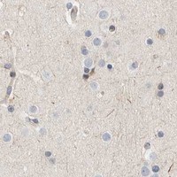
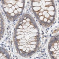


Comprehensive Human Protein Atlas IHC scoring per tissue (reliability: Enhanced — Medium consistency between antibody staining and RNA expression data. Pending external verification.). Rows are taken directly from the HPA tissue chart — click any row's HPA link to view the source.
| Tissue | Cell type | Level | Evidence | Source |
|---|---|---|---|---|
| Duodenum | Glandular cells | High | Protein (IHC) | HPA → |
| Endometrium | Glandular cells | High | Protein (IHC) | HPA → |
| Gallbladder | Glandular cells | High | Protein (IHC) | HPA → |
| Kidney | Proximal tubules (microvilli) | High | Protein (IHC) | HPA → |
| Pancreas | Pancreatic endocrine cells | High | Protein (IHC) | HPA → |
| Tissue | Cell type | Level | Evidence | Source |
|---|---|---|---|---|
| Adipose tissue | Adipocytes | Not detected | Protein (IHC) | HPA → |
| Bone marrow | Hematopoietic cells | Not detected | Protein (IHC) | HPA → |
| Breast | Adipocytes | Not detected | Protein (IHC) | HPA → |
| Cerebellum | Cells in granular layer | Not detected | Protein (IHC) | HPA → |
| Cerebral cortex | Endothelial cells | Not detected | Protein (IHC) | HPA → |
Use the catalog antibody’s paraffin section evidence as a starting point, then judge SUMF2 staining by compartment, cell type and controls.
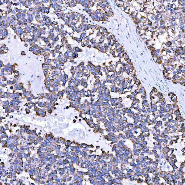
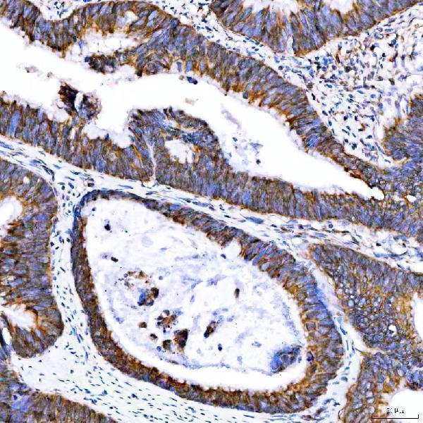
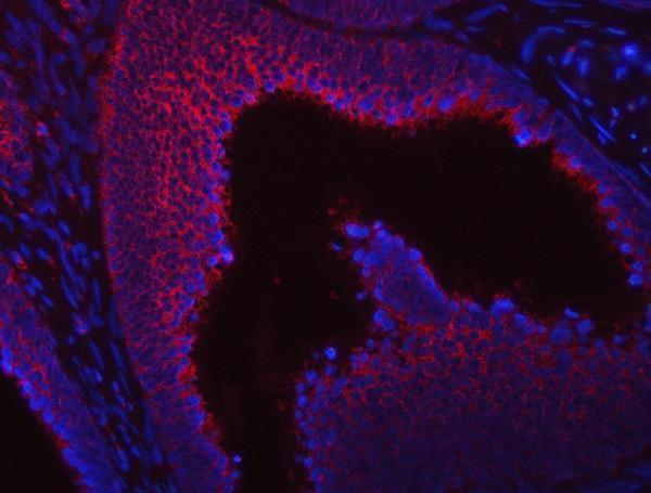
A08890 has IHC data from human paraffin breast cancer sections and IF data from human paraffin colon cancer, ovarian cancer and placenta sections (catalog: A08890 image captions).
A08890 is a rabbit antibody listed for human IHC and IF (catalog: A08890 host, applications and reactivity). Its captions show IHC in human paraffin breast cancer, colon adenocarcinoma, ovarian tumor and placenta sections, and IF in human paraffin colon cancer, ovarian cancer and placenta sections (catalog: A08890 image captions).
Which to pick: For tissue IHC, choose A08890: its paraffin breast cancer image uses EDTA retrieval at pH 8.0 and 2 μg/ml primary antibody, within the listed 2–5 μg/ml IHC range (A08890 IHC image caption; catalog: A08890 IHC dilution). For IF, A08890 is listed at 5 μg/ml and shown in human paraffin sections; the captions do not report a fixative (catalog: A08890 IF dilution and image captions). ICC and cross-species validation are not listed, so this payload does not support a recommendation for either use (catalog: A08890 applications and human reactivity).