SUN2 / SUN domain-containing protein 2 · IHC design guide

Design Immunohistochemistry for SUN2

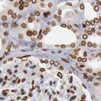
Plan SUN2 paraffin IHC around nuclear membrane staining seen across tissues (HPA tissue IHC). Start with the catalog antibody at 2–5 μg/ml (datasheet A03291-1) and use high-staining breast glandular cells as a reference (HPA tissue IHC).

Evidence assembled Oct 2026 · For research use; verify linked source records and product datasheet before use
Immunohistochemistry protocol sheet for SUN2 (IHC for SUN2): expected localisation Nuclear membrane in tissue sections (HPA tissue IHC), antibody A03291-1, validated IHC image, and IHC protocol steps
Printable SUN2 IHC protocol sheet — expected localisation Nuclear membrane in tissue sections (HPA tissue IHC), antibody A03291-1, controls and protocol steps. Open the full SUN2 IHC guide →

SUN2 Immunohistochemistry Experimental Design Guide

Expected localisation, validated protocols, controls and antibodies — the at-a-glance facts below, then the full design guide.

Must know before staining
Expected localisation Nuclear membrane in tissue sections (HPA tissue IHC)
Staining pattern Ubiquitous nuclear membranous staining (HPA tissue IHC)
Antigen retrieval EDTA pH 8.0 HIER, heat-mediated (datasheet A03291-1)
Positive control ⓘ Appendix+4 more · see all
Negative control ⓘ None in HPA (detected in all 44 tissues); use no-primary + isotype controls
Important caveats
Reasons your staining may differ from the expected pattern.
Fixation Keep fixation consistent (standard IHC practice; not target-specific); Matched tissue-IHC evidence does not establish the fixation claim. Validate the specimen-specific method before use. (selected-SKU IHC image A03291-1)
Caveat Widespread staining limits tissue-negative controls (HPA tissue IHC)
Regulation Slightly higher in some congenital hearts (UniProt)
Isoform / epitope 3 isoforms; epitope coverage needs checking (UniProt)
Section 1

Recommended SUN2 IHC & IF Protocols

The catalog antibody’s IHC-P protocol is accompanied by published SUN2 protocols for liver and breast tissue (PMC6170444; PMC4618625).

Recommended immunohistochemistry (IHC-P) protocol parameters
SampleParaffin-embedded human liver cancer tissue; fixative not specified (datasheet A03291-1)
FixationImage fixative and duration unreported (datasheet A03291-1); verify before use.
Sectioning4–5 µm sections on charged slides (standard)
DeparaffinisationXylene, graded ethanol series to water (standard)
Antigen retrievalHeat retrieval: EDTA pH 8.0 (datasheet A03291-1); 20 min, 95–100 °C (standard)
Peroxidase block3% H2O2, 10 min, room temperature (standard)
Blocking10% goat serum (datasheet A03291-1)
Primary antibodyRabbit anti-SUN2, 2-5 μg/ml (datasheet A03291-1)
Primary incubationOvernight at 4 °C (datasheet A03291-1)
DetectionStreptavidin-biotin complex (SABC), DAB chromogen (datasheet A03291-1)
CounterstainHematoxylin, blue, dehydrate and mount (standard)
Expected resultSUN2-positive staining in glandular cells of appendix (HPA tissue IHC: High). HPA tissue profile: Ubiquitous nuclear membranous expression. No signal in the no-primary control.
💡Decision noteStart with heat-mediated EDTA at pH 8.0 for the catalog antibody (datasheet: A03291-1); the published retrieval conditions used other antibodies (PMC6170444; PMC4618625).
Section 2

What Is the Expected SUN2 Staining Pattern?

SUN2 is an inner nuclear membrane protein with a nuclear segment at residues 1–212 and a perinuclear segment at 234–717 (UniProt Q9UH99 topology). In paraffin section IHC, expect nuclear membranous staining across many cell types: HPA describes a ubiquitous pattern with Supported reliability based on agreement between antibody staining and RNA expression (HPA tissue IHC).

What am I looking at on my slide?
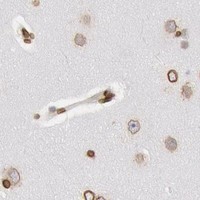
Distinct staining outlines nuclei in glandular, epithelial, hematopoietic or other represented cells.This matches the expected nuclear membrane compartment (HPA tissue IHC; UniProt Q9UH99). HPA reports High staining in appendix and breast glandular cells, bronchial respiratory epithelial cells and bone marrow hematopoietic cells (HPA tissue IHC). Judge the rim within identifiable cells; a positive field alone does not establish that every stained structure is a nucleus (general IHC interpretation).
Strong diffuse cytoplasmic or uniform intranuclear color appears without a discernible nuclear rim.That distribution does not match HPA's main nuclear membrane pattern (HPA tissue IHC). UniProt also lists an endosome membrane location, so an isolated nonnuclear signal is not by itself proof of cross-reactivity (UniProt Q9UH99). Review morphology and controls before assigning it to SUN2; the nuclear rim remains the clearest IHC interpretation (HPA tissue IHC; general IHC interpretation).
An unexpected cell population stains strongly while identifiable expected cells lack nuclear rim staining.Consider nonspecific antibody binding or endogenous detection activity (general IHC practice). HPA calls tissue expression ubiquitous and supplies no negative cell category, so staining of a particular cell type cannot be declared false solely because of its identity (HPA tissue IHC). Compare compartment, adjacent cells and detection controls before making that call (general IHC interpretation).
Color spreads across tissue, extracellular space or the slide, obscuring nuclear boundaries.This is background rather than an interpretable nuclear membrane pattern (HPA tissue IHC; general IHC interpretation). Check whether the signal persists when the primary antibody is omitted, and assess blocking, washing and chromogen development under the laboratory's standard IHC workflow (general IHC practice). A broadly dark section cannot support cell level scoring.
No nuclear rim is visible in a section expected to provide a positive comparison.Target-specific fixation effects are not established by the supplied assay evidence. Verify with a matched IHC source before attributing a result to fixation.
💡Expected SUN2 appearanceCall a result positive when identifiable cells show a defined nuclear membrane rim, especially in HPA High cell populations (HPA tissue IHC); diffuse field color or strong staining confined to the wrong compartment is suspect (HPA tissue IHC; general IHC interpretation).
How each factor affects the staining
Compartment and topologySUN2 spans the inner nuclear membrane at residues 213–233; its nuclear and perinuclear portions lie on opposite sides (UniProt Q9UH99 topology). This supports rim centered interpretation, but topology alone cannot predict which epitope a particular IHC antibody detects.
Tissue and cell contextHPA describes ubiquitous nuclear membranous expression and low RNA tissue specificity (HPA tissue IHC). Its High IHC examples are useful positive comparisons, but the supplied HPA record lists no negative or low staining group (HPA tissue IHC).
Antibody evidenceThe listed antibody, HPA001209, is a rabbit polyclonal with Supported IHC and ICC validation (HPA antibodies). Supported status helps frame the expected pattern; it does not validate every staining event in a new section.
Isoforms and epitope limitsUniProt lists three SUN2 isoforms and a full length chain of residues 1–717 (UniProt Q9UH99). The supplied record gives no antibody epitope or isoform coverage, so do not infer that the catalog antibody detects each isoform equally.
IF/ICC Q&A: what should be seen?A nuclear membrane signal is expected; HPA calls that location Supported in ICC/IF and lists images for A-431, U-251MG and U2OS (HPA subcellular; HPA antibodies). Fluorescence interpretation belongs to the separate IF/ICC guide; this IHC section supplies no IF protocol.
Why is my staining missing, weak or wrong?
SituationLikely causeNext action
A positive comparison has no visible nuclear rims.Section handling, detection or insufficient usable signal may be involved (general IHC practice); the supplied sources establish no SUN2 specific fixation effect.Confirm the expected cells are present using HPA High examples, then review the laboratory's IHC detection and control results (HPA tissue IHC; general IHC practice).
Nuclei stain throughout rather than at their edges.The distribution conflicts with HPA's nuclear membrane profile; heavy background or poorly resolved boundaries may obscure the rim (HPA tissue IHC; general IHC interpretation).Inspect well preserved cells at higher magnification and compare a detection control before scoring a compartment specific result (general IHC practice).
Cytoplasm is dark but nuclear rims are faint.Background or off target binding is possible (general IHC practice). UniProt's additional endosome membrane annotation does not establish that diffuse cytoplasmic IHC is a specific SUN2 pattern (UniProt Q9UH99).Prioritize the nuclear rim, compare expected cells and controls, and leave cytoplasmic staining unassigned if its specificity remains unclear (HPA tissue IHC; general IHC interpretation).
The whole section develops a similar brown haze.Nonspecific detection activity or excess chromogen development can reduce contrast (general chromogenic IHC practice).Check a section processed without primary antibody and review blocking, washes and development against the laboratory's established controls (general IHC practice).
Only one unexpected cell population is strongly positive.Cell identity alone is inconclusive because HPA reports ubiquitous expression and provides no negative population; nonspecific staining remains possible (HPA tissue IHC; general IHC interpretation).Verify morphology and nuclear rim localization, then compare nearby expected cells and a detection control before calling the population positive (HPA tissue IHC; general IHC practice).
Results differ between sections or staining runs.Variable section quality or detection conditions may alter interpretability (general IHC practice); no target specific retrieval or fixation sensitivity is supplied.Compare matched positive sections and control results, and document the conditions used before attributing a difference to SUN2 biology (HPA tissue IHC; general IHC practice).

Sample controls for SUN2 IHC & IF

🧪Run appendix first and expect glandular cells to stain at the nuclear envelope (HPA: High in appendix glandular cells; UniProt Q9UH99: nuclear envelope). HPA detects SUN2 in all 44 scored tissues, so there is no supported negative tissue; use no-primary and isotype slides for the negative comparison, and do not designate any cell population on the positive slide as an established internal negative (HPA: no negative tissues).
Positive control tissue: Appendix (Glandular cells, HPA High)
Negative control tissue: None in HPA: SUN2 is detected in all 44 scored tissues. Use a no-primary (secondary-only) and an isotype control instead.
ICC-IF cell lines (HPA subcellular resource): HPA ICC-IF images show SUN2 in A-431, U-251MG, U2OS, with annotated localisation: Nuclear membrane (supported) (HPA subcellular).
Technical controls: Include a secondary-only slide, a nonimmune rabbit IgG control matched to the primary antibody’s clonality and concentration where known, and SUN2-knockout material processed alongside the test sample as a biological negative (selected IHC caption: rabbit primary antibody). For chromogenic detection, quench endogenous peroxidase and check background from endogenous biotin if using the caption’s avidin–biotin detection system (selected IHC caption: SABC with DAB).
⚠️Feasibility: No target-specific fixation window or fixation effect is reported, and the selected A03291-1 paraffin-section caption does not state a fixative (selected IHC caption). The caption reports heat-mediated retrieval in EDTA at pH 8.0; it does not establish whether retrieval is required under other conditions (selected IHC caption). Frozen-section feasibility and whether IF is easier are unreported; HPA supports nuclear-membrane localization by ICC-IF, while endogenous biotin can complicate interpretation with the caption’s SABC detection in tissue sections (HPA: subcellular localization; selected IHC caption: SABC with DAB).

HPA tissue IHC evidence for SUN2

Comprehensive Human Protein Atlas IHC scoring per tissue (reliability: Supported — High consistency between antibody staining and RNA expression data.). Rows are taken directly from the HPA tissue chart — click any row's HPA link to view the source.

Positive expression · recommended positive controls

TissueCell typeLevelEvidenceSource
Appendix Glandular cells High Protein (IHC) HPA →
Bone marrow Hematopoietic cells High Protein (IHC) HPA →
Breast Glandular cells High Protein (IHC) HPA →
Bronchus Respiratory epithelial cells High Protein (IHC) HPA →
Caudate Glial cells High Protein (IHC) HPA →

Undetected expression · recommended negative controls

TissueCell typeLevelEvidenceSource
None in HPA: SUN2 is detected in all 44 scored tissues. Use a no-primary (secondary-only) and an isotype control instead.
Section 3

Advanced SUN2 IHC Tips

Troubleshoot SUN2 chromogenic IHC by checking nuclear envelope staining, section processing, detection controls and consistent scoring.

How should I adjust retrieval when SUN2 nuclear rims stain weakly?
Start with heat mediated retrieval in EDTA at pH 8.0 for paraffin sections (datasheet A03291-1). The selected tissue IHC example used that retrieval before 2 μg/ml primary antibody overnight at 4°C (datasheet A03291-1). If rims remain weak, adjust heating duration in small increments while holding antibody concentration and detection conditions constant; compare sections processed together. Include a consistently stained control section, because SUN2 is expected at the nuclear membrane (HPA subcellular: supported nuclear membrane; HPA tissue IHC: ubiquitous nuclear membranous expression). Excessive heating can damage morphology, so judge retrieval by both rim clarity and intact nuclei (standard IHC practice).
Could fixation explain weak or uneven SUN2 staining?
Target specific fixation sensitivity is unknown: the selected paraffin tissue caption does not report its fixative (datasheet A03291-1). Record fixative, fixation duration, tissue thickness and processing history for each case, then compare matched sections using the same retrieval and staining run (standard IHC practice). Inspect nuclear preservation before interpreting a faint or absent rim, since damaged nuclei can undermine compartment based scoring (standard IHC practice; HPA subcellular: supported nuclear membrane). If a fixation change is necessary, test it on matched material with a positive control rather than attributing improvement to SUN2 biology. Keep the documented EDTA pH 8.0 retrieval constant during that comparison (datasheet A03291-1).
What staining pattern should count as SUN2 positive in tissue?
Score a distinct rim around intact nuclei as the primary pattern: tissue IHC reports ubiquitous nuclear membranous expression, and subcellular data support nuclear membrane localisation (HPA tissue IHC; HPA subcellular). SUN2 is an inner nuclear membrane protein with a transmembrane segment at residues 213–233 (UniProt Q9UH99 topology). Its record also lists endosome membrane localisation, so document any punctate cytoplasmic signal separately rather than combining it with nuclear rim scores (UniProt Q9UH99 subcellular). Review thin, crowded or tangentially cut nuclei at higher magnification before calling a rim absent (standard IHC practice). Compare the compartment pattern with a concurrently stained control section (standard IHC practice).
Can epitope position change the apparent SUN2 staining pattern?
Map the antibody epitope before comparing staining across retrieval conditions or antibodies; its position is not supplied in the selected caption (datasheet A03291-1). SUN2 has 3 annotated isoforms and a transmembrane segment at residues 213–233, with residues 1–212 nuclear facing and 234–717 facing the perinuclear space (UniProt Q9UH99 isoforms and topology). The SUN domain spans residues 555–716, and a glycosylation site is annotated at residue 636 (UniProt Q9UH99 domains and glycosylation). These features identify questions to check against the immunogen sequence; they do not establish which isoforms this antibody detects. Interpret differing staining only after confirming epitope coverage and comparable processing (standard IHC practice).
How should I investigate a questionable SUN2 rim by IF?
Use IF as an orthogonal localisation check while keeping chromogenic paraffin IHC as this page’s primary readout. Multiplex SUN2 with a marker for the expected cell population, such as a glandular cell marker when examining appendix glandular cells, which show high SUN2 staining (HPA tissue IHC: high in appendix glandular cells). Choose fluorophores and filter sets after checking tissue autofluorescence, and include single stain controls to assess spectral bleed through (standard IF practice). If the epitope maps to residues 1–212, optimise access to the nuclear facing side; residues 234–717 face the perinuclear space, so permeabilisation requirements may differ (UniProt Q9UH99 topology). Do not infer a validated IF fixation or permeabilisation condition from the IHC caption.
How can I reduce diffuse DAB signal without losing nuclear rims?
First compare the full stain with a section omitting primary antibody to assess detection related background (standard IHC practice). The selected workflow used 10% goat serum block, biotinylated goat anti rabbit secondary, a streptavidin biotin complex and DAB (datasheet A03291-1). Check the peroxidase block and consider endogenous biotin controls when troubleshooting this detection format; treat both as general IHC controls (standard IHC practice). Titrate primary incubation or detection development one variable at a time, preserving a matched control section (standard IHC practice). Retain signal only when intact nuclear rims remain distinct from diffuse cytoplasmic or extracellular deposit (HPA subcellular: supported nuclear membrane; standard IHC practice).
How should I score SUN2 across sections with different staining intensity? ⚠ ANSWER MARKED FOR VERIFICATION
Define positivity as a discernible nuclear rim in an intact cell before examining study groups (HPA tissue IHC: ubiquitous nuclear membranous expression; standard IHC practice). Report the percentage of rim positive cells and, when intensity matters, a prespecified nuclear rim H score using consistent intensity bins (standard IHC practice). Normalise counts to the number of evaluable nuclei in the same annotated cell population, or report positive cell density per mm² of evaluable tissue (standard IHC practice). Exclude folds, torn edges and necrotic areas by the same rules in every section (standard IHC practice). Keep retrieval, imaging, chromogen development and scoring thresholds consistent across batches (standard IHC practice).
When is apparent SUN2 positivity more likely to be artefact?
A reproducible rim around intact nuclei is consistent with the supported nuclear membrane localisation of SUN2 (HPA subcellular: supported nuclear membrane). Diffuse staining confined to section edges, folds or necrotic regions should trigger review of processing and background controls before it is scored (standard IHC practice). Check whether staining lies in the annotated cell population rather than assuming every brown deposit belongs to the intended cells (standard IHC practice). A primary omitted control helps identify detection background, including endogenous enzyme signal in a DAB workflow (standard IHC practice). Interpret cytoplasmic puncta separately: endosome membrane localisation is annotated, but the tissue IHC profile is nuclear membranous (UniProt Q9UH99 subcellular; HPA tissue IHC).
Boster reagents

Best SUN2 / SUN domain-containing protein 2 IHC Antibodies

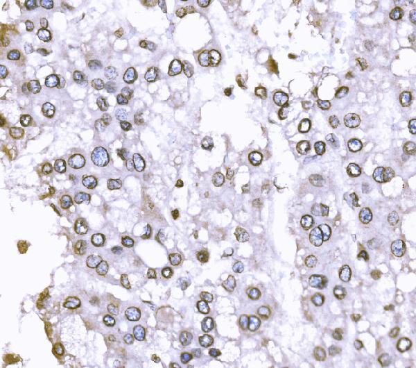
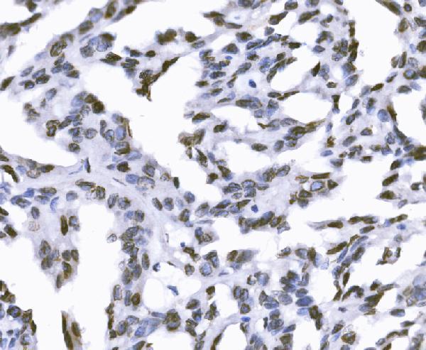
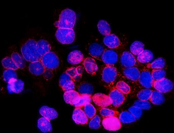
A03291-1 has human paraffin-section IHC images and IF imaging in A431 cells (catalog image captions); M03291-1 lists human, mouse, and rat reactivity (catalog reactivity).

Real IHC data IHC analysis of SUN2 using anti-SUN2 antibody (A03291-1). SUN2 was detected in a paraffin-embedded section of human liver cancer tissue. Heat mediated antigen retrieval was performed in EDTA buffer (pH 8.0, epitope retrieval solution). The tissue section was blocked with 10% goat serum. The tissue section was then incubated with 2 μg/ml rabbit anti-SUN2 Antibody (A03291-1) overnight at 4°C. Biotinylated goat anti-rabbit IgG was used as secondary antibody and incubated for 30 minutes at 37°C. The tissue section was developed using Strepavidin-Biotin-Complex (SABC) (Catalog # SA1022) with DAB as the chromogen.
Anti-SUN2 Antibody ®
Cat # A03291-1

A03291-1 has IHC images from human liver, renal, lung, and breast cancer paraffin sections, plus an IF image from A431 cells (A03291-1 image captions). M03291-1 lists IHC and ICC/IF applications for human, mouse, and rat, but has no IHC or IF images in the payload (M03291-1 applications, reactivity, and image fields).

Which to pick: Choose A03291-1 for tissue IHC: its own captions document human paraffin sections, EDTA retrieval at pH 8.0, and 2 μg/ml primary antibody; the fixative is unreported (A03291-1 IHC image captions). For IF/ICC, A03291-1 has an image from A431 cells at 5 μg/ml (A03291-1 IF image caption). For mouse or rat samples, M03291-1 is the listed option because it is a rabbit monoclonal with IHC and ICC/IF applications and human, mouse, and rat reactivity; the payload provides no IHC or IF images for it (M03291-1 catalog fields).

Each figure is that product's own IHC / IF validation image from its datasheet.

References

  1. UniProt Consortium. UniProt entry Q9UH99 (SUN2_HUMAN, SUN domain-containing protein 2).
  2. Human Protein Atlas. SUN2 tissue IHC expression (reliability: Supported).
  3. Human Protein Atlas. SUN2 subcellular location (ICC-IF): Localized to the nuclear membrane..
  4. Human Protein Atlas. SUN2 antibody validation summary (1 antibodies).
  5. Suppression of SUN2 by DNA methylation is associated with HSCs activation and hepatic fibrosis. Cell death & disease 2018 — PMC6170444.
  6. Loss of Sun2 promotes the progression of prostate cancer by regulating fatty acid oxidation. Oncotarget 2017 — PMC5685696.
  7. Global loss of a nuclear lamina component, lamin A/C, and LINC complex components SUN1, SUN2, and nesprin-2 in breast cancer. Cancer medicine 2015 — PMC4618625.
  8. Activation of Pyramidal Neurons in Mouse Medial Prefrontal Cortex Enhances Food-Seeking Behavior While Reducing Impulsivity in the Absence of an Effect on Food Intake. Frontiers in behavioral neuroscience 2016 — PMC4813092.
  9. PubMed PMID:10818110 — UniProt-cited evidence.
  10. PubMed PMID:17132086 — UniProt-cited evidence.
  11. PubMed PMID:9734811 — UniProt-cited evidence.