This website uses cookies to ensure you get the best experience on our website.


- Table of Contents
Plan chromogenic IHC for SUPT5H using its widespread nuclear tissue pattern (HPA tissue IHC). Compare nuclear staining across sections with consistent fixation, and interpret intensity in light of the medium consistency between antibody staining and RNA data (HPA tissue IHC).

Expected localisation, validated protocols, controls and antibodies — the at-a-glance facts below, then the full design guide.
| Expected localisation | Nuclear staining across tissues (HPA tissue IHC) | |
| Staining pattern | Widespread nuclear staining across cell types (HPA tissue IHC) | |
| Antigen retrieval | EDTA pH 8.0 HIER, heat-mediated (datasheet A03426-2) | |
| Positive control | Placenta+4 more · see all | |
| Negative control | None in HPA (detected in all 45 tissues); use no-primary + isotype controls |
| Fixation | Keep fixation conditions consistent across sections. (standard IHC practice; not target-specific) | |
| Caveat | Antibody staining has medium consistency with RNA data (HPA tissue IHC) | |
| Regulation | Ubiquitously expressed (UniProt) | |
| Isoform / epitope | 2 isoforms; epitope differences are unspecified (UniProt) |
The catalog antibody's IHC-P protocol (datasheet A03426-2) and one published SUPT5H IHC protocol (PMC4741733) provide starting conditions for paraffin sections.
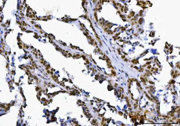
| Sample | Paraffin-embedded human ovarian serous carcinoma tissue; fixative not specified (datasheet A03426-2) |
| Fixation | Image fixative and duration unreported (datasheet A03426-2); verify before use. |
| Sectioning | 4–5 µm sections on charged slides (standard) |
| Deparaffinisation | Xylene, graded ethanol series to water (standard) |
| Antigen retrieval | Heat retrieval: EDTA pH 8.0 (datasheet A03426-2); 20 min, 95–100 °C (standard) |
| Peroxidase block | 3% H2O2, 10 min, room temperature (standard) |
| Blocking | 10% goat serum (datasheet A03426-2) |
| Primary antibody | Rabbit anti-SUPT5H, 2-5 μg/ml (datasheet A03426-2) |
| Primary incubation | Overnight at 4 °C (datasheet A03426-2) |
| Detection | HRP-conjugated secondary, DAB chromogen (datasheet A03426-2) |
| Counterstain | Hematoxylin, blue, dehydrate and mount (standard) |
| Expected result | SUPT5H-positive staining in cytotrophoblasts of placenta (HPA tissue IHC: High). HPA tissue profile: Ubiquitous nuclear expression. No signal in the no-primary control. |
SUPT5H is a nuclear transcription elongation factor with no transmembrane segment (UniProt O00267: location, topology). Expect nuclear staining across many cell types, with prominent staining in placental cytotrophoblasts (HPA: ubiquitous nuclear expression; cytotrophoblasts High). HPA rates the tissue staining pattern Supported, with medium consistency between antibody staining and RNA expression; interpret intensity differences with that qualification (HPA: tissue IHC reliability).
| Nuclear staining in placental cytotrophoblasts, with weaker or variable nuclear staining elsewhere (HPA: cytotrophoblasts High; ubiquitous nuclear profile). | This matches the expected compartment and a documented strong tissue example. Compare nuclei within each cell population; broad expression does not require every nucleus to have identical intensity (UniProt O00267: nucleus, ubiquitous expression; HPA: tissue IHC profile). |
| Predominantly cytoplasmic, membranous, or extracellular color while nuclei remain pale (UniProt O00267: nucleus, no transmembrane segment). | Treat this as a discordant pattern requiring controls, rather than evidence of SUPT5H redistribution. Check whether staining persists without primary antibody and review the detection system (UniProt O00267: location, topology; general IHC practice). |
| Intense signal in a cell population expected to stain only weakly, especially with little corresponding nuclear signal (HPA: glial cells in hippocampus and caudate Low). | Consider cross-reactivity or endogenous detection activity; low HPA staining is not proof that these cells lack SUPT5H. Resolve the signal's compartment and control dependence before calling it specific (HPA: Low, not negative; general IHC practice). |
| Uniform color over cells and tissue spaces that obscures nuclear boundaries (HPA: expected nuclear profile). | This is background rather than an interpretable nuclear pattern. Examine a no-primary control and review blocking, washing, and chromogen development for the chosen detection system (HPA: tissue IHC profile; general IHC practice). |
| No nuclear signal in placental cytotrophoblasts on an otherwise interpretable section (HPA: cytotrophoblasts High). | A false negative is plausible. Confirm that the antibody and detection reagents worked, then review the antibody's IHC-P instructions and the laboratory's retrieval settings; HPA does not establish SUPT5H-specific fixation sensitivity (HPA: cytotrophoblasts High; general IHC practice). |
| Compartment | SUPT5H is nuclear and lacks a transmembrane segment; score nuclear color, and investigate dominant membrane or cytoplasmic color before assigning specificity (UniProt O00267: location, topology). |
| Tissue and cell intensity | HPA reports High staining in placental cytotrophoblasts, Medium in several other listed populations, and Low in some glial and other cells. These are observed levels, not guaranteed results for every section (HPA: tissue IHC). |
| IHC antibody evidence | HPA lists IHC as Supported for HPA029273 and CAB034370, and rates the tissue profile Supported with medium RNA–staining consistency. Check the validation status of the antibody actually used (HPA: antibodies; tissue IHC reliability). |
| Isoforms and modifications | UniProt lists two isoforms and multiple modified residues. Their effect on staining cannot be assigned without an antibody epitope and recognition data; avoid interpreting intensity as an isoform or modification readout (UniProt O00267: isoforms, modified residues). |
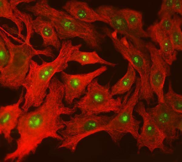
| IF/ICC question: where should signal appear? | In the nucleoplasm (HPA: subcellular ICC-IF, supported). HPA shows ICC-IF images in A-431, U-251MG, and U2OS and lists ICC validation as Enhanced for HPA029273 and Supported for CAB034370 (HPA: subcellular; antibodies). |
| Situation | Likely cause | Next action |
|---|---|---|
| Placental cytotrophoblast nuclei have no visible signal (HPA: cytotrophoblasts High). | The run may have failed, or the chosen conditions may not yield detectable staining (general IHC practice). | Verify detection reagents and a run control; then check the catalog antibody's IHC-P instructions and the laboratory's retrieval settings. Do not infer SUPT5H fixation sensitivity from this result (general IHC practice; HPA: tissue IHC scope). |
| Cytoplasm or cell borders stain more strongly than nuclei (UniProt O00267: nucleus). | The distribution conflicts with SUPT5H's reported location and may reflect nonspecific binding or detection activity (UniProt O00267: location; general IHC practice). | Compare a no-primary control and inspect nuclear counterstain alignment; assess whether the color truly lies within nuclei before scoring (general IHC practice). |
| Weakly profiled cells stain as intensely as placental cytotrophoblasts (HPA: selected cells Low; cytotrophoblasts High). | Cross-reactivity or endogenous activity is possible, but HPA intensity levels do not define a negative cell population (HPA: tissue IHC; general IHC practice). | Check compartment and no-primary staining, and compare matched regions under the same detection conditions; report the discrepancy without declaring those cells SUPT5H-negative (HPA: ubiquitous nuclear profile; general IHC practice). |
| Brown or other chromogenic color spreads across tissue spaces and masks nuclei (HPA: expected nuclear profile). | Excess background may arise from detection activity, inadequate washing, or excessive chromogen development (general IHC practice). | Review the no-primary control, blocking and washes; adjust development within the detection system's instructions, then reassess nuclear contrast (general IHC practice). |
| Staining appears only in a few cell types despite an adequate run control (HPA: ubiquitous nuclear expression). | Technical loss of detectable signal is possible; HPA's Supported profile has only medium consistency with RNA expression (HPA: reliability; general IHC practice). | Compare expected nuclear populations in the same section and check antibody and detection conditions before treating the restricted pattern as biological (HPA: tissue IHC profile; general IHC practice). |
| A low-staining HPA tissue is proposed as a negative control (HPA: listed Low populations; negative list empty). | Low staining means weak observed signal, not established absence of SUPT5H (HPA: tissue IHC). | Use a no-primary control to assess detection background, and interpret low-staining populations as relative intensity comparisons only (HPA: tissue IHC; general IHC practice). |


Comprehensive Human Protein Atlas IHC scoring per tissue (reliability: Supported — Medium consistency between antibody staining and RNA expression data.). Rows are taken directly from the HPA tissue chart — click any row's HPA link to view the source.
| Tissue | Cell type | Level | Evidence | Source |
|---|---|---|---|---|
| Placenta | Cytotrophoblasts | High | Protein (IHC) | HPA → |
| Adipose tissue | Adipocytes | Medium | Protein (IHC) | HPA → |
| Adrenal gland | Glandular cells | Medium | Protein (IHC) | HPA → |
| Appendix | Glandular cells | Medium | Protein (IHC) | HPA → |
| Bone marrow | Hematopoietic cells | Medium | Protein (IHC) | HPA → |
| Tissue | Cell type | Level | Evidence | Source |
|---|---|---|---|---|
| None in HPA: SUPT5H is detected in all 45 scored tissues. Use a no-primary (secondary-only) and an isotype control instead. | ||||
Troubleshoot SUPT5H staining in paraffin sections by prioritising nuclear signal, matched controls and the conditions documented for the IHC-validated antibody.
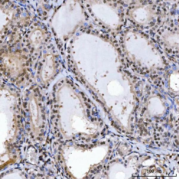
A03426-2 has paraffin-section IHC images from human tissue and an IF image from human cells (catalog image captions); M03426 lists IHC and IF applications with human, mouse and rat reactivity (catalog applications/reactivity).
A03426-2 shows IHC in paraffin sections of human ovarian serous carcinoma, thyroiditis and tonsil, plus IF in A549 cells (catalog IHC/IF image captions). M03426 lists IHC and ICC/IF applications and human, mouse and rat reactivity; no IHC or IF image is supplied (catalog applications/reactivity; catalog image listings).
Which to pick: Choose A03426-2 for tissue IHC because its own images document paraffin sections and EDTA retrieval at pH 8.0; the fixative is unreported (A03426-2 IHC image captions). For IF/ICC, A03426-2 has an A549 cell image, while M03426 lists ICC/IF without an image (catalog IF image caption; catalog applications/image listings). Consider M03426 when rat reactivity matters because it lists rat and is monoclonal; establish performance in the intended specimen before use (catalog reactivity/clone; catalog image listings).