SV2A / Synaptic vesicle glycoprotein 2A · IHC design guide

Design Immunohistochemistry for SV2A

Plan SV2A chromogenic IHC on paraffin sections using 2 µg/mL catalog antibody with overnight incubation at 4°C (datasheet A03752-3). Compare neuropil, Purkinje cell, retinal and pancreatic endocrine staining with the tissue profile (HPA tissue IHC).

Evidence assembled Oct 2026 · For research use; verify linked source records and product datasheet before use
Immunohistochemistry protocol sheet for SV2A (IHC for SV2A): expected localisation Neuropil cytoplasm (HPA tissue IHC); synaptic vesicle membrane (UniProt), antibody A03752-3, validated IHC image, and IHC protocol steps
Printable SV2A IHC protocol sheet — expected localisation Neuropil cytoplasm (HPA tissue IHC); synaptic vesicle membrane (UniProt), antibody A03752-3, controls and protocol steps. Open the full SV2A IHC guide →

SV2A Immunohistochemistry Experimental Design Guide

Expected localisation, validated protocols, controls and antibodies — the at-a-glance facts below, then the full design guide.

Must know before staining
Expected localisation Neuropil cytoplasm (HPA tissue IHC); synaptic vesicle membrane (UniProt)
Staining pattern Cytoplasmic neuropil, Purkinje cells, retinal plexiform layer and islets (HPA tissue IHC)
Antigen retrieval EDTA pH 8.0 HIER, heat-mediated (datasheet A03752-3)
Positive control ⓘ Cerebellum+4 more · see all
Negative control ⓘ Adipose tissue+4 more · see all
Important caveats
Reasons your staining may differ from the expected pattern.
Fixation Keep fixation consistent (standard IHC practice; not target-specific); Matched tissue-IHC evidence does not establish the fixation claim. Validate the specimen-specific method before use. (selected-SKU IHC image A03752-3)
Caveat Rectal endocrine cells stain, but appendix endocrine cells do not (HPA tissue IHC)
Regulation Expression regulation unannotated (UniProt)
Isoform / epitope 2 isoforms; epitope coverage and loop location need mapping (UniProt)
Section 1

Recommended SV2A IHC & IF Protocols

Compare the catalog antibody’s IHC-P protocol (datasheet: A03752-3) with published staining of rat hippocampus (PMC4893657) and human tumors (PMC7960782).

Recommended immunohistochemistry (IHC-P) protocol parameters
SampleParaffin-embedded mouse brain tissue; fixative not specified (datasheet A03752-3)
FixationImage fixative and duration unreported (datasheet A03752-3); verify before use.
Sectioning4–5 µm sections on charged slides (standard)
DeparaffinisationXylene, graded ethanol series to water (standard)
Antigen retrievalHeat retrieval: EDTA pH 8.0 (datasheet A03752-3); 20 min, 95–100 °C (standard)
Peroxidase block3% H2O2, 10 min, room temperature (standard)
Blocking10% goat serum (datasheet A03752-3)
Primary antibodyRabbit anti-SV2A, 2μg/ml (datasheet A03752-3)
Primary incubationOvernight at 4 °C (datasheet A03752-3)
DetectionStreptavidin-biotin complex (SABC), DAB chromogen (datasheet A03752-3)
CounterstainHematoxylin, blue, dehydrate and mount (standard)
Expected resultSV2A-positive staining in gLUC cells - cytoplasm/membrane of cerebellum (HPA tissue IHC: High). HPA tissue profile: Cytoplasmic expression in neuropil, Purkinje cells, inner plexiform layer of retina and cells in islets of Langerhans. No signal in the no-primary control.
💡Decision noteStart with heat-mediated EDTA pH 8.0 retrieval for the catalog antibody (datasheet: A03752-3); the human tumor protocol used pH 9 retrieval (PMC7960782).
Section 2

What Is the Expected SV2A Staining Pattern?

SV2A is a protein of the synaptic vesicle membrane with 12 transmembrane segments (UniProt Q7L0J3 topology). In paraffin-section IHC, expect cytoplasmic or membrane-associated staining in neuropil, cerebellar cells, retinal inner plexiform layer, and endocrine cells at the sites reported by HPA (HPA tissue IHC). HPA rates its tissue IHC pattern Enhanced, while its profile remains pending RNA-based expert annotation (HPA tissue IHC).

What am I looking at on my slide?
Strong staining in cerebral-cortex neuropil, with comparatively little signal in unrelated cell populations.This matches the reported High neuropil staining and SV2A's presynaptic vesicle location (HPA tissue IHC; UniProt Q7L0J3 subcellular location). Interpret signal within the tissue structure, rather than treating every stained area as a positive cell body.
Cytoplasmic or membrane-associated signal in pancreatic endocrine cells, rectal endocrine cells, or the retinal inner plexiform layer.These are reported High IHC sites (HPA tissue IHC). The pancreatic finding is consistent with the reported association of SV2A with insulin granules and synaptic-like microvesicles in insulin-secreting cells, an annotation made by similarity (UniProt Q7L0J3 subcellular location).
Predominantly nuclear staining, or a uniform outline of all cells in a known-positive section.Neither appearance follows the reported vesicle-membrane location or the cell-selective HPA tissue pattern (UniProt Q7L0J3 topology; HPA tissue IHC). Consider nonspecific detection and assess a known-positive area alongside a control lacking the primary antibody (general IHC practice).
Strong staining of adipocytes or respiratory epithelial cells while expected positive structures are weak.HPA reports SV2A as not detected in adipose-tissue adipocytes and bronchial respiratory epithelium (HPA tissue IHC). Assess nonspecific antibody binding or endogenous chromogenic activity before calling those cells SV2A-positive (general IHC practice).
No signal in cerebral-cortex neuropil or pancreatic endocrine cells.Both are reported High sites, so a blank result warrants a technical check before a biological interpretation (HPA tissue IHC). Review section integrity, staining reagents, antibody conditions, and retrieval using an appropriate known-positive section (general IHC practice).
💡Expected SV2A appearanceCall a result positive when the expected structures show clear cytoplasmic or membrane-associated staining, especially High cerebral-cortex neuropil or pancreatic endocrine-cell signal; isolated strong nuclear or widespread unrelated-cell staining is suspect (HPA tissue IHC; UniProt Q7L0J3 topology).
How each factor affects the staining
Vesicle location and topologySV2A is annotated at the synaptic vesicle membrane with 12 transmembrane segments (UniProt Q7L0J3 topology). Read staining in its cellular and tissue context: a vesicle-associated protein need not produce a continuous plasma-membrane rim.
Tissue and cell selectionHPA reports High signal in cerebral-cortex neuropil, pancreatic endocrine cells, rectal endocrine cells, and retinal inner plexiform layer (HPA tissue IHC). Choose a control containing an identified positive structure; whole-tissue averages can obscure cell-specific staining.
Strength of IHC evidenceThe HPA tissue pattern is rated Enhanced, and both listed antibodies have Enhanced IHC status (HPA tissue IHC; HPA antibodies). The tissue profile is still pending RNA-based expert annotation (HPA tissue IHC), so interpret unexpected sites cautiously.
Antibody epitope and retrievalUniProt maps cytoplasmic, extracellular, and membrane-spanning regions, but the supplied record does not map the antibodies' epitopes (UniProt Q7L0J3 topology; HPA antibodies). If retrieval needs adjustment, compare conditions on the same known-positive tissue (general IHC practice); no SV2A-specific fixation effect is established here.
Isoforms and modificationUniProt lists 2 isoforms and glycosylation sites at residues 498, 548, and 573 (UniProt Q7L0J3). Their effect on these antibodies' IHC staining is unreported in the supplied evidence; do not attribute a weak result to either without separate validation.
Why is my staining missing, weak or wrong?
SituationLikely causeNext action
Known-positive neuropil is blank.The expected High cerebral-cortex neuropil result is absent (HPA tissue IHC); a failed staining step or unsuitable assay condition is possible (general IHC practice).Confirm the positive structure is present, then check reagent order, detection, antibody condition, and retrieval with a known-positive section processed in the same run (general IHC practice).
Diffuse brown signal obscures cell boundaries.Diffuse signal does not resolve the cell-selective pattern reported by HPA (HPA tissue IHC). Nonspecific binding or excess chromogen development can obscure interpretation (general IHC practice).Compare with a section lacking the primary antibody; review blocking, wash steps, antibody concentration, and chromogen development (general IHC practice). Score only structures distinguishable from background.
Unexpected cells stain strongly.A strong result in a population reported as not detected may reflect nonspecific binding or endogenous detection activity (HPA tissue IHC; general IHC practice).Check the cell identity on the counterstained section and compare primary-antibody-omission and endogenous-activity controls (general IHC practice). Confirm an expected positive structure stains in the same run (HPA tissue IHC).
Only nuclei stain in a positive-control tissue.A nuclear-only result conflicts with SV2A's reported synaptic vesicle membrane location and HPA's cytoplasmic tissue profile (UniProt Q7L0J3 subcellular location; HPA tissue IHC).Treat the nuclear signal as suspect; inspect the control lacking primary antibody and reassess the antibody and detection conditions before scoring the section (general IHC practice).
Retinal or cerebellar staining seems weak when the whole section is judged at once.The expected sites are specific: the retinal inner plexiform layer is High, and the cerebellar entry identifies GLUC-cell cytoplasm or membrane as High (HPA tissue IHC).Locate and score the named structure or cell population at appropriate magnification (general IHC practice). Compare its signal with background in adjacent structures before changing assay conditions.
Does an ICC-IF cytosol result predict the same appearance in paraffin IHC?HPA lists cytosol as its approved ICC-IF location, while its tissue IHC profile reports neuropil, retinal, and endocrine patterns (HPA subcellular ICC-IF; HPA tissue IHC).Interpret each application against its own HPA evidence (HPA subcellular ICC-IF; HPA tissue IHC). For this paraffin IHC result, assess the named tissue structures and cell types (HPA tissue IHC).

Sample controls for SV2A IHC & IF

🧪Run cerebral cortex first: its neuropil should stain strongly (HPA: High in cerebral cortex neuropil). Use adipose tissue as the negative tissue (HPA: Not detected in adipocytes); nuclei within the cortex section should show counterstain without SV2A signal, consistent with SV2A’s synaptic vesicle membrane location (UniProt Q7L0J3 subcellular location).
Positive control tissue: Cerebellum (GLUC cells - cytoplasm/membrane, HPA High)
Negative control tissue: Adipose tissue (HPA Not detected)
ICC-IF cell lines (HPA subcellular resource): HPA ICC-IF images show SV2A in A-431, U-251MG, U2OS, with annotated localisation: Cytosol (approved) (HPA subcellular).
Technical controls: Include no-primary (secondary-only) and species- and clonality-matched rabbit isotype controls; use SV2A knockout tissue, if available, as a biological negative (selected IHC caption: rabbit primary; standard IHC practice). Check endogenous peroxidase and biotin background when using the caption’s biotin-based DAB detection in brain tissue (selected IHC caption; standard IHC practice).
⚠️Feasibility: A target-specific fixation window or fixation effect is unreported, and the exact A03752-3 paraffin-section caption does not state a fixative (selected IHC caption). That caption reports heat retrieval in EDTA at pH 8.0 before chromogenic detection, but does not establish that retrieval is required; the supplied evidence also cannot establish whether frozen sections or IF are easier (selected IHC caption). For cerebral cortex IF, assess tissue autofluorescence with an unstained section (standard IF practice).

HPA tissue IHC evidence for SV2A

Comprehensive Human Protein Atlas IHC scoring per tissue (reliability: Enhanced — Pending RNA-based expert annotation.). Rows are taken directly from the HPA tissue chart — click any row's HPA link to view the source.

Positive expression · recommended positive controls

TissueCell typeLevelEvidenceSource
Cerebellum GLUC cells - cytoplasm/membrane High Protein (IHC) HPA →
Cerebral cortex Neuropil High Protein (IHC) HPA →
Pancreas Pancreatic endocrine cells High Protein (IHC) HPA →
Rectum Endocrine cells High Protein (IHC) HPA →
Retina Inner plexiform layer High Protein (IHC) HPA →

Undetected expression · recommended negative controls

TissueCell typeLevelEvidenceSource
Adipose tissue Adipocytes Not detected Protein (IHC) HPA →
Adrenal gland Glandular cells Not detected Protein (IHC) HPA →
Appendix Endocrine cells Not detected Protein (IHC) HPA →
Bone marrow Hematopoietic cells Not detected Protein (IHC) HPA →
Breast Adipocytes Not detected Protein (IHC) HPA →
Section 3

Advanced SV2A IHC Tips

Troubleshoot SV2A chromogenic IHC by checking retrieval, cellular pattern and controls before comparing signal across sections (datasheet A03752-3; HPA tissue IHC).

What retrieval should I try first when SV2A staining is weak in paraffin sections?
Start with heat-mediated retrieval in EDTA pH 8.0 for paraffin sections (datasheet A03752-3). The selected mouse brain example then used 10% goat serum blocking and 2 µg/mL antibody overnight at 4°C (datasheet A03752-3). If signal remains weak, vary heating duration on matched sections while keeping antibody concentration and detection conditions constant; record tissue damage alongside signal (standard IHC practice). Because SV2A spans the vesicle membrane 12 times, retrieval performance cannot identify which side of the membrane this antibody recognizes without epitope information (UniProt Q7L0J3 topology; standard IHC interpretation).
How should I troubleshoot fixation-related loss of SV2A signal?
Target-specific fixation sensitivity is unknown: the selected paraffin-section caption does not state its fixative (datasheet A03752-3). Record the fixative, fixation duration and processing history for each specimen, then compare matched sections processed together with the same EDTA pH 8.0 retrieval (standard IHC practice; datasheet A03752-3). Include a known positive region on each run, such as cerebral cortex neuropil, to distinguish a specimen issue from a run-wide detection failure (HPA tissue IHC; standard IHC practice). If fixation histories differ, interpret weaker staining cautiously until a controlled comparison separates processing effects from biological variation (standard IHC practice).
Where should convincing SV2A staining appear in chromogenic IHC?
Expect signal in neuropil and selected endocrine cells rather than a uniform nuclear pattern (HPA tissue IHC; UniProt Q7L0J3 subcellular location). HPA reports high staining in cerebral cortex neuropil, the retinal inner plexiform layer and pancreatic endocrine cells (HPA tissue IHC). SV2A is associated with synaptic vesicle membranes, and the record also places it with insulin granules and synaptic-like microvesicles in pancreatic insulin-secreting cells by similarity (UniProt Q7L0J3 subcellular location). Examine cell type and local distribution at higher magnification; diffuse chromogen over the entire section warrants comparison with a no-primary control before calling it SV2A (standard IHC practice).
Could epitope position explain inconsistent SV2A staining?
Check the antibody's documented immunogen or epitope against both annotated SV2A isoforms before interpreting a negative section (UniProt Q7L0J3 isoforms; standard IHC practice). The record lists 2 isoforms and 12 transmembrane segments, but the selected caption does not identify this antibody's epitope (UniProt Q7L0J3 isoforms and topology; datasheet A03752-3). Cytoplasmic regions include residues 1–169 and 356–447, while the annotated extracellular region 469–598 contains glycosylation sites at 498, 548 and 573 (UniProt Q7L0J3 topology and glycosylation). Treat effects of epitope position or modification on staining as hypotheses until tested with mapped antibodies and matched retrieval conditions (standard IHC practice).
How can IF help assess a questionable SV2A IHC pattern?
Use IF on a parallel specimen to compare SV2A with a validated marker for the expected neural or endocrine cell population (HPA tissue IHC; standard IF practice). Choose spectrally separated fluorophores and check an unstained section for tissue autofluorescence before judging apparent colocalisation (standard IF practice). Plan permeabilisation around the mapped epitope: an antibody to a cytoplasmic region may require access across membranes, whereas an accessible extracellular epitope calls for separate optimisation (UniProt Q7L0J3 topology; standard IF practice). Compare the IF distribution with chromogenic staining in neuropil or pancreatic endocrine cells, while treating differences between preparations as unresolved until controls support either pattern (HPA tissue IHC; standard IHC/IF practice).
What should I check when SV2A DAB staining looks widespread?
Run a no-primary control and inspect whether brown signal follows tissue edges, damaged areas or endogenous pigment rather than expected SV2A-rich structures (standard IHC practice; HPA tissue IHC). The selected method uses a biotinylated secondary antibody, streptavidin–biotin complex and DAB, so assess background from the detection system with appropriate controls (datasheet A03752-3; standard IHC practice). Check peroxidase blocking as a general chromogenic IHC step, then titrate primary antibody around the documented 2 µg/mL condition if staining remains excessive (standard IHC practice; datasheet A03752-3). Compare cerebral cortex neuropil with an appropriate low-signal compartment in the same run before assigning widespread staining to SV2A (HPA tissue IHC; standard IHC practice).
How should I quantify SV2A staining across paraffin sections? ⚠ ANSWER MARKED FOR VERIFICATION
Define the anatomical region and cell population before scoring, because neuropil staining and endocrine-cell staining require different denominators (HPA tissue IHC; standard IHC quantification). For discrete endocrine cells, report the percentage positive and an intensity-weighted H-score from 0–300, with the scoring threshold fixed across sections (standard IHC quantification). For neuropil, measure positive area or optical density per mm² of annotated neuropil after consistent background subtraction, rather than counting nuclei as positive units (HPA tissue IHC; standard IHC quantification). Normalize to the sampled region or eligible cell count, and keep retrieval, imaging and DAB development conditions comparable across groups (standard IHC practice).
How do I distinguish true SV2A positivity from artefact?
Judge a positive result by its compartment, cell type and reproducibility across sections, with cerebral cortex neuropil or retinal inner plexiform staining as useful reference patterns (HPA tissue IHC; standard IHC interpretation). Strong nuclear staining alone is discordant with SV2A's synaptic vesicle membrane assignment and the reported tissue pattern (UniProt Q7L0J3 subcellular location; HPA tissue IHC). Exclude tissue-edge deposits, necrotic areas and residual endogenous enzyme signal using morphology and no-primary or detection controls before scoring DAB (standard IHC practice). HPA reports no detection in adipocytes of adipose tissue and glandular cells of adrenal gland; evaluate unexpected staining there against controls and specimen context (HPA tissue IHC; standard IHC interpretation).
Boster reagents

Best SV2A / Synaptic vesicle glycoprotein 2A IHC Antibodies

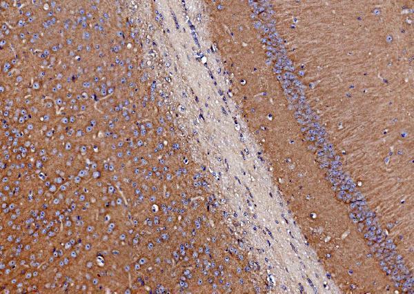
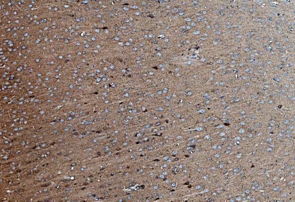
The IHC-validated antibody A03752-3 has paraffin-section images from mouse and rat brain and human liver and lung cancer tissue (catalog image captions); no IF/ICC image is provided (catalog payload).

Real IHC data IHC analysis of SV2A using anti-SV2A antibody (A03752-3). SV2A was detected in paraffin-embedded section of mouse brain tissue. Heat mediated antigen retrieval was performed in EDTA buffer (pH8.0, epitope retrieval solution). The tissue section was blocked with 10% goat serum. The tissue section was then incubated with 2μg/ml rabbit anti-SV2A Antibody (A03752-3) overnight at 4°C. Biotinylated goat anti-rabbit IgG was used as secondary antibody and incubated for 30 minutes at 37°C. The tissue section was developed using Strepavidin-Biotin-Complex (SABC) (Catalog # SA1022) with DAB as the chromogen.
Anti-SV2A Antibody ®
Cat # A03752-3

A03752-3 is the only SKU listed for rendering (catalog payload). Its IHC captions report staining in paraffin sections of mouse and rat brain and human liver and lung cancer tissue (catalog image captions).

Which to pick: Choose A03752-3 for paraffin-section IHC: its own captions show EDTA pH 8 retrieval and chromogenic DAB detection (catalog image captions). It is a rabbit antibody listed as reactive with human, mouse and rat, making it the listed cross-species option (catalog payload). No listed antibody has IF/ICC validation, and the IHC captions do not report the fixative (catalog payload; catalog image captions).

Each figure is that product's own IHC / IF validation image from its datasheet.

References

  1. UniProt Consortium. UniProt entry Q7L0J3 (SV2A_HUMAN, Synaptic vesicle glycoprotein 2A).
  2. Human Protein Atlas. SV2A tissue IHC expression (reliability: Enhanced).
  3. Human Protein Atlas. SV2A subcellular location (ICC-IF): Localized to the cytosol..
  4. Human Protein Atlas. SV2A antibody validation summary (2 antibodies).
  5. The long noncoding RNA RMRP-miR-3135a-SV2A axis promotes the development of hepatocellular carcinoma. Journal of gastrointestinal oncology 2025 — PMC12260943.
  6. Synaptic vesicle glycoprotein 2A (SV2A) regulates kindling epileptogenesis via GABAergic neurotransmission. Scientific reports 2016 — PMC4893657.
  7. Neuropathological Mapping of Synaptic Vesicle Protein 2A in Human Brains: Ground Truth for PET Interpretation in Neurodegenerative Diseases. Alzheimer's & Dementia 2024 — PMC11710421.
  8. Repurposing of the Antiepileptic Drug Levetiracetam to Restrain Neuroendocrine Prostate Cancer and Inhibit Mast Cell Support to Adenocarcinoma. Frontiers in immunology 2021 — PMC7960782.
  9. PubMed PMID:9872452 — UniProt-cited evidence.
  10. PubMed PMID:16303743 — UniProt-cited evidence.
  11. PubMed PMID:17974005 — UniProt-cited evidence.