This website uses cookies to ensure you get the best experience on our website.


- Table of Contents
Plan chromogenic IHC for SV2C in paraffin sections using brain staining as a reference (HPA tissue IHC). Start with the catalog antibody at 1:100 (datasheet: 1:100), then assess cytoplasmic and neuropil staining against the documented tissue pattern (HPA tissue IHC).

Expected localisation, validated protocols, controls and antibodies — the at-a-glance facts below, then the full design guide.
| Expected localisation | Cytoplasmic in brain and peripheral nerves (HPA tissue IHC) | |
| Staining pattern | Caudate and cerebellar cytoplasm; cortical neuropil (HPA tissue IHC) | |
| Antigen retrieval | Citrate pH 6.0 HIER, 95–98 °C, 20 min (rule: cytoplasmic / membrane antigen) | |
| Positive control | Cerebellum+1 more · see all | |
| Negative control | Adipose tissue+4 more · see all |
| Fixation | Keep paraffin-section fixation consistent across samples. (standard IHC practice; not target-specific) | |
| Caveat | Adrenal nerves stain; glandular cells are undetected (HPA tissue IHC) | |
| Regulation | Condition-dependent regulation not annotated (UniProt) | |
| Isoform / epitope | No isoforms annotated; cytoplasmic vs extracellular epitope affects access (UniProt) |
The catalog antibody’s IHC-P protocol is accompanied by two published SV2C mouse brain IHC protocols (PMC6013333; PMC5073372).
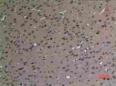
| Sample | Paraffin-embedded Rat Brain tissue; fixative not specified (datasheet A10958) |
| Fixation | Image fixative and duration unreported (datasheet A10958); verify before use. |
| Sectioning | 4–5 µm sections on charged slides (standard) |
| Deparaffinisation | Xylene, graded ethanol series to water (standard) |
| Antigen retrieval | Heat-induced epitope retrieval in citrate buffer, pH 6.0, 20 min at 95–98 °C (standard rule: cytoplasmic / membrane antigen) |
| Peroxidase block | 3% H2O2, 10 min, room temperature (standard) |
| Blocking | 10% normal serum of the secondary host, 30 min, room temperature (standard) |
| Primary antibody | Rabbit anti-SV2C, 1:100 (datasheet A10958) |
| Primary incubation | Overnight at 4 °C (standard) |
| Detection | HRP-polymer secondary, DAB chromogen 5–10 min (standard) |
| Counterstain | Hematoxylin, blue, dehydrate and mount (standard) |
| Expected result | SV2C-positive staining in gLUC cells - cytoplasm/membrane of cerebellum (HPA tissue IHC: High). HPA tissue profile: Distinct cytoplasmic expression in brain, mainly in caudate nucleus, granular layer in cerebellum and in peripheral nerves in pancreas and adrenal gland. No signal in the no-primary control. |
SV2C is a 12-pass protein of synaptic vesicle membranes, so IHC should favor cytoplasmic or neuropil-associated staining rather than a smooth cell-surface rim (UniProt Q496J9 topology and localization). HPA reports distinct brain staining, especially in caudate nucleus and the cerebellar granular layer, plus peripheral nerves in pancreas and adrenal gland (HPA tissue IHC). Tissue IHC reliability is Supported, with medium agreement between staining and RNA data (HPA tissue IHC).
| Strong cytoplasmic or membrane-associated staining in cerebellar GLUC cells (HPA tissue IHC: High). | This matches an HPA positive tissue and is consistent with a vesicle-associated protein (HPA tissue IHC; UniProt Q496J9 localization). Judge the stained cell population as well as intensity: HPA's High rating applies to the annotated GLUC cells, not every cell in the section (HPA tissue IHC). |
| Moderate staining in cerebral-cortex neuropil, or distinct cytoplasmic staining in caudate (HPA tissue IHC). | Both fit the reported brain distribution: cortical neuropil is rated Medium, and caudate appears in the tissue-profile summary (HPA tissue IHC). Caudate glial cells are separately listed as Not detected; their staining should not be counted as the expected caudate pattern (HPA tissue IHC). |
| Predominantly nuclear staining in paraffin-section IHC, with little expected cytoplasmic or neuropil signal. | Recheck compartment assignment and controls before calling this the expected tissue pattern: UniProt places SV2C on vesicle membranes, while HPA tissue IHC describes cytoplasmic staining (UniProt Q496J9; HPA tissue IHC). HPA separately reports nucleoplasm in ICC-IF, so the discrepancy does not by itself prove an artifact (HPA subcellular ICC-IF). |
| Staining in adrenal glandular cells, adipocytes, or caudate glial cells (HPA tissue IHC: Not detected). | These cell-specific results conflict with HPA's tissue IHC annotations, even where nearby structures may stain: peripheral nerves are reported in adrenal gland (HPA tissue IHC). Consider cross-reactivity or endogenous detection activity, then compare the signal with a no-primary control (general IHC practice). |
| Diffuse chromogen across cells and stroma, or no discernible signal in cerebellar GLUC cells. | Diffuse signal lacks the cell and compartment pattern reported by HPA; check background controls before scoring it as SV2C (HPA tissue IHC; general IHC practice). An absent signal in the annotated High cerebellar cells warrants a run-control check, although HPA rates tissue IHC reliability Supported rather than conclusive (HPA tissue IHC). |
| Tissue and cell selection (HPA tissue IHC) | Use the annotated cerebellar GLUC cells as a positive reference: HPA rates them High, versus Medium cortical neuropil (HPA tissue IHC). Adrenal gland requires cell-level reading because its glandular cells are Not detected although peripheral nerves appear in the tissue-profile summary (HPA tissue IHC). |
| Vesicle topology and unknown antibody epitope (UniProt Q496J9 topology) | SV2C has 12 transmembrane segments and annotated cytoplasmic and extracellular regions (UniProt Q496J9 topology). The supplied record does not identify the antibody epitope; topology alone cannot establish whether a particular antigen-retrieval condition will expose it. |
| Glycosylation and processing (UniProt Q496J9) | UniProt lists 5 glycosylation sites and a full-length chain spanning residues 1–727, with no annotated signal peptide or propeptide (UniProt Q496J9). These annotations do not establish an IHC intensity shift, an epitope-masking effect, or a need for a special retrieval step. |
| Tissue IHC evidence and antibody validation (HPA tissue IHC; HPA antibodies) | HPA calls the tissue pattern Supported and notes medium consistency with RNA, including staining in cells or structures it has not annotated (HPA tissue IHC). Of the listed antibodies, HPA040770 is IHC Supported; HPA040722 has an ICC Approved designation without a listed IHC status (HPA antibodies). |
| IF/ICC Q&A: should nuclear signal be expected in this IHC guide? (HPA subcellular ICC-IF) | HPA ICC-IF reports supported nucleoplasm and approved vesicle localization in SH-SY5Y and U-251MG images (HPA subcellular ICC-IF). That is assay-specific evidence; score paraffin-section IHC against the HPA tissue cytoplasmic and neuropil pattern, and consult the separate IF/ICC guide for that application (HPA tissue IHC). |
| Situation | Likely cause | Next action |
|---|---|---|
| No signal in cerebellar GLUC cells (HPA tissue IHC: High). | The expected positive cell population may have been missed, or the IHC detection run may have failed (HPA tissue IHC; general IHC practice). | Confirm the cell region, inspect a known-positive control from the same run, and verify primary incubation, detection reagents, and counterstain before interpreting a negative sample (general IHC practice). |
| Weak cortical neuropil signal beside strong cerebellar signal. | This difference may reflect HPA's Medium rating for cortical neuropil versus High for cerebellar GLUC cells (HPA tissue IHC). | Score each tissue against its own annotated level and cell compartment; do not require cortex to match cerebellar intensity (HPA tissue IHC). |
| Broad, diffuse chromogen obscures cellular detail. | Nonspecific reagent binding or endogenous detection activity can obscure the expected tissue distribution (general IHC practice; HPA tissue IHC). | Compare with a no-primary control, check blocking and detection steps, and adjust staining conditions using an annotated positive tissue (general IHC practice; HPA tissue IHC). |
| Adrenal glandular cells stain, while nearby nerve profiles are difficult to distinguish. | HPA lists glandular cells as Not detected but reports peripheral nerves in adrenal gland; a section-level positive call could merge those structures (HPA tissue IHC). | Inspect cell morphology and location at higher magnification; use a no-primary control if glandular signal persists (HPA tissue IHC; general IHC practice). |
| Paraffin-section IHC shows mostly nuclear signal. | That differs from HPA's tissue cytoplasmic profile, although HPA separately reports nucleoplasm in ICC-IF (HPA tissue IHC; HPA subcellular ICC-IF). | Check positive and no-primary controls, then report the IHC compartment actually observed; do not transfer the ICC-IF localization call to tissue IHC without support (general IHC practice; HPA tissue IHC). |
| A negative result is attributed to formalin sensitivity or failed antigen retrieval. | Target-specific fixation effects are not established by the supplied assay evidence. Verify with a matched IHC source before attributing a result to fixation. | Check the positive run control and documented IHC retrieval conditions; if optimizing retrieval, compare conditions on the same annotated positive tissue (general IHC practice; HPA tissue IHC). |


Comprehensive Human Protein Atlas IHC scoring per tissue (reliability: Supported — Medium consistency between antibody staining and RNA expression data. Antibody staining in cells/structures not annotated, view images.). Rows are taken directly from the HPA tissue chart — click any row's HPA link to view the source.
| Tissue | Cell type | Level | Evidence | Source |
|---|---|---|---|---|
| Cerebellum | GLUC cells - cytoplasm/membrane | High | Protein (IHC) | HPA → |
| Cerebral cortex | Neuropil | Medium | Protein (IHC) | HPA → |
| Tissue | Cell type | Level | Evidence | Source |
|---|---|---|---|---|
| Adipose tissue | Adipocytes | Not detected | Protein (IHC) | HPA → |
| Adrenal gland | Glandular cells | Not detected | Protein (IHC) | HPA → |
| Appendix | Endocrine cells | Not detected | Protein (IHC) | HPA → |
| Bone marrow | Hematopoietic cells | Not detected | Protein (IHC) | HPA → |
| Breast | Adipocytes | Not detected | Protein (IHC) | HPA → |
Troubleshoot SV2C chromogenic IHC in paraffin sections using the selected antibody image, tissue staining references, and SV2C topology.
A10958 has paraffin-embedded rat brain IHC imagery (catalog IHC image caption). IF is listed without an image (catalog applications; IF image alts). Listed reactivity covers human, mouse, and rat (catalog reactivity).
A10958 is shown in paraffin-embedded rat brain IHC at 1:100 (catalog IHC image caption). A10958 also lists IF as an application and human, mouse, and rat reactivity, but provides no IF image (catalog applications; catalog reactivity; IF image alts).
Which to pick: Choose A10958 for paraffin-section tissue IHC: its own image shows rat brain at 1:100; the fixative is unreported (catalog IHC image caption). For IF/ICC planning, A10958 lists IF at 1:50, but provides no IF image or ICC validation (catalog applications; catalog IF dilution; IF image alts). For cross-species work, A10958 is a rabbit antibody listed as reactive with human, mouse, and rat; its direct IHC image evidence is limited to rat brain (catalog host; catalog reactivity; catalog IHC image caption).