SYCP3 / Synaptonemal complex protein 3 · IHC design guide

Design Immunohistochemistry for SYCP3

Plan chromogenic SYCP3 IHC in paraffin sections using pachytene spermatocyte nuclei as the expected positive pattern (HPA tissue IHC). This guide covers fixation, staining controls and interpretation of stage-dependent staining (UniProt; HPA tissue IHC).

Evidence assembled Oct 2026 · For research use; verify linked source records and product datasheet before use
Immunohistochemistry protocol sheet for SYCP3 (IHC for SYCP3): expected localisation Nuclear in spermatocytes (HPA tissue IHC), antibody A05718-3, validated IHC image, and IHC protocol steps
Printable SYCP3 IHC protocol sheet — expected localisation Nuclear in spermatocytes (HPA tissue IHC), antibody A05718-3, controls and protocol steps. Open the full SYCP3 IHC guide →

SYCP3 Immunohistochemistry Experimental Design Guide

Expected localisation, validated protocols, controls and antibodies — the at-a-glance facts below, then the full design guide.

Must know before staining
Expected localisation Nuclear in spermatocytes (HPA tissue IHC)
Staining pattern Selective nuclear staining in pachytene spermatocytes (HPA tissue IHC)
Antigen retrieval EDTA pH 8.0 HIER, heat-mediated (datasheet A05718-3)
Positive control ⓘ Testis
Negative control ⓘ Adipose tissue+4 more · see all
Important caveats
Reasons your staining may differ from the expected pattern.
Fixation Keep tissue fixation consistent across sections. (standard IHC practice; not target-specific)
Caveat Positive cells depend on the meiotic stage sampled (UniProt; HPA tissue IHC)
Regulation Enriched in testicular spermatocytes (HPA tissue IHC)
Isoform / epitope No annotated isoforms or precursor processing (UniProt)
Section 1

Recommended SYCP3 IHC & IF Protocols

The catalog antibody’s IHC-P protocol is accompanied by published SYCP3 chromogenic IHC methods for mouse sections (PMC12136779; PMC3727218; PMC9242989).

Recommended immunohistochemistry (IHC-P) protocol parameters
SampleParaffin-embedded human testicular cancer tissue; fixative not specified (datasheet A05718-3)
FixationImage fixative and duration unreported (datasheet A05718-3); verify before use.
Sectioning4–5 µm sections on charged slides (standard)
DeparaffinisationXylene, graded ethanol series to water (standard)
Antigen retrievalHeat retrieval: EDTA pH 8.0 (datasheet A05718-3); 20 min, 95–100 °C (standard)
Peroxidase block3% H2O2, 10 min, room temperature (standard)
Blocking10% goat serum (datasheet A05718-3)
Primary antibodyRabbit anti-SYCP3, 2-5 μg/ml (datasheet A05718-3)
Primary incubationOvernight at 4 °C (datasheet A05718-3)
DetectionStreptavidin-biotin complex (SABC), DAB chromogen (datasheet A05718-3)
CounterstainHematoxylin, blue, dehydrate and mount (standard)
Expected resultSYCP3-positive staining in pachytene spermatocytes of testis (HPA tissue IHC: High). HPA tissue profile: Selective nuclear expression in spermatocytes. No signal in the no-primary control.
💡Decision noteStart with heat-mediated EDTA retrieval at pH 8.0 for the catalog antibody (datasheet A05718-3); adjust using the published section and detection details below.
Section 2

What Is the Expected SYCP3 Staining Pattern?

SYCP3 localises to meiotic chromosome axes and centromeres within the nucleus (UniProt Q8IZU3 localisation). In paraffin-section IHC, expect selective nuclear staining of pachytene spermatocytes in testis, where HPA reports high expression and an Enhanced tissue-IHC reliability rating (HPA: testis IHC). SYCP3 has no transmembrane segment (UniProt Q8IZU3 topology); membrane staining is therefore outside the expected pattern.

What am I looking at on my slide?
Strong nuclear chromogen in pachytene spermatocytes, with little staining in neighboring cells.This matches HPA's high, selective nuclear signal in testis (HPA: testis IHC). At routine IHC resolution, score the nuclear cell pattern; chromosome-axis detail described by UniProt may not be distinguishable (UniProt Q8IZU3 localisation; general IHC practice).
Predominantly cytoplasmic or membrane staining, without the expected spermatocyte nuclear signal.The compartment conflicts with SYCP3's nuclear and chromosome localisation and its lack of a transmembrane segment (UniProt Q8IZU3). Review morphology and staining controls; a discordant compartment alone does not establish genuine SYCP3 expression (general IHC practice).
Prominent staining in cells outside the expected spermatocyte population or in an HPA-negative control tissue.That disagrees with selective spermatocyte staining; HPA reports no detection in the listed negative tissues, including adipose-tissue adipocytes (HPA: tissue IHC). Consider antibody cross-reactivity or endogenous detection activity, then compare with controls (general IHC practice).
Diffuse chromogen across cells, connective areas, or much of the section.A widespread deposit obscures the selective nuclear pattern reported in spermatocytes (HPA: testis IHC). Check background and detection controls before scoring it as SYCP3; diffuse staining can reflect nonspecific antibody binding or detection background (general IHC practice).
No nuclear signal in morphologically identifiable pachytene spermatocytes in testis.This is discordant with HPA's high staining in that cell population (HPA: testis IHC). First confirm that the section contains the relevant cells; then assess antibody and detection controls. Absence in an unverified section cannot establish absent target expression (general IHC practice).
💡Expected SYCP3 appearanceCall positive when pachytene spermatocytes show high nuclear staining (HPA: testis IHC); predominantly cytoplasmic, membrane, or broadly distributed staining is discordant with the expected localisation (UniProt Q8IZU3 localisation; HPA: testis IHC).
How each factor affects the staining
Cell population and meiotic stageHPA identifies pachytene spermatocytes as the high-staining population (HPA: testis IHC). UniProt places SYCP3 on meiotic chromosome structures that change through prophase and division (UniProt Q8IZU3 localisation). Interpret a section against its identifiable germ-cell populations, rather than treating every unstained nucleus as a failed reaction (general IHC practice).
Tissue choiceTestis is the supported positive tissue (HPA: high in pachytene spermatocytes; UniProt Q8IZU3: testis-specific). The listed HPA negative tissues support controls for unexpected staining, but a negative result in those tissues does not test whether the assay detects SYCP3 in spermatocytes (HPA: tissue IHC; general IHC practice).
Antibody evidenceHPA rates tissue IHC Enhanced overall and lists HPA039635 and HPA043938 as Enhanced for IHC; CAB032978 is Supported (HPA: tissue IHC and antibody validation). Those ratings support the reported pattern, but do not validate every catalog antibody or every staining condition (HPA: antibody validation; general IHC practice).
Protein form and compartmentUniProt lists one 1–236 chain, no signal peptide, no propeptide, no annotated isoforms, and no transmembrane segment (UniProt Q8IZU3). These annotations support interpreting nuclear staining as the expected compartment; they provide no basis for predicting secreted, membrane, or isoform-specific IHC patterns (UniProt Q8IZU3).
Can the IHC pattern be assumed for IF/ICC?Use nuclear chromosome localisation as a biological expectation (UniProt Q8IZU3 localisation), but HPA supplies no ICC-IF images, subcellular location call, or ICC validation for the listed antibodies (HPA: subcellular and antibody records). The tissue-IHC result alone cannot establish an IF/ICC staining pattern (HPA: tissue IHC; general assay interpretation).
Why is my staining missing, weak or wrong?
SituationLikely causeNext action
Testis control is blank.The relevant spermatocytes may be absent from the section, or the IHC reaction may have failed (HPA: high in pachytene spermatocytes; general IHC practice).Identify pachytene spermatocytes by morphology and review the assay's positive control, antibody dilution, detection steps, and antigen retrieval conditions (general IHC practice). No SYCP3-specific retrieval condition is supplied by HPA or UniProt.
Signal is mainly cytoplasmic or membrane associated.That pattern conflicts with nuclear chromosome localisation and the absence of a transmembrane segment (UniProt Q8IZU3). Background or misidentified cell boundaries are possible (general IHC practice).Recheck the nuclear counterstain and cell morphology; compare with a no-primary control and the expected spermatocyte nuclear pattern (HPA: testis IHC; general IHC practice).
Negative-control tissue stains strongly.HPA reports no detection in its listed negative tissue and cell combinations (HPA: tissue IHC). Nonspecific binding or endogenous detection activity may explain a discordant deposit (general IHC practice).Confirm the sampled tissue and cell type against the HPA entry; inspect no-primary and detection controls, then review blocking and wash steps (HPA: tissue IHC; general IHC practice).
Diffuse background makes nuclei hard to score.Widespread deposit masks HPA's selective nuclear spermatocyte pattern (HPA: testis IHC). Excess antibody or detection background is possible (general IHC practice).Compare a no-primary control, check wash and blocking steps, and adjust antibody dilution only against a positive testis control (general IHC practice; HPA: high in pachytene spermatocytes).
Only some tubule profiles show convincing nuclear staining.The expected signal is restricted to pachytene spermatocytes, while SYCP3 associates with chromosome structures across meiotic stages (HPA: testis IHC; UniProt Q8IZU3 localisation). Tubules may differ in their sampled germ-cell populations (general histology practice).Score identifiable spermatocyte nuclei within each profile and document the cells present; avoid calling the entire section negative solely because some profiles lack signal (HPA: testis IHC; general IHC practice).
IF/ICC gives a different localisation from the paraffin-section IHC result.HPA has no ICC-IF image or ICC validation for the listed antibodies, so its IHC reliability does not establish the IF/ICC result (HPA: subcellular and antibody records).Assess IF/ICC controls and cell identity within that assay, then compare any specific signal with UniProt's nuclear chromosome localisation (UniProt Q8IZU3; general IF practice).

Sample controls for SYCP3 IHC & IF

🧪Run testis first; pachytene spermatocytes should show nuclear or chromosomal staining (HPA: High in pachytene spermatocytes; UniProt Q8IZU3: nuclear and chromosomal localization). Use appendix glandular cells as the negative tissue, and expect nonmeiotic somatic cells within the testis section to lack specific staining (HPA: Not detected in appendix glandular cells; UniProt Q8IZU3: meiotic function and testis specificity).
Positive control tissue: Testis (Pachytene spermatocytes, HPA High)
Negative control tissue: Adipose tissue (HPA Not detected)
ICC-IF cell lines (HPA subcellular resource): HPA carries no ICC-IF cell line for SYCP3; derive a cell-line control from the positive tissue's cell type (Pachytene spermatocytes) and confirm it by RNA or western blot first.
Technical controls: Include a no-primary, secondary-only control and an isotype control matched to the rabbit primary antibody; use SYCP3-knockout tissue as a biological negative if available (catalog antibody caption: rabbit primary; standard IHC practice). For chromogenic detection, quench endogenous peroxidase and check for endogenous biotin when using the caption’s biotin-based detection system (catalog antibody caption: biotinylated secondary, streptavidin–biotin complex and DAB; standard IHC practice).
⚠️Feasibility: A target-specific fixation window or fixation effect is unreported, and the selected A05718-3 tissue-IHC caption does not state the fixative (catalog antibody caption: fixative unreported). The paraffin-section example uses heat retrieval in EDTA at pH 8.0; whether retrieval is required across other preparations, or whether frozen sections or IF are easier, is unreported (catalog antibody caption: EDTA heat retrieval; HPA: no ICC-IF images). In testis, interpret staining by meiotic cell type and nuclear or chromosomal pattern rather than expecting every cell to stain (HPA: High in pachytene spermatocytes; UniProt Q8IZU3: meiotic chromosomal localization).

HPA tissue IHC evidence for SYCP3

Comprehensive Human Protein Atlas IHC scoring per tissue (reliability: Enhanced — High consistency between antibody staining and RNA expression data.). Rows are taken directly from the HPA tissue chart — click any row's HPA link to view the source.

Positive expression · recommended positive controls

TissueCell typeLevelEvidenceSource
Testis Pachytene spermatocytes High Protein (IHC) HPA →

Undetected expression · recommended negative controls

TissueCell typeLevelEvidenceSource
Adipose tissue Adipocytes Not detected Protein (IHC) HPA →
Adrenal gland Glandular cells Not detected Protein (IHC) HPA →
Appendix Glandular cells Not detected Protein (IHC) HPA →
Bone marrow Hematopoietic cells Not detected Protein (IHC) HPA →
Breast Adipocytes Not detected Protein (IHC) HPA →
Section 3

Advanced SYCP3 IHC Tips

Troubleshoot SYCP3 staining in paraffin section IHC by checking retrieval, nuclear localisation, cell identity and assay controls.

What should I change when SYCP3 staining is weak after retrieval?
Start with heat mediated antigen retrieval in EDTA at pH 8.0 (datasheet A05718-3). The catalog antibody detected SYCP3 in a paraffin section of human testicular cancer tissue after this retrieval, followed by 2 μg/ml primary antibody overnight at 4°C (datasheet A05718-3). Check that sections remain attached, the retrieval solution reaches the intended temperature, and runs use consistent heating and cooling (standard IHC practice). Compare a testis section containing spermatocytes with a negative control before changing conditions (HPA: high in pachytene spermatocytes; standard IHC practice). If staining remains weak, trial an alternative retrieval buffer on adjacent sections while keeping detection conditions fixed (standard IHC practice).
Could fixation explain weak or uneven SYCP3 staining?
SYCP3 specific sensitivity to fixation is unknown from the supplied evidence; the catalog image describes a paraffin section but does not state its fixative (datasheet A05718-3). Record the actual fixative, fixation interval, section thickness and processing history for each block before comparing staining intensity (standard IHC practice). Run blocks with matched processing alongside a testis control containing spermatocytes, which are the expected positive cells (HPA: high in pachytene spermatocytes; standard IHC practice). Keep EDTA retrieval at pH 8.0 and the 2 μg/ml primary incubation fixed during that comparison (datasheet A05718-3). Interpret any fixation effect as an experimental finding, not an established SYCP3 property (standard IHC practice).
Where should convincing SYCP3 staining appear in a tissue section?
Look for staining in spermatocyte nuclei: tissue IHC shows selective nuclear expression and high signal in pachytene spermatocytes (HPA: tissue IHC). SYCP3 associates with chromosome cores, synaptonemal complex lateral domains and meiotic centromeres at specific stages (UniProt Q8IZU3: subcellular location). A 4°C overnight primary incubation was used for the catalog paraffin section, but that image alone does not establish every staining pattern as specific (datasheet A05718-3). Compare nuclear signal with morphology and a negative control on the same run (standard IHC practice). Diffuse cytoplasmic or extracellular color without the expected nuclear pattern warrants a review of background, counterstain and tissue preservation (HPA: tissue IHC; standard IHC practice).
Could an isoform or modified epitope account for discordant staining?
The supplied record lists one SYCP3 chain spanning residues 1–236 and no annotated isoforms, so an isoform explanation is unsupported here (UniProt Q8IZU3: processing and isoforms). It lists phosphoserines at positions 36, 38 and 59, but gives no antibody epitope or evidence that phosphorylation alters binding (UniProt Q8IZU3: modified residues; datasheet A05718-3). Ask for the immunogen or epitope information before attributing a failed stain to epitope masking (standard IHC practice). Compare adjacent sections with the same EDTA pH 8.0 retrieval and consistent detection to isolate procedural variation (datasheet A05718-3; standard IHC practice). Use a positive testis control to judge whether the expected nuclear staining is preserved (HPA: tissue IHC; standard IHC practice).
How should I investigate SYCP3 with multiplex immunofluorescence?
Treat IF as a separate assay requiring its own validation; the supplied catalog example documents chromogenic staining in a paraffin section (datasheet A05718-3). Pair SYCP3 with an independently validated spermatocyte marker and a nuclear counterstain to check cell identity and compartment (HPA: high in pachytene spermatocytes; standard IF practice). Choose fluorophores after inspecting tissue autofluorescence and assign the dimmer expected signal to a cleaner detection channel (standard IF practice). SYCP3 is nuclear and has no transmembrane segment, so optimise permeabilisation for access to intracellular nuclear epitopes while checking tissue integrity (UniProt Q8IZU3: location and topology; standard IF practice). Include single color and secondary only controls when assessing overlap (standard IF practice).
How can I reduce widespread brown staining around SYCP3 positive cells?
First check whether brown signal follows spermatocyte nuclei rather than many unrelated cells, because tissue IHC reports selective nuclear staining in spermatocytes (HPA: tissue IHC). The catalog example used 10% goat serum blocking, 2 μg/ml primary antibody overnight at 4°C, and biotinylated secondary detection with DAB (datasheet A05718-3). Use a no primary control to assess secondary reagent, biotin system and endogenous enzyme contributions (standard IHC practice). Apply a peroxidase block and control DAB development consistently across slides (standard chromogenic IHC practice). If background persists, compare adjacent sections while changing one blocking, washing or antibody concentration variable at a time (standard IHC practice).
How should I score SYCP3 staining across tissue sections? ⚠ ANSWER MARKED FOR VERIFICATION
Define spermatocytes morphologically and score nuclear staining within that compartment, because high expression is reported in pachytene spermatocytes (HPA: tissue IHC). Report the percentage of positive spermatocytes and, where intensity is reproducible, an H-score using the same scoring thresholds across cases (standard IHC practice). For spatial comparisons, report positive nuclear density per mm² of evaluable tissue and exclude damaged or necrotic regions (standard IHC practice). Normalise counts to the number of eligible spermatocytes or the measured evaluable area, and record how many sections and fields contributed (standard IHC practice). Keep retrieval at EDTA pH 8.0 and detection conditions consistent within the series (datasheet A05718-3; standard IHC practice).
When is a SYCP3 positive result likely to be artefactual?
A convincing result aligns with nuclear staining in spermatocytes, particularly pachytene spermatocytes, and with suitable tissue morphology (HPA: tissue IHC). SYCP3 is associated with meiotic chromosome structures, so staining restricted to extracellular deposits or diffuse cytoplasm does not match its reported location (UniProt Q8IZU3: subcellular location). Compare suspicious staining with a no primary control and examine section edges, folds and necrotic regions for uneven DAB deposition (standard IHC practice). Check endogenous peroxidase contribution when brown color appears broadly despite a plausible nuclear pattern elsewhere (standard chromogenic IHC practice). The catalog paraffin example used EDTA at pH 8.0; replicate that retrieval before assigning a discordant pattern biological meaning (datasheet A05718-3).
Boster reagents

Best SYCP3 / Synaptonemal complex protein 3 IHC Antibodies

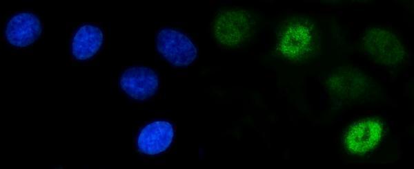
A05718-3 has IHC images from human paraffin tissue sections and an IF image from A549 cells (catalog image captions); the catalog lists Human, Mouse, and Rat reactivity (catalog reactivity).

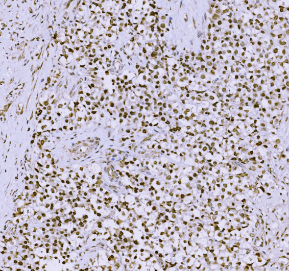
Real IHC data IHC analysis of SCP3/SYCP3 using anti-SCP3/SYCP3 antibody (A05718-3). SCP3/SYCP3 was detected in a paraffin-embedded section of human testicular cancer tissue. Heat mediated antigen retrieval was performed in EDTA buffer (pH 8.0, epitope retrieval solution). The tissue section was blocked with 10% goat serum. The tissue section was then incubated with 2 μg/ml rabbit anti-SCP3/SYCP3 Antibody (A05718-3) overnight at 4°C. Biotinylated goat anti-rabbit IgG was used as secondary antibody and incubated for 30 minutes at 37°C. The tissue section was developed using Strepavidin-Biotin-Complex (SABC) (Catalog # SA1022) with DAB as the chromogen.
Anti-SCP3/SYCP3 Antibody ®
Cat # A05718-3

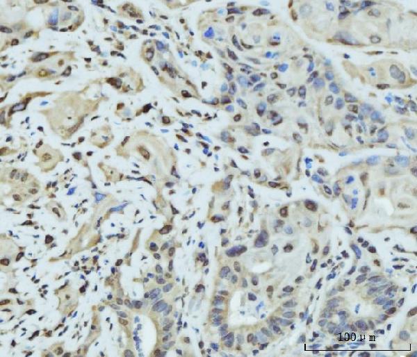
A05718-3 has IHC images from paraffin sections of human testicular cancer, gall bladder adenosquamous carcinoma, and placenta (catalog IHC image captions). Its IF image shows A549 cells, and the catalog lists IHC, IF, and ICC applications (catalog IF image caption; catalog applications).

Which to pick: Choose A05718-3 for paraffin-section IHC: its rendered figure shows human testicular cancer tissue with EDTA pH 8.0 retrieval and 2 μg/ml primary antibody (A05718-3 IHC figure caption). Choose the same SKU for IF/ICC based on its A549 IF image and listed IF/ICC applications (catalog IF image caption; catalog applications). For Mouse or Rat samples, its listed reactivity supports considering it, but the supplied IHC and IF images show human samples; the tissue fixative is unreported (catalog reactivity; catalog image captions).

Each figure is that product's own IHC / IF validation image from its datasheet.

References

  1. UniProt Consortium. UniProt entry Q8IZU3 (SYCP3_HUMAN, Synaptonemal complex protein 3).
  2. Human Protein Atlas. SYCP3 tissue IHC expression (reliability: Enhanced).
  3. Human Protein Atlas. SYCP3 subcellular location (ICC-IF): Highest expression in RPTEC/TERT1: 0.4 nTPM.
  4. Human Protein Atlas. SYCP3 antibody validation summary (3 antibodies).
  5. Melatonin ameliorates heat stress-induced oxidative apoptosis in mouse spermatocytes via autophagy and ferroptosis pathways. Cell stress & chaperones 2025 — PMC12136779.
  6. Adenine nucleotide translocase 4 deficiency leads to early meiotic arrest of murine male germ cells. Reproduction (Cambridge, England) 2009 — PMC3727218.
  7. Etoposide damages female germ cells in the developing ovary. BMC cancer 2016 — PMC4980800.
  8. TCFL5 deficiency impairs the pachytene to diplotene transition during spermatogenesis in the mouse. Scientific reports 2022 — PMC9242989.
  9. PubMed PMID:12213195 — UniProt-cited evidence.
  10. PubMed PMID:15489334 — UniProt-cited evidence.
  11. PubMed PMID:14643120 — UniProt-cited evidence.