This website uses cookies to ensure you get the best experience on our website.


- Table of Contents
Plan SYNGR1 paraffin IHC using high staining in cerebral cortical neuropil as a reference (HPA tissue IHC). Start catalog antibody A10148 at 2.5 μg/mL (datasheet) and compare sections using consistent fixation.

Expected localisation, validated protocols, controls and antibodies — the at-a-glance facts below, then the full design guide.
| Expected localisation | Cerebral cortical neuropil (HPA tissue IHC); synaptic vesicle membrane (UniProt) | |
| Staining pattern | Selective staining in cerebral cortical neuropil (HPA tissue IHC) | |
| Antigen retrieval | Citrate pH 6.0 HIER, 95–98 °C, 20 min (rule: cytoplasmic / membrane antigen) | |
| Positive control | Cerebral cortex+4 more · see all | |
| Negative control | Duodenum+4 more · see all |
| Fixation | Keep fixation consistent across paraffin sections. (standard IHC practice; not target-specific) | |
| Caveat | Staining and RNA expression show medium consistency (HPA tissue IHC) | |
| Regulation | Expression regulation not reported (UniProt) | |
| Isoform / epitope | 3 isoforms; cytoplasmic versus lumenal epitope coverage is unknown (UniProt) |
The catalog antibody’s IHC-P protocol is accompanied by one published chromogenic SYNGR1 IHC protocol (PMC6944231).
| Sample | Tissue sections; selected-image fixative not specified (standard IHC workflow) |
| Fixation | Image fixative and duration unreported (datasheet A10148); verify before use. |
| Sectioning | 4–5 µm sections on charged slides (standard) |
| Deparaffinisation | Xylene, graded ethanol series to water (standard) |
| Antigen retrieval | Heat-induced epitope retrieval in citrate buffer, pH 6.0, 20 min at 95–98 °C (standard rule: cytoplasmic / membrane antigen) |
| Peroxidase block | 3% H2O2, 10 min, room temperature (standard) |
| Blocking | 10% normal serum of the secondary host, 30 min, room temperature (standard) |
| Primary antibody | Rabbit anti-SYNGR1, 2.5 μg/mL (datasheet A10148) |
| Primary incubation | Overnight at 4 °C (standard) |
| Detection | HRP-polymer secondary, DAB chromogen 5–10 min (standard) |
| Counterstain | Hematoxylin, blue, dehydrate and mount (standard) |
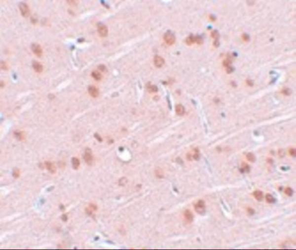
| Expected result | SYNGR1-positive staining in neuropil of cerebral cortex (HPA tissue IHC: High). HPA tissue profile: Selective expression in neuropil. No signal in the no-primary control. |
SYNGR1 is a four-pass membrane protein of synaptic vesicles and other cytoplasmic vesicles (UniProt O43759 topology and subcellular location). In paraffin sections, expect the strongest reported staining in cerebral cortex neuropil, with medium staining in cerebellar granular-layer cells and selected non-neural cell types (HPA tissue IHC). HPA rates the tissue profile Enhanced, while noting medium consistency between antibody staining and RNA expression (HPA reliability).
| Strong staining in cerebral cortex neuropil, with little nuclear staining. | This matches the reported High neuropil signal (HPA tissue IHC) and SYNGR1’s vesicle-membrane location (UniProt O43759 subcellular location). Read staining against tissue architecture: neuropil signal should be assessed separately from nearby cell bodies, because a strong regional signal does not establish that every cell in the region expresses SYNGR1. |
| Moderate staining in selected cells outside the cortex. | Medium staining is reported in cerebellar granular-layer cells, kidney tubular cells, lung macrophages, and glandular cells in several named tissues (HPA tissue IHC). These are plausible positives when the stained cell type matches the reference; staining of every cell throughout such a tissue would exceed the reported selective pattern (HPA tissue IHC). |
| Predominantly nuclear staining or an unexpected uniform compartmental pattern. | Treat this as discordant with the vesicle-membrane assignment (UniProt O43759 subcellular location and topology) and the selective neuropil tissue pattern (HPA tissue IHC). Recheck morphology, counterstain, and detection controls before interpreting the signal as SYNGR1; paraffin-section IHC cannot resolve individual synaptic vesicles by routine light microscopy (standard IHC practice). |
| Strong signal in a cell type reported as undetected. | For example, hippocampal glial cells and duodenal glandular cells are listed as Not detected (HPA tissue IHC). Investigate antibody cross-reactivity or endogenous detection activity as possible explanations (standard IHC practice). An unexpected positive alone cannot distinguish those causes or prove that the HPA reference applies to every specimen. |
| Diffuse stain across cells and extracellular spaces, or no stain in cortex neuropil. | Widespread diffuse signal is difficult to reconcile with selective neuropil expression (HPA tissue IHC); assess background with a no-primary control (standard IHC practice). Absent signal in a known-positive cortex section conflicts with the reported High neuropil level (HPA tissue IHC), but first check tissue preservation and the IHC workflow (standard IHC practice). |
| Antibody validation and reference pattern | The tissue profile is Enhanced, with medium antibody–RNA consistency (HPA reliability). CAB034277 has Enhanced IHC validation; HPA029673 has Approved IHC validation (HPA antibodies). Compare the staining pattern with the cell-type reference and include controls; these labels do not guarantee specificity in a new preparation (standard IHC practice). |
| Epitope position and membrane topology | SYNGR1 has 4 transmembrane segments, with cytoplasmic and lumenal intervals (UniProt O43759 topology). Whether a particular antibody recognizes an accessible epitope depends on its binding site and the preparation (standard IHC practice); no epitope location or target-specific retrieval effect is supplied. |
| Isoforms and processing | UniProt lists 3 isoforms, 1A, 1B, and 1C, and a single 1–233 chain without an annotated signal peptide or propeptide (UniProt O43759 isoforms and processing). The supplied evidence does not identify which isoforms an antibody detects; do not infer isoform specificity or tissue-dependent shedding from this record. |
| IF/ICC: what pattern is reported? | HPA reports supported cytosol localization in ICC-IF images from A-431, U-251MG, and U2OS (HPA subcellular). This is a cell-imaging observation, while the paraffin-section reference is selective neuropil staining (HPA tissue IHC). Use the separate IF/ICC guide for that application; the ICC observation does not establish an IHC protocol. |
| Antigen retrieval and fixation evidence | Target-specific fixation effects are not established by the supplied assay evidence. Verify with a matched IHC source before attributing a result to fixation. |
| Situation | Likely cause | Next action |
|---|---|---|
| No staining in a cerebral cortex positive control. | The result conflicts with High cortical neuropil staining (HPA tissue IHC); inadequate detection, retrieval, or tissue preservation are possible workflow causes (standard IHC practice). | Check that the positive-control section contains intact neuropil, then verify primary antibody application, retrieval, detection reagents, and counterstain using the validated IHC workflow (standard IHC practice). Avoid calling the sample negative until the control works. |
| Nuclear signal dominates the section. | A nuclear-dominant pattern conflicts with the annotated vesicle-membrane location (UniProt O43759 subcellular location). Nonspecific binding or misleading counterstain interpretation are possibilities (standard IHC practice). | Compare with a no-primary slide and inspect nuclei under the same illumination and colour settings (standard IHC practice). If nuclear colour persists independently of the primary antibody, troubleshoot the detection or counterstain steps before scoring. |
| Diffuse colour obscures the neuropil pattern. | Excess background can result from nonspecific binding, overly concentrated reagents, or incomplete washing (standard IHC practice). A uniform field is unlike HPA’s selective neuropil profile (HPA tissue IHC). | Compare a no-primary control, review blocking and washes, and titrate the IHC-validated antibody within its documented IHC conditions (standard IHC practice). Judge improvement by recovery of the anatomical pattern, rather than by colour intensity alone. |
| A nominal negative cell population stains strongly. | Duodenal glandular cells, hippocampal glia, and several other specified populations are Not detected in the reference (HPA tissue IHC). Cross-reactivity or endogenous detection activity may produce an apparent positive (standard IHC practice). | Confirm the stained cell type morphologically, compare with a no-primary control, and assess endogenous enzyme activity where relevant to the chromogen (standard IHC practice). Record any persistent discrepancy as specimen-specific until independently supported. |
| Expected positive cells stain, but the whole tissue also appears positive. | HPA reports selective neuropil expression and cell-type-specific signals (HPA tissue IHC); broad colour may reflect background or loss of spatial discrimination during detection (standard IHC practice). | Score compartments and cell types separately, review the no-primary control, and adjust antibody concentration or detection development using the documented workflow (standard IHC practice). Preserve enough counterstain to identify the reported cells without obscuring the chromogen. |
| ICC-IF appears cytosolic, while tissue IHC highlights neuropil. | These are different reported readouts: supported cytosol localization in cultured-cell ICC-IF (HPA subcellular) and High cerebral cortex neuropil staining in tissue IHC (HPA tissue IHC). | Interpret each preparation against its own HPA reference and confirm that the antibody has validation for the chosen application (HPA antibodies). Do not use the ICC-IF description as evidence that a paraffin-section retrieval or detection step should change. |
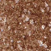


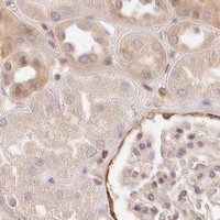


Comprehensive Human Protein Atlas IHC scoring per tissue (reliability: Enhanced — Medium consistency between antibody staining and RNA expression data.). Rows are taken directly from the HPA tissue chart — click any row's HPA link to view the source.
| Tissue | Cell type | Level | Evidence | Source |
|---|---|---|---|---|
| Cerebral cortex | Neuropil | High | Protein (IHC) | HPA → |
| Adrenal gland | Glandular cells | Medium | Protein (IHC) | HPA → |
| Appendix | Glandular cells | Medium | Protein (IHC) | HPA → |
| Breast | Glandular cells | Medium | Protein (IHC) | HPA → |
| Cerebellum | Cells in granular layer | Medium | Protein (IHC) | HPA → |
| Tissue | Cell type | Level | Evidence | Source |
|---|---|---|---|---|
| Duodenum | Glandular cells | Not detected | Protein (IHC) | HPA → |
| Esophagus | Squamous epithelial cells | Not detected | Protein (IHC) | HPA → |
| Hippocampus | Glial cells | Not detected | Protein (IHC) | HPA → |
| Oral mucosa | Squamous epithelial cells | Not detected | Protein (IHC) | HPA → |
| Ovary | Ovarian stroma cells | Not detected | Protein (IHC) | HPA → |
Troubleshoot SYNGR1 staining by comparing the observed pattern with neuropil enrichment and the protein’s vesicle membrane topology (HPA tissue IHC; UniProt O43759 topology).
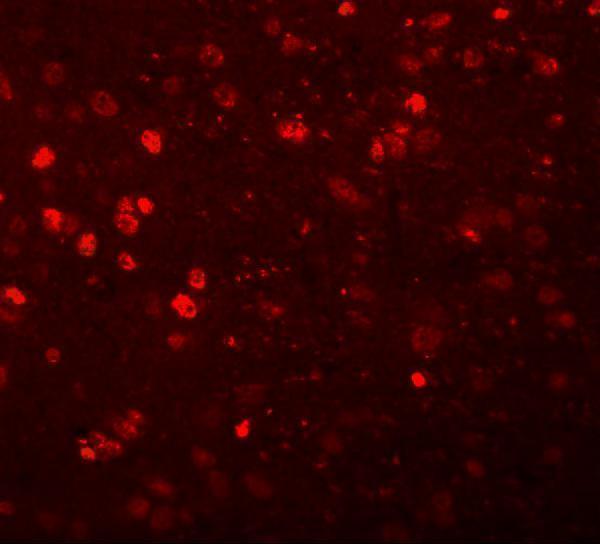
Anti-SYNGR1 antibodies have rat brain IHC and IF images (A10148 image captions) and a human A549 ICC/IF image (A10148-1 image caption); both list Human, Mouse and Rat reactivity (catalog applications/reactivity).
The rendering SKU, A10148, has rat brain IHC at 2.5 μg/mL and IF at 20 μg/mL (A10148 image captions). A10148-1 has an A549 ICC/IF image, but no IHC image in the payload (A10148-1 image captions).
Which to pick: Choose A10148 for tissue IHC: it lists IHC-P and shows rat brain staining at 2.5 μg/mL; its fixative is unreported (A10148 applications; A10148 IHC image caption). For IF/ICC, A10148-1 shows A549 cell staining at 5 μg/mL, while A10148 shows rat brain IF at 20 μg/mL (A10148-1 IF image caption; A10148 IF image caption). Both list Human, Mouse and Rat reactivity, but cross-species IHC performance needs verification for the intended sample; both are rabbit antibodies with clonality unreported (catalog reactivity/host/clone; image captions).