This website uses cookies to ensure you get the best experience on our website.


- Table of Contents
Plan chromogenic SYNGR4 IHC-P with testis as a positive tissue and high staining in elongated or late spermatids (HPA tissue IHC). This guide covers fixation consistency, retrieval, controls and antibody titration from 5 μg/mL (datasheet; standard IHC practice).

Expected localisation, validated protocols, controls and antibodies — the at-a-glance facts below, then the full design guide.
| Expected localisation | Cytoplasmic staining in seminiferous ducts (HPA tissue IHC) | |
| Staining pattern | High cytoplasmic staining in elongated or late spermatids (HPA tissue IHC) | |
| Antigen retrieval | Citrate pH 6.0 HIER, 95–98 °C, 20 min (rule: cytoplasmic / membrane antigen) | |
| Positive control | Testis | |
| Negative control | Adipose tissue+4 more · see all |
| Fixation | Keep fixation consistent across sections (standard IHC practice; not target-specific); assess its effects empirically. | |
| Caveat | Antibody staining and RNA show medium consistency (HPA tissue IHC) | |
| Regulation | Expression regulation is unreported (UniProt) | |
| Isoform / epitope | No isoforms; extracellular versus cytoplasmic epitope is unmapped (UniProt) |
The catalog antibody’s IHC-P protocol is paired with one published SYNGR4 staining workflow (PMC11788336).
| Sample | Tissue sections; selected-image fixative not specified (standard IHC workflow) |
| Fixation | Image fixative and duration unreported (datasheet A19415); verify before use. |
| Sectioning | 4–5 µm sections on charged slides (standard) |
| Deparaffinisation | Xylene, graded ethanol series to water (standard) |
| Antigen retrieval | Heat-induced epitope retrieval in citrate buffer, pH 6.0, 20 min at 95–98 °C (standard rule: cytoplasmic / membrane antigen) |
| Peroxidase block | 3% H2O2, 10 min, room temperature (standard) |
| Blocking | 10% normal serum of the secondary host, 30 min, room temperature (standard) |
| Primary antibody | Rabbit anti-SYNGR4, 5 μg/mL (datasheet A19415) |
| Primary incubation | Overnight at 4 °C (standard) |
| Detection | HRP-polymer secondary, DAB chromogen 5–10 min (standard) |
| Counterstain | Hematoxylin, blue, dehydrate and mount (standard) |
| Expected result | SYNGR4-positive staining in elongated or late spermatids of testis (HPA tissue IHC: High). HPA tissue profile: Cytoplasmic expression in seminiferous ducts. No signal in the no-primary control. |
SYNGR4 is a four-pass membrane protein (UniProt O95473 topology). In tissue IHC, the expected signal is cytoplasmic staining within seminiferous ducts, strongest in elongated or late spermatids (HPA: testis High; tissue profile). HPA rates the tissue IHC evidence Enhanced, while noting medium consistency between antibody staining and RNA expression (HPA: reliability description). Interpret the observed cytoplasmic pattern as an IHC finding; the membrane annotation alone does not specify a resolvable membrane outline in paraffin sections (UniProt O95473; HPA: tissue profile).
| Strong cytoplasmic signal in elongated or late spermatids within seminiferous ducts. | This matches the reported positive tissue and cell type (HPA: testis High; tissue profile). Check that the signal follows cells and duct architecture rather than appearing uniformly across the section (general IHC practice). HPA's Enhanced rating supports this tissue pattern, subject to its stated medium staining–RNA consistency (HPA: reliability description). |
| Predominantly nuclear signal, or staining confined to an unrelated compartment. | Treat this as a localization mismatch: HPA describes cytoplasmic tissue staining, and UniProt annotates a membrane protein (HPA: tissue profile; UniProt O95473). Review morphology, counterstain, and control sections before calling such staining SYNGR4 (general IHC practice). A punctate or perinuclear appearance alone is insufficient to override the tissue IHC reference pattern. |
| Strong signal in cells other than elongated or late spermatids, especially in a reference-negative tissue. | Question specificity when the staining departs from the reported High testis cell population (HPA: testis High). Adipocytes in adipose tissue and glandular cells in adrenal gland are listed as Not detected (HPA: tissue IHC). Cross-reactivity or residual endogenous detection activity are possible technical explanations; compare controls before assigning either cause (general IHC practice). |
| Diffuse staining across ducts, stroma, or most of the section, with poor cell boundaries. | A widespread haze does not reproduce the cell-restricted cytoplasmic profile (HPA: tissue profile; testis High). It can reflect nonspecific binding, incomplete blocking, or detection background (general IHC practice). Compare a no-primary control and a reference-negative tissue, then assess whether any cell-specific signal remains above the background (general IHC practice). |
| No convincing signal in a testis section containing elongated or late spermatids. | This conflicts with the reported High staining in that cell population (HPA: testis High). First confirm the relevant cells are present on the section; then check control performance, antibody compatibility, and detection steps (general IHC practice). An absent result in one run does not by itself overturn the HPA tissue pattern. |
| Protein topology and apparent compartment | Four transmembrane segments place SYNGR4 in membranes (UniProt O95473 topology); tissue IHC reports cytoplasmic expression (HPA: tissue profile). Score the observed cell-associated pattern without requiring a crisp plasma-membrane rim (general IHC practice). |
| Tissue and cell selection | Testis elongated or late spermatids are High (HPA: tissue IHC). Duodenal, small-intestinal, colonic, and rectal glandular cells and hepatocytes are Low; these are weaker comparison tissues, not equivalent positive controls (HPA: tissue IHC). |
| Reference-negative cells | Adipose adipocytes, adrenal glandular cells, and several other sampled cells are Not detected (HPA: tissue IHC). Use a listed negative cell population to assess background while retaining the cell-level scope of each HPA call (general IHC practice). |
| Antibody evidence | HPA030075 and HPA059286 each have Enhanced IHC status (HPA: antibody validation). This supports the reference pattern, while HPA reports only medium consistency between tissue staining and RNA expression (HPA: reliability description). |
| IF/ICC: what localization is reported? | Golgi apparatus is the approved main ICC-IF location; images are listed for A-549 and HEK293, and HPA059286 has Approved ICC status (HPA: subcellular; antibody validation). This is an IF/ICC reference, not a paraffin IHC localization requirement. |
| Situation | Likely cause | Next action |
|---|---|---|
| Testis positive control is blank despite visible late spermatids. | The expected High cell population is present (HPA: testis High), so the run or reagent chain may have failed (general IHC practice). | Check primary-antibody use, detection reagents, and the run control; review the retrieval conditions used for this assay before repeating (general IHC practice). No SYNGR4-specific retrieval condition is supplied. |
| Signal is weak and difficult to distinguish from counterstain. | The reported testis signal is High (HPA: testis High); weak contrast may reflect staining or imaging conditions (general IHC practice). | Compare the testis control with its no-primary control; check chromogen development and counterstain intensity, then adjust the assay using validated control sections (general IHC practice). |
| Most tissue compartments stain evenly. | Uniform signal does not match cytoplasmic staining in seminiferous ducts (HPA: tissue profile); background from binding or detection is possible (general IHC practice). | Run a no-primary control, review blocking and washes, and compare with a listed Not detected cell population (HPA: tissue IHC; general IHC practice). |
| Reference-negative cells show strong chromogenic staining. | Staining in cells listed as Not detected raises a specificity or endogenous-detection concern (HPA: tissue IHC; general IHC practice). | Compare no-primary and other assay controls; check the detection chemistry's endogenous-activity block where applicable before interpreting the signal as SYNGR4 (general IHC practice). |
| Only nuclei appear positive in the testis section. | Nuclear dominance conflicts with the reported cytoplasmic tissue pattern (HPA: tissue profile) and membrane annotation (UniProt O95473). | Recheck cell identification and counterstain, then compare antibody-stained and no-primary sections for a reproducible cytoplasmic signal (general IHC practice). |
| An IF/ICC Golgi pattern seems different from the IHC slide. | HPA reports approved Golgi localization by ICC-IF and cytoplasmic expression by tissue IHC (HPA: subcellular; tissue profile). | Assess each application against its own reference observation; use cell type and cytoplasmic signal for the paraffin IHC call (HPA: tissue IHC; general IHC practice). |


Comprehensive Human Protein Atlas IHC scoring per tissue (reliability: Enhanced — Medium consistency between antibody staining and RNA expression data.). Rows are taken directly from the HPA tissue chart — click any row's HPA link to view the source.
| Tissue | Cell type | Level | Evidence | Source |
|---|---|---|---|---|
| Testis | Elongated or late spermatids | High | Protein (IHC) | HPA → |
| Tissue | Cell type | Level | Evidence | Source |
|---|---|---|---|---|
| Adipose tissue | Adipocytes | Not detected | Protein (IHC) | HPA → |
| Adrenal gland | Glandular cells | Not detected | Protein (IHC) | HPA → |
| Appendix | Glandular cells | Not detected | Protein (IHC) | HPA → |
| Bone marrow | Hematopoietic cells | Not detected | Protein (IHC) | HPA → |
| Breast | Adipocytes | Not detected | Protein (IHC) | HPA → |
Use cell type, compartment and matched controls to troubleshoot SYNGR4 staining in paraffin sections; interpret the rat brain image with its fixation details unresolved.
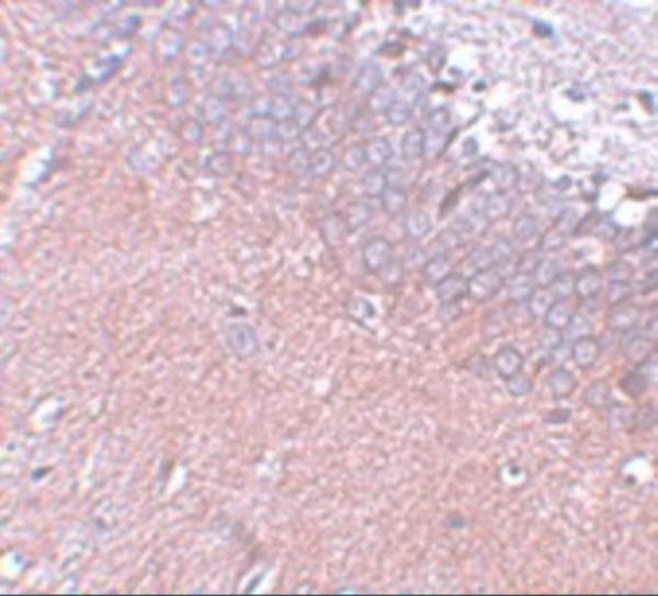
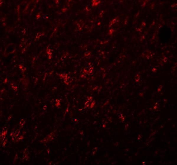
A19415 has IHC and IF images from rat brain tissue (catalog image captions); its listed reactivity covers human, mouse and rat (catalog: reactivity).
A19415 is listed for IHC-P and IF (catalog: applications). Its IHC and IF images show rat brain tissue at 5 μg/mL and 20 μg/mL, respectively (catalog image captions).
Which to pick: For paraffin-section IHC, choose A19415: IHC-P is listed, and its IHC image shows rat brain tissue at 5 μg/mL (catalog: applications; A19415 IHC image caption); the fixative is unreported (A19415 IHC image caption). For IF, A19415 has a rat brain tissue image at 20 μg/mL (A19415 IF image caption); ICC validation and clonality are unreported (catalog: applications; clone field). For human, mouse or rat samples, A19415 lists reactivity with all three species (catalog: reactivity); the supplied IHC and IF images show rat tissue only (catalog image captions).