This website uses cookies to ensure you get the best experience on our website.


- Table of Contents
Plan SYNPO2 chromogenic IHC-P around cytoplasmic myocyte staining (HPA tissue IHC). Start the catalog antibody at 5 μg/mL (datasheet: IHC-P), and use skeletal or heart muscle as positive tissue (HPA tissue IHC).

Expected localisation, validated protocols, controls and antibodies — the at-a-glance facts below, then the full design guide.
| Expected localisation | Cytoplasm in myocytes (HPA tissue IHC) | |
| Staining pattern | Mainly cytoplasmic staining in myocytes (HPA tissue IHC) | |
| Antigen retrieval | Tris-EDTA pH 9.0 HIER, 95–98 °C, 20 min (rule: nuclear antigen) | |
| Positive control | Endometrium+4 more · see all | |
| Negative control | Adipose tissue+4 more · see all |
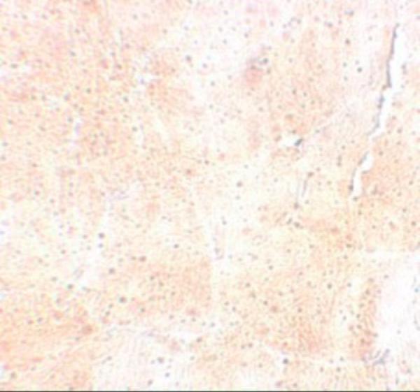
| Fixation | Keep fixation consistent across sections (standard IHC practice; not target-specific); Matched tissue-IHC evidence does not establish the fixation claim. Validate the specimen-specific method before use. (selected-SKU IHC image A07616) | |
| Caveat | Stress can shift cytoplasmic signal to nuclei (UniProt) | |
| Regulation | Isoform 5 is skeletal muscle-specific (UniProt) | |
| Isoform / epitope | 5 isoforms; epitope coverage is unspecified (UniProt) |
The catalog antibody protocol is followed by published SYNPO2 IHC methods for gastric cancer, nasopharyngeal carcinoma, and bladder cancer samples (PMC10660625; PMC10378812; PMC10758676).
| Sample | Tissue sections; selected-image fixative not specified (standard IHC workflow) |
| Fixation | Image fixative and duration unreported (datasheet A07616); verify before use. |
| Sectioning | 4–5 µm sections on charged slides (standard) |
| Deparaffinisation | Xylene, graded ethanol series to water (standard) |
| Antigen retrieval | Heat-induced epitope retrieval in Tris-EDTA buffer, pH 9.0, 20 min at 95–98 °C (standard rule: nuclear antigen) |
| Peroxidase block | 3% H2O2, 10 min, room temperature (standard) |
| Blocking | 10% normal serum of the secondary host, 30 min, room temperature (standard) |
| Primary antibody | Rabbit anti-SYNPO2, 5 μg/mL (datasheet A07616) |
| Primary incubation | Overnight at 4 °C (standard) |
| Detection | HRP-polymer secondary, DAB chromogen 5–10 min (standard) |
| Counterstain | Hematoxylin, blue, dehydrate and mount (standard) |
| Expected result | SYNPO2-positive staining in smooth muscle cells of endometrium (HPA tissue IHC: High). HPA tissue profile: Cytoplasmic expression mainly in myocytes. No signal in the no-primary control. |
SYNPO2 is mainly cytoplasmic in myocytes, with high IHC staining in skeletal and smooth muscle myocytes and cardiomyocytes (HPA: tissue IHC). Nuclear, actin-associated and Z-line localization can occur according to cell state (UniProt Q9UMS6: subcellular location). SYNPO2 has no transmembrane segment, so a membrane-only pattern is unexpected (UniProt Q9UMS6: topology). HPA rates the tissue pattern Enhanced but notes that external verification is pending (HPA: tissue IHC reliability).
| Strong cytoplasmic staining in myocytes, especially in skeletal muscle, smooth muscle or heart (HPA: High in these cells). | This matches the principal tissue pattern. Score the relevant myocytes separately from adjacent cells; a positive area alone does not establish which cell type stained (HPA: cytoplasmic expression mainly in myocytes). |
| Actin-associated or Z-line staining, with some nuclear signal in muscle cells (UniProt Q9UMS6: subcellular location). | These compartments are plausible: SYNPO2 associates with actin and can move between cytoplasm and nucleus with differentiation or stress (UniProt Q9UMS6: localization and function). Interpret nuclear staining alongside cell identity and morphology. |
| Only sharp plasma-membrane outlines appear, without a myocyte cytoplasmic pattern (HPA: tissue IHC; UniProt Q9UMS6: topology). | Treat this as an unexpected compartment pattern requiring review. SYNPO2 has no transmembrane segment; a membrane-only result does not match the reported predominant cytoplasmic IHC pattern (UniProt Q9UMS6: topology; HPA: tissue IHC). |
| Adipocytes or appendix glandular cells stain strongly, while nearby expected-positive muscle cells do not (HPA: Not detected in those cell types; High in myocytes). | Investigate antibody cross-reactivity or endogenous detection activity before calling those cells SYNPO2-positive (standard IHC practice). HPA's negative entries describe particular cells, not every cell in the tissue (HPA: tissue IHC). |
| A uniform haze covers many cell types, or a known-positive myocyte region has no signal (HPA: cytoplasmic expression mainly in myocytes). | Haze obscures compartment and cell-type scoring; absent signal in a positive control makes a negative study result inconclusive. Check background and detection controls, then repeat the affected staining run (standard IHC practice). |
| Cell type and tissue used as controls (HPA: tissue IHC). | Skeletal muscle myocytes, cardiomyocytes and smooth muscle cells are High; breast myoepithelial cells are Medium. Choose a documented positive cell population and score it by cell type (HPA: tissue IHC). |
| Differentiation and stress state (UniProt Q9UMS6: subcellular location). | SYNPO2 can shift between nucleus and cytoplasm; differentiated myotubes can show Z-line localization. A nuclear component is therefore context dependent, not automatically an artefact (UniProt Q9UMS6: subcellular location). |
| Isoform coverage (UniProt Q9UMS6: isoforms and tissue specificity). | UniProt lists 5 isoforms and reports isoform 5 specifically in skeletal muscle. Without a supplied antibody epitope, isoform recognition cannot be inferred from tissue staining alone (UniProt Q9UMS6: isoforms; HPA: tissue IHC). |
| Antibody evidence (HPA: antibody validation). | HPA030665 and HPA068563 have Enhanced IHC evidence; CAB037231 is Supported for IHC. These ratings guide confidence in the observed pattern but do not validate every new specimen or staining run (HPA: antibody validation). |
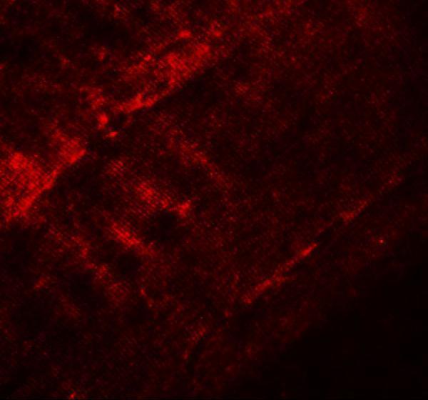
| IF/ICC Q: What localization is reported? (HPA: subcellular ICC-IF). | A: HPA reports mainly vesicles and actin filaments, both Enhanced, in ICC-IF; UniProt also describes nuclear shuttling. Interpret IF/ICC with its own guide, since these observations do not supply an IHC-P protocol (HPA: subcellular ICC-IF; UniProt Q9UMS6: localization). |
| Situation | Likely cause | Next action |
|---|---|---|
| No staining in a skeletal muscle myocyte positive control (HPA: High in skeletal muscle myocytes). | A failed IHC run or a missed positive cell population is possible (standard IHC practice). | Verify muscle-cell morphology and control performance; review the antibody's supplied IHC-P instructions, retrieval and detection steps, then repeat the run (standard IHC practice). |
| Myocytes are faint, but background is also low (HPA: High in listed muscle myocytes). | Signal may be below the assay's useful range; this result alone cannot establish a SYNPO2-specific fixation effect (standard IHC practice). | Check positive-control staining and the supplied IHC-P antibody dilution and detection instructions before adjusting the assay (standard IHC practice). |
| Diffuse chromogen masks cell borders or cytoplasmic detail (HPA: cytoplasmic expression mainly in myocytes). | Nonspecific binding, inadequate blocking or excessive chromogen development can obscure localization (standard IHC practice). | Inspect the negative detection control; review blocking, washes and development time, then score only a run with interpretable cell detail (standard IHC practice). |
| Staining appears only as a plasma-membrane rim (UniProt Q9UMS6: no transmembrane segment). | The compartment disagrees with the predominant cytoplasmic IHC profile (HPA: tissue IHC). | Recheck tissue morphology and detection controls; compare with a documented myocyte positive control before assigning SYNPO2 localization (standard IHC practice; HPA: tissue IHC). |
| Strong signal appears in adipocytes or appendix glandular cells (HPA: Not detected in those cells). | Cross-reactivity or endogenous chromogenic detection activity is possible (standard IHC practice). | Run an appropriate negative detection control, assess background and confirm the stained cell identity; interpret other cells in that tissue separately (standard IHC practice; HPA: tissue IHC). |
| Nuclear staining accompanies myocyte cytoplasmic staining (UniProt Q9UMS6: nuclear shuttling). | Differentiation or stress can change SYNPO2 distribution, although the slide alone does not identify the cause (UniProt Q9UMS6: subcellular location). | Record nuclear and cytoplasmic staining separately and compare the pattern across the same cell type and controls (standard IHC practice). |


![HPA ICC-IF BJ [Human fibroblast] — SYNPO2 (HPA030665)](https://images.proteinatlas.org/30665/1368_A12_2_blue_red_green.jpg)
![HPA ICC-IF BJ [Human fibroblast] — SYNPO2 (HPA030665)](https://images.proteinatlas.org/30665/1368_A12_4_blue_red_green.jpg)
Comprehensive Human Protein Atlas IHC scoring per tissue (reliability: Enhanced — Antibody staining in cells/structures not annotated, view images. Pending external verification.). Rows are taken directly from the HPA tissue chart — click any row's HPA link to view the source.
| Tissue | Cell type | Level | Evidence | Source |
|---|---|---|---|---|
| Endometrium | Smooth muscle cells | High | Protein (IHC) | HPA → |
| Heart muscle | Cardiomyocytes | High | Protein (IHC) | HPA → |
| Placenta | Decidual cells | High | Protein (IHC) | HPA → |
| Skeletal muscle | Myocytes | High | Protein (IHC) | HPA → |
| Smooth muscle | Smooth muscle cells | High | Protein (IHC) | HPA → |
| Tissue | Cell type | Level | Evidence | Source |
|---|---|---|---|---|
| Adipose tissue | Adipocytes | Not detected | Protein (IHC) | HPA → |
| Appendix | Glandular cells | Not detected | Protein (IHC) | HPA → |
| Bone marrow | Hematopoietic cells | Not detected | Protein (IHC) | HPA → |
| Bronchus | Respiratory epithelial cells | Not detected | Protein (IHC) | HPA → |
| Caudate | Glial cells | Not detected | Protein (IHC) | HPA → |
Troubleshoot SYNPO2 staining in paraffin sections by checking retrieval, tissue context, subcellular pattern, and matched controls before scoring chromogenic signal.
A07616 has IHC and IF images of mouse skeletal muscle (catalog image captions) and lists Human and Mouse reactivity (catalog reactivity).
A07616 lists IHC-P and has a mouse skeletal muscle IHC image at 5 μg/mL (catalog applications; A07616 IHC image caption). A07616 also lists IF and has a mouse skeletal muscle IF image at 20 μg/mL (catalog applications; A07616 IF image caption).
Which to pick: Choose A07616 for paraffin-section tissue IHC (catalog: IHC-P); its own IHC image shows mouse skeletal muscle, while the fixative is unreported (A07616 IHC image caption). For IF, choose A07616 based on its mouse skeletal muscle image; ICC validation is unreported (A07616 IF image caption; catalog applications). For Human or Mouse samples, A07616 lists reactivity in both species, but its IHC and IF images show Mouse samples only; clonality is unreported (catalog reactivity; A07616 image captions; catalog clone field).