This website uses cookies to ensure you get the best experience on our website.
- Table of Contents
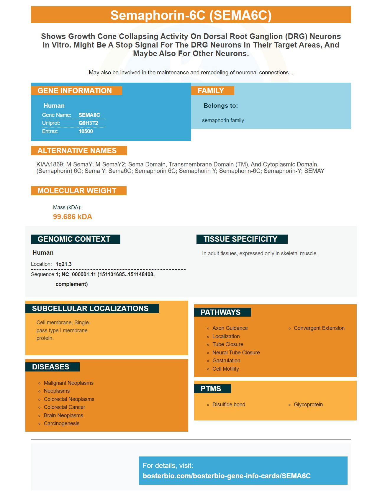
Facts about Semaphorin-6C.
May also be involved in the maintenance and remodeling of neuronal connections. .
Human | |
---|---|
Gene Name: | SEMA6C |
Uniprot: | Q9H3T2 |
Entrez: | 10500 |
Belongs to: |
---|
semaphorin family |
KIAA1869; m-SemaY; m-SemaY2; sema domain, transmembrane domain (TM), and cytoplasmic domain, (semaphorin) 6C; Sema Y; Sema6C; Semaphorin 6C; Semaphorin Y; semaphorin-6C; semaphorin-Y; SEMAY
Mass (kDA):
99.686 kDA
Human | |
---|---|
Location: | 1q21.3 |
Sequence: | 1; NC_000001.11 (151131685..151148408, complement) |
In adult tissues, expressed only in skeletal muscle.
Cell membrane; Single-pass type I membrane protein.
What are the best uses to the SEMA6C markers? This marker is used in many ways, including for research in the area of cancer. Here are some examples:
The SEMA6C Gene has been identified as a key factor in the development of oocytes. The difficulty of detecting the target gene mRNA limits the use of this gene in experiments. This article will cover the various uses of SEMA6C. It will also provide some guidelines on how to use this marker in research and clinical practice.
SEMA6C can be found in the cytoplasmic oocytes of neonatal mouse ovary oocytes. Although there was some staining within interstitial or granulosa oocytes, the intensity was not the same in different primordial foollicles. As shown in Figure 1A_d, two distinct oocytes were present in one primordial foollicle. In addition, SEMA6C expression was lower on Day 7 when compared with Day 3 and 5 ovaries. The difference was not statistically significant. Further research is needed to determine the effect of SEMA6C upon survival.
The SEMA6C gene has been shown to be linked with early primordial follicle activation and dormancy. The process of follicular activation was accelerated by reduced SEMA6C expression in neonatal mouse eggs. It is possible that SEMA6C plays a role in regulating the PI3K/AKT/rpS6 pathway.
SEMA6C is an important gene in the activation primordial follicles. When SEMA6C is expressed in a low level, it regulates the PI3K-AKT-rpS6 pathway. LY294002 reverses SEMA6C deficiency on primordial follicles. SEMA6C works upstream from PI3K and regulates rpS6/AKT phosphorylation. SEMA6C might be used to determine if premature ovarian-ageing is occurring.
SEMA6C, a nuclear transcription factor found in the nucleus, is known as The normal development and maintenance embryonic growth are dependent on this gene. Its expression in the ovary's nucleus determines if a woman has a uniparent follicle. LY294002 inhibits PI3K, a signaling system that regulates gene expression.
Boster Bio's SEMA6C is a novel biomarker for cancer. Its activity is controlled by PTEN (a gene that regulates RTK dependent PI3K/Akt signaling). Mutated PTEN can be found in hepatocellular tumors. These biomarkers are useful in helping physicians design personalized treatments. For more information, visit bosterbio.com
The SEMA6C protein is a non-coding RNA (non-coding RNA) that regulates cellular proliferation. It is also involved with the progression of cancer. This research suggests that SEMA6C may be a therapeutic target. Its impact on tumor growth is discussed in the article, "MALAT1 Regulates Hepatocellular Cancer": Targeting this Noncoding RNA
The PI3K pathway plays a central role in cancer development, and inhibiting PI3K signaling has great potential as a cancer treatment. It is however hampered with intrinsic and acquired immunity, making it difficult clinically to use PI3K antagonists. It is imperative to continue research to ensure that the PI3K pathway continues to function.
Potentially therapeutic targets for cancer have emerged in the PI3K/AKT/mTOR Signaling pathway. Hyperactivity of PI3K signaling is associated with human tumor progression, increased tumor microvessel density, enhanced chemotaxis, and enhanced invasive capabilities of cancer cells. A wide range PI3K inhibitions targeting PI3K signaling have been developed and are currently in clinical trial.
CY3 fluorescent dye can be detected with the naked eyes in both solution or gels. It can be excited to seventy five percent of its maximum fluorescence using a mercury lamp or helium-neon light laser. It was traditionally used with fluorescein and Cy5 fluorescent dyes. However, it has been found useful in flow cytometry and imaging.
The Theory of antibody stocks and the fluorescence intensities of the cell-bound fraction according Eqs were used to calculate the intensity. 9 and 10. The total antibody level was set to 20mg/mL. Cells were normalized according their lowest DOL. The dashed lines indicate the dependence on DOL. The middle graphs represented the degree labeling. The data in Figure 1a are used to interpret the results.
Atto dyes' fluorescence lifetime is much longer than that of carbocyanine and is therefore suitable to measure aqueous solution. It can be measured by a time-gated detector system using pulsed light excitation. Atto dyes have higher signal lifetimes than background fluorescence. They also have shorter life expectancies than fluorophores. They also reduce Rayleigh- and Raman-light scattering, which is common in the biosciences.
Sudan black B (UHSS) is a synthetic dye that reduces the autofluorescence of murine kidney tissue. It is manufactured by Beyotime Biotechnology, China. Autofluorescence in paraffin embedded tissue is reduced by Sudan black. Sudan black B reduces autofluorescence in paraffinembedded and formaldehyde fix tissues.
CY3 Immunofluorescence is based upon the interaction between antibodies, antigens, and a cell's distribution of the target molecule. This technique is widely used in clinical immunology labs to diagnose and monitor patients suffering from autoimmune diseases. They also track the progress of treatment. This technology is compatible and suitable for many applications. Although there are not many direct antibody conjugates currently on the marketplace, they are readily accessible.
PMID: 12110693 by Qu X., et al. Identification, characterization, and functional study of the two novel human members of the semaphorin gene family.