TAB2 / TGF-beta-activated kinase 1 and MAP3K7-binding protein 2 · IHC design guide

Design Immunohistochemistry for TAB2

Plan chromogenic TAB2 IHC in paraffin sections using the observed cytoplasmic tissue pattern (HPA tissue IHC). Compare staining across cell types, including strongly stained lung macrophages (HPA tissue IHC), while keeping fixation consistent across sections (standard IHC practice).

Evidence assembled Oct 2026 · For research use; verify linked source records and product datasheet before use
Immunohistochemistry protocol sheet for TAB2 (IHC for TAB2): expected localisation General cytoplasmic staining (HPA tissue IHC), antibody A02499-1, validated IHC image, and IHC protocol steps
Printable TAB2 IHC protocol sheet — expected localisation General cytoplasmic staining (HPA tissue IHC), antibody A02499-1, controls and protocol steps. Open the full TAB2 IHC guide →

TAB2 Immunohistochemistry Experimental Design Guide

Expected localisation, validated protocols, controls and antibodies — the at-a-glance facts below, then the full design guide.

Must know before staining
Expected localisation General cytoplasmic staining (HPA tissue IHC)
Staining pattern Cytoplasmic staining across multiple cell types (HPA tissue IHC)
Antigen retrieval EDTA pH 8.0 HIER, heat-mediated (datasheet A02499-1)
Positive control ⓘ Bone marrow+4 more · see all
Negative control ⓘ None in HPA (detected in all 45 tissues); use no-primary + isotype controls
Important caveats
Reasons your staining may differ from the expected pattern.
Fixation Keep formalin fixation consistent across sections (standard IHC practice; not target-specific)
Caveat Lung macrophages stain strongly and may dominate the field (HPA tissue IHC)
Regulation IL1 shifts location; abundance effect unknown (UniProt)
Isoform / epitope Two isoforms; epitope coverage is unspecified (UniProt)
Section 1

Recommended TAB2 IHC & IF Protocols

The catalog antibody’s IHC-P protocol is accompanied by published TAB2 staining protocols for adult mouse cryosections and cervical carcinoma sections (PMC2770139; PMC8266450).

Recommended immunohistochemistry (IHC-P) protocol parameters
SampleParaffin-embedded human rectal cancer tissue; fixative not specified (datasheet A02499-1)
FixationImage fixative and duration unreported (datasheet A02499-1); verify before use.
Sectioning4–5 µm sections on charged slides (standard)
DeparaffinisationXylene, graded ethanol series to water (standard)
Antigen retrievalHeat retrieval: EDTA pH 8.0 (datasheet A02499-1); 20 min, 95–100 °C (standard)
Peroxidase block3% H2O2, 10 min, room temperature (standard)
Blocking10% goat serum (datasheet A02499-1)
Primary antibodyRabbit anti-TAB2, 2-5μg/ml (datasheet A02499-1)
Primary incubationOvernight at 4 °C (datasheet A02499-1)
DetectionStreptavidin-biotin complex (SABC), DAB chromogen (datasheet A02499-1)
CounterstainHematoxylin, blue, dehydrate and mount (standard)
Expected resultTAB2-positive staining in hematopoietic cells of bone marrow (HPA tissue IHC: High). HPA tissue profile: General cytoplasmic expression. No signal in the no-primary control.
💡Decision noteStart with heat-mediated EDTA, pH 8.0, for the catalog antibody (datasheet A02499-1). Sodium citrate, pH 6.0, was used for cervical carcinoma sections (PMC8266450).
Section 2

What Is the Expected TAB2 Staining Pattern?

TAB2 is reported in the cytosol and at membranes, including endosome and lysosome membranes; it has no transmembrane segment (UniProt Q9NYJ8). In tissue sections, expect predominantly cytoplasmic staining across many cell types, with particularly strong staining reported in lung macrophages, bone marrow hematopoietic cells and several epithelia (HPA: tissue IHC). HPA rates its tissue profile Approved, with medium consistency between antibody staining and RNA expression (HPA: tissue IHC).

What am I looking at on my slide?
Cytoplasmic staining in lung macrophages or bone marrow hematopoietic cells, with some cell-to-cell variation.This fits HPA's general cytoplasmic profile and High staining in these cells (HPA: tissue IHC). Read intensity relative to nearby cells and the section's background; High is an HPA category, not a required score for every specimen.
Cytoplasmic staining in respiratory epithelium, glandular epithelium or placental trophoblasts.These are plausible positive cells: HPA reports High staining in bronchus and nasopharynx respiratory epithelium, endometrium and epididymis glands, pancreatic exocrine glands, and placental trophoblasts (HPA: tissue IHC). Their appearance can differ by tissue and assay conditions.
Predominantly nuclear staining, or a sharp cell-surface rim without convincing cytoplasmic staining.This is discordant with the reported cytosolic and membrane-associated locations (UniProt Q9NYJ8). Check morphology, counterstain, detection controls and antibody specificity before calling it TAB2; membrane association alone does not predict a continuous surface rim (UniProt Q9NYJ8 topology).
Strong signal concentrated in cells outside the expected positive population, especially when the rest of the section is weak.Consider cross-reactivity or endogenous detection activity (standard IHC practice). TAB2 is widely expressed (UniProt Q9NYJ8), and HPA reports low tissue specificity (HPA: tissue IHC), so an unlisted cell type is not automatically a false positive. Check its compartment and controls.
Diffuse haze across tissue and empty spaces, or no staining in an HPA High tissue.Diffuse haze does not match HPA's cellular cytoplasmic profile (HPA: tissue IHC) and suggests background (standard IHC practice). An absent signal in an HPA High cell population warrants a run check, but HPA categories do not guarantee positivity in every specimen (HPA: tissue IHC).
💡Expected TAB2 appearanceCall a convincing positive when identifiable cells show cytoplasmic signal above local background, especially HPA High populations such as lung macrophages or bronchial respiratory epithelium (HPA: tissue IHC); isolated nuclear staining or diffuse haze is suspect against the reported TAB2 locations (UniProt Q9NYJ8; standard IHC practice).
How each factor affects the staining
Cellular location and stimulationTAB2 is reported in cytosol and on membranes; IL1 stimulation can move it from membrane to cytosol, while TRIM38 interaction can move it toward endosome and lysosome (UniProt Q9NYJ8). A static tissue section cannot establish which event occurred.
Tissue and cell populationHPA reports Low tissue specificity and a general cytoplasmic profile, with High and Low staining in different listed cell populations (HPA: tissue IHC). Choose a clearly identified High population for a comparison control; interpret weaker cells in their tissue context.
IHC evidence and IF limitTwo listed rabbit polyclonal antibodies, HPA065436 and HPA070137, have IHC Approved status (HPA: antibodies). HPA provides no main ICC-IF location or ICC-IF images here (HPA: subcellular), so this evidence does not establish an IF pattern.
Protein forms and topologyUniProt lists two isoforms, one chain spanning residues 1–693, no signal peptide, no propeptide and no transmembrane segment (UniProt Q9NYJ8). Without an antibody epitope in the supplied record, isoform coverage and the effect of epitope location cannot be predicted.
Antigen retrieval and detectionNo TAB2-specific retrieval condition, dilution or fixation sensitivity is supplied (UniProt Q9NYJ8; HPA: tissue IHC). Retrieval, blocking and detection controls can be optimized as general paraffin IHC practice; their effects should be established for the actual antibody and tissue.
Why is my staining missing, weak or wrong?
SituationLikely causeNext action
Expected lung macrophages or bone marrow hematopoietic cells show no convincing stain (HPA: High in these cells).The staining run, antibody conditions or tissue preservation may be unsuitable (standard IHC practice); specimen variation remains possible despite an HPA High category (HPA: tissue IHC).Confirm the positive cell population on the counterstain, inspect the run's positive control and review the antibody's IHC-P instructions. Optimize retrieval and detection using paired sections (standard IHC practice).
The whole section, including spaces between cells, appears brown.Diffuse deposition or nonspecific background is more likely than the reported cellular cytoplasmic profile (HPA: tissue IHC; standard IHC practice).Check the no-primary control and wash, blocking and chromogen steps; compare cellular signal with adjacent background before scoring (standard IHC practice).
Pigmented or blood-rich areas appear positive despite little matching cellular signal.Endogenous pigment or detection activity can mimic chromogenic staining (standard IHC practice). HPA's TAB2 profile alone cannot identify the deposit (HPA: tissue IHC).Compare a no-primary section, examine pigment in an unstained or counterstained section, and verify the detection system's endogenous-activity block (standard IHC practice).
Nuclei dominate the signal while cytoplasm is weak.A nuclear-dominant result conflicts with TAB2's reported cytosolic and membrane-associated locations (UniProt Q9NYJ8); counterstain overlap or nonspecific staining is possible (standard IHC practice).Inspect the no-primary control and cell boundaries, then compare a known HPA High cell population on the same run before accepting the compartment call (HPA: tissue IHC; standard IHC practice).
Strong staining appears only in an unexpected cell type.Cross-reactivity or endogenous detection activity is possible (standard IHC practice), but wide expression means an unlisted positive cell is not excluded (UniProt Q9NYJ8; HPA: low tissue specificity).Identify the cell type morphologically, check whether staining is cytoplasmic, and compare controls and a listed HPA High population before assigning TAB2 specificity (HPA: tissue IHC; standard IHC practice).
An IF/ICC image does not match the tissue IHC result.The supplied HPA subcellular record has no main location or ICC-IF images, while the antibody entries list IHC approval without ICC status (HPA: subcellular; HPA: antibodies).Treat IF/ICC as a separate validation question; assess its own controls and antibody documentation. Do not infer an IF protocol or validated IF compartment from the tissue IHC profile (HPA: tissue IHC; HPA: subcellular).

Sample controls for TAB2 IHC & IF

🧪Run lung first and score its macrophages for TAB2 staining (HPA: High in lung macrophages). HPA detects TAB2 in all 45 scored tissues, so there is no supported negative tissue; use no-primary and isotype controls, and treat other cells on the slide as background comparators rather than validated TAB2-negative cells (HPA: no negative tissue rows; TAB2 detected in all 45 scored tissues).
Positive control tissue: Bone marrow (Hematopoietic cells, HPA High)
Negative control tissue: None in HPA: TAB2 is detected in all 45 scored tissues. Use a no-primary (secondary-only) and an isotype control instead.
ICC-IF cell lines (HPA subcellular resource): HPA carries no ICC-IF cell line for TAB2; derive a cell-line control from the positive tissue's cell type (Hematopoietic cells) and confirm it by RNA or western blot first.
Technical controls: Include a no-primary, secondary-only section and a concentration-matched rabbit IgG isotype control alongside a TAB2 knockout specimen or a validated immunizing-peptide block, if available (selected-SKU caption: rabbit primary antibody; standard IHC control practice). For lung sections, check endogenous peroxidase and endogenous biotin background before interpreting the biotin-based DAB signal (selected-SKU caption: biotinylated secondary, SABC and DAB; HPA: High in lung macrophages).
⚠️Feasibility: A target-specific fixation window or fixation effect is unreported, and the exact A02499-1 tissue-IHC caption does not state a fixative (selected-SKU caption: fixative not stated). Heat retrieval in EDTA at pH 8.0 is a documented starting condition for paraffin sections, but its necessity has not been established by the supplied evidence (selected-SKU caption: heat retrieval in EDTA, pH 8.0). Neither frozen sections nor IF are established as easier here; lung macrophage peroxidase and biotin background should be checked when using the caption's SABC/DAB detection (HPA: High in lung macrophages; selected-SKU caption: SABC/DAB).

HPA tissue IHC evidence for TAB2

Comprehensive Human Protein Atlas IHC scoring per tissue (reliability: Approved — Medium consistency between antibody staining and RNA expression data.). Rows are taken directly from the HPA tissue chart — click any row's HPA link to view the source.

Positive expression · recommended positive controls

TissueCell typeLevelEvidenceSource
Bone marrow Hematopoietic cells High Protein (IHC) HPA →
Bronchus Respiratory epithelial cells High Protein (IHC) HPA →
Endometrium Glandular cells High Protein (IHC) HPA →
Epididymis Glandular cells High Protein (IHC) HPA →
Lung Macrophages High Protein (IHC) HPA →

Undetected expression · recommended negative controls

TissueCell typeLevelEvidenceSource
None in HPA: TAB2 is detected in all 45 scored tissues. Use a no-primary (secondary-only) and an isotype control instead.
Section 3

Advanced TAB2 IHC Tips

Use the documented paraffin-section workflow as a starting point, then assess TAB2 staining by cell type, compartment and section quality.

Which retrieval should I try first for weak TAB2 staining in paraffin sections?
Start with heat-mediated retrieval in EDTA, pH 8.0, for paraffin sections (datasheet A02499-1). The selected TAB2 tissue image used that retrieval before overnight incubation at 4°C with 2 μg/mL antibody (datasheet A02499-1). If staining remains weak, vary heating duration on adjacent sections while keeping the antibody and detection conditions fixed, and check whether tissue morphology deteriorates. Compare each run with a documented positive cell population, such as lung macrophages or pancreatic exocrine glandular cells (HPA: High in lung macrophages; High in pancreatic exocrine glandular cells).
Could fixation explain inconsistent TAB2 staining between paraffin specimens?
Yes, fixation can change epitope accessibility in IHC, but TAB2-specific fixation sensitivity is unknown from the supplied evidence. The selected image documents a paraffin-embedded human rectal cancer section and does not state its fixative (datasheet A02499-1); do not treat it as a formalin-fixed validation. Record each specimen’s fixative and fixation history, then compare sections processed with the same EDTA, pH 8.0 retrieval and 2 μg/mL primary antibody conditions (datasheet A02499-1). If a batch loses signal, inspect tissue preservation and include a previously responsive section in the same staining run before changing retrieval.
Where should TAB2 appear, and when is a compartment shift plausible?
Expect predominantly cytoplasmic staining in tissue sections (HPA: general cytoplasmic expression), with membrane-associated, endosomal and lysosomal pools also consistent with the protein record (UniProt Q9NYJ8: subcellular location). TAB2 has no transmembrane segment, so a membrane-adjacent signal alone does not establish membrane insertion (UniProt Q9NYJ8: topology). Following IL1 stimulation, TAB2 can move from membrane to cytosol; interaction with TRIM38 can redirect it toward endosomes and lysosomes (UniProt Q9NYJ8: subcellular location). Score compartment and cell type separately, and require a matched biological comparison before attributing a staining shift to either mechanism.
How should isoforms and epitope location affect interpretation of TAB2 IHC?
TAB2 has 2 listed isoforms, and the supplied antibody caption does not identify its epitope (UniProt Q9NYJ8: isoforms; datasheet A02499-1: caption). Consequently, staining cannot be assigned to one isoform without antibody epitope information and evidence that the epitope differs between isoforms. The protein has a CUE domain at residues 8–51 and reported modified residues including phosphoserines at 372, 450, 482 and 524 (UniProt Q9NYJ8: domains; modified residues). If a condition changes staining, check epitope documentation before interpreting the difference as altered abundance or a particular modification.
How can I investigate TAB2 cell types and localisation by IF?
Treat IF as a separate application: the selected antibody evidence describes chromogenic IHC in a paraffin section, and no ICC/IF images are listed in the supplied subcellular record (datasheet A02499-1; HPA: subcellular record). Multiplex TAB2 with a marker for the expected cell population, such as CD68 when examining macrophages, which show high TAB2 tissue staining in lung (HPA: High in lung macrophages). Choose a far-red fluorophore after checking tissue autofluorescence and include single-color controls. For intracellular TAB2, assess detergent permeabilisation against morphology and marker retention; its cytosolic and endosomal or lysosomal locations inform that choice (UniProt Q9NYJ8: subcellular location).
How do I separate diffuse TAB2 staining from chromogenic background?
The selected tissue image used 10% goat-serum blocking, a biotinylated secondary antibody, an avidin–biotin detection complex and DAB (datasheet A02499-1). If brown signal spreads across cell-free areas or follows section edges, inspect reagent deposits, drying and uneven coverage before calling it TAB2. Include a primary-antibody omission control to assess secondary and detection-system signal, and use a peroxidase block as a general DAB workflow step. Interpret cytoplasmic staining in defined cells against the section’s negative-control signal; HPA reports general cytoplasmic TAB2 expression, but its tissue staining has only medium RNA–protein consistency (HPA: profile; Approved reliability description).
What scoring scheme captures TAB2 staining without confusing cell mixture with expression? ⚠ ANSWER MARKED FOR VERIFICATION
Define the cell population and compartment before scoring, because HPA reports high staining in several distinct populations, including bone-marrow hematopoietic cells and lung macrophages (HPA: positive tissue IHC). For a defined population, record the percentage of positive cells and an H-score that combines percentage with intensity; keep the intensity categories constant across slides. If cell abundance is the endpoint, report TAB2-positive cell density per mm² of viable tissue and separately record total cells or area in the same compartment. Normalise comparisons to the same cell type, viable area, staining batch and exposure or scan settings.
What evidence distinguishes real TAB2 signal from a misleading positive?
A credible result shows reproducible staining in identifiable cells with predominantly cytoplasmic localisation, consistent with the HPA tissue profile (HPA: general cytoplasmic expression). High staining in lung macrophages or bronchial respiratory epithelial cells can support a run control, but HPA reports medium consistency between staining and RNA expression, so a positive pattern is not proof of specificity (HPA: positive tissue IHC; Approved reliability description). Scrutinise strong nuclear-only signal, section-edge staining, necrotic areas and DAB deposits for artefact. Use adjacent-section controls, including primary omission and a previously responsive specimen, before assigning a compartment change to TAB2 biology.
Boster reagents

Best TAB2 / TGF-beta-activated kinase 1 and MAP3K7-binding protein 2 IHC Antibodies

Two human-reactive anti-TAB2 antibodies have IHC images from paraffin-embedded rectal cancer tissue and human brain tissue (catalog image captions; reactivity). Neither lists IF/ICC validation (catalog applications).

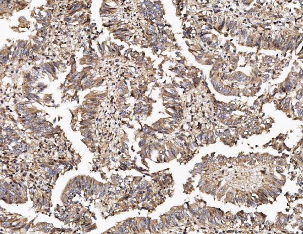
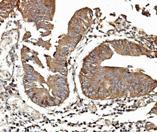
Real IHC data IHC analysis of TAB2 using anti-TAB2 antibody (A02499-1). TAB2 was detected in paraffin-embedded section of human rectal cancer tissue. Heat mediated antigen retrieval was performed in EDTA buffer (pH8.0, epitope retrieval solution). The tissue section was blocked with 10% goat serum. The tissue section was then incubated with 2μg/ml rabbit anti-TAB2 Antibody (A02499-1) overnight at 4°C. Biotinylated goat anti-rabbit IgG was used as secondary antibody and incubated for 30 minutes at 37°C. The tissue section was developed using Strepavidin-Biotin-Complex (SABC) (Catalog # SA1022) with DAB as the chromogen.
Anti-TAB2 Antibody ®
Cat # A02499-1
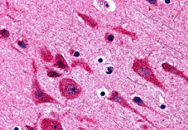
Real IHC data Immunohistochemistry of TAB2 in human brain tissue with TAB2 antibody at 5 μg/mL.
Anti-TAB2 Antibody
Cat # A02499

A02499-1 has an IHC image from a paraffin-embedded human rectal cancer section (A02499-1 image caption). A02499 lists human IHC-P and has an IHC image from human brain tissue (A02499 applications; image caption).

Which to pick: For paraffin-section IHC, choose A02499-1 when its documented retrieval and detection conditions are useful; its caption reports 2 μg/mL antibody after EDTA retrieval at pH 8.0 (A02499-1 image caption). A02499 is another human IHC-P option, shown in brain tissue at 5 μg/mL; its catalog calls it polyclonal, while A02499-1 has no clone specified (A02499 applications; image caption; dilution record; A02499-1 catalog clone field). Neither is a validated choice for IF/ICC or other species, and the fixative is unreported for both pictured tissues (catalog applications; reactivity; image captions).

Each figure is that product's own IHC / IF validation image from its datasheet.

References

  1. UniProt Consortium. UniProt entry Q9NYJ8 (TAB2_HUMAN, TGF-beta-activated kinase 1 and MAP3K7-binding protein 2).
  2. Human Protein Atlas. TAB2 tissue IHC expression (reliability: Approved).
  3. Human Protein Atlas. TAB2 subcellular location (ICC-IF): Highest expression in HDLM-2: 96.5 nTPM.
  4. Human Protein Atlas. TAB2 antibody validation summary (2 antibodies).
  5. Expression analysis of the TAB2 protein in adult mouse tissues. Inflammation research : official journal of the European Histamine Research Society ... [et al.] 2007 — PMC2770139.
  6. TAB2 Promotes the Stemness and Biological Functions of Cervical Squamous Cell Carcinoma Cells. Stem cells international 2021 — PMC8266450.
  7. Hepatitis B Surface Antigen Suppresses the Activation of Nuclear Factor Kappa B Pathway via Interaction With the TAK1-TAB2 Complex. Frontiers in immunology 2021 — PMC7947203.
  8. PubMed PMID:10882101 — UniProt-cited evidence.
  9. PubMed PMID:9872452 — UniProt-cited evidence.
  10. PubMed PMID:12168954 — UniProt-cited evidence.