This website uses cookies to ensure you get the best experience on our website.


- Table of Contents
This guide covers paraffin section IHC for TACR1 using the IHC-validated M01006 antibody at 1:50 (datasheet M01006). Assess cytoplasmic staining in squamous epithelial cells (HPA tissue IHC), while considering receptor movement to early endosomes after substance P binding (UniProt).

Expected localisation, validated protocols, controls and antibodies — the at-a-glance facts below, then the full design guide.
| Expected localisation | Epithelial cytoplasm (HPA tissue IHC) | |
| Staining pattern | Squamous epithelial cells: cytoplasmic staining (HPA tissue IHC) | |
| Antigen retrieval | EDTA pH 8.0 HIER, heat-mediated (datasheet M01006) | |
| Positive control | Bronchus+4 more · see all | |
| Negative control | Adipose tissue+4 more · see all |
| Fixation | Keep fixation consistent across sections (standard IHC practice; not target-specific) | |
| Caveat | Substance P can shift receptor into early endosomes (UniProt) | |
| Regulation | No expression regulator annotated (UniProt) | |
| Isoform / epitope | 2 isoforms; map extracellular versus cytoplasmic epitopes (UniProt) |
The catalog antibody uses heat-mediated EDTA pH 8.0 retrieval (datasheet: M01006). The published rat spinal cord IHC protocol uses citric acid pH 6.0 and chromogenic detection (PMC10954158).
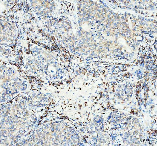
| Sample | Paraffin-embedded human lung cancer tissue; fixative not specified (datasheet M01006) |
| Fixation | Image fixative and duration unreported (datasheet M01006); verify before use. |
| Sectioning | 4–5 µm sections on charged slides (standard) |
| Deparaffinisation | Xylene, graded ethanol series to water (standard) |
| Antigen retrieval | Heat retrieval: EDTA pH 8.0 (datasheet M01006); 20 min, 95–100 °C (standard) |
| Peroxidase block | 3% H2O2, 10 min, room temperature (standard) |
| Blocking | 10% goat serum (datasheet M01006) |
| Primary antibody | Rabbit monoclonal (clone AFBH-20) anti-TACR1, 1:50 (datasheet M01006) |
| Primary incubation | Overnight at 4 °C (datasheet M01006) |
| Detection | HRP-conjugated secondary, DAB chromogen (datasheet M01006) |
| Counterstain | Hematoxylin, blue, dehydrate and mount (standard) |
| Expected result | TACR1-positive staining in respiratory epithelial cells of bronchus (HPA tissue IHC: Medium). HPA tissue profile: Cytoplasmic expression in squamous epithelia. No signal in the no-primary control. |
TACR1 is a seven-pass receptor at the cell membrane that can move to early endosomes after substance P binding (UniProt P25103 topology and subcellular location). In paraffin IHC, expect staining in squamous epithelial cells of cervix, esophagus and vagina, and in skin keratinocytes; HPA reports medium staining in these cells (HPA tissue IHC). Its tissue profile has Enhanced reliability, with medium agreement between staining and RNA and external verification pending (HPA tissue IHC).
| Membrane-associated staining, with some cytoplasmic signal, in the expected epithelial cells. | This fits the membrane location and possible endosomal internalization of TACR1 (UniProt P25103 subcellular location). HPA describes cytoplasmic staining in squamous epithelia and medium staining in cervix, esophagus, vagina and skin keratinocytes (HPA tissue IHC). Score the relevant cells and compartments rather than requiring a continuous membrane outline. |
| Predominantly nuclear staining, without convincing membrane-associated or epithelial cytoplasmic staining. | A nuclear pattern is outside the supplied TACR1 localization evidence: UniProt places the receptor at the membrane and early endosomes, while HPA describes cytoplasmic staining in squamous epithelia (UniProt P25103 subcellular location; HPA tissue IHC). Treat this pattern as suspect and compare it with a known-positive section and the detection control before assigning TACR1 positivity (general IHC practice). |
| Staining concentrates in a cell population listed as not detected by HPA. | For example, HPA reports TACR1 as not detected in adipocytes of adipose tissue and hematopoietic cells of bone marrow (HPA tissue IHC). Unexpected staining raises cross-reactivity or endogenous detection activity as possibilities (general IHC practice). Check the named cell population, since those HPA observations do not declare every cell in either tissue negative. |
| Diffuse color covers cells and surrounding tissue without a clear cellular pattern. | This is difficult to interpret as TACR1 when the expected evidence points to membrane-associated and epithelial cytoplasmic localization (UniProt P25103 subcellular location; HPA tissue IHC). Review background on a section processed without primary antibody, then optimize the chromogenic workflow if background persists (general IHC practice). |
| No staining appears in the expected epithelial cells of a positive reference section. | HPA reports medium staining in cervix, esophagus and vagina squamous epithelial cells, and skin keratinocytes (HPA tissue IHC). A blank reference section makes a negative result in the experimental section inconclusive. Check antibody and detection performance, tissue preservation and the assay's routine retrieval conditions (general IHC practice); the supplied sources do not establish TACR1-specific fixation sensitivity. |
| Seven-pass topology and epitope access | UniProt maps extracellular and cytoplasmic regions around seven transmembrane segments (UniProt P25103 topology). If an antibody's epitope is known, its position can guide interpretation of accessibility after section processing; no epitope position or TACR1-specific retrieval requirement is supplied here. Follow the validated IHC procedure for the antibody used (general IHC practice). |
| Receptor trafficking | Substance P binding can move TACR1 from the cell surface to early endosomes, followed by recycling (UniProt P25103 subcellular location). Thus, some intracellular signal can be biologically plausible. That mechanism alone cannot prove that cytoplasmic chromogen in an individual section is specific; compare its distribution with the HPA epithelial pattern and controls. |
| Tissue-pattern confidence | HPA labels the tissue IHC profile Enhanced, yet describes only medium consistency with RNA and says external verification is pending (HPA tissue IHC). Use its named cell types and staining levels as reference observations, while keeping an independent positive reference and detection control in the assay (general IHC practice). |
| Isoforms and glycosylation | UniProt records two TACR1 isoforms and glycosylation sites at residues 14 and 18 (UniProt P25103 isoforms and glycosylation). The supplied record gives no antibody epitope or isoform selectivity. Do not assign different staining patterns to isoforms or infer a retrieval effect from the glycosylation sites. |
| IF/ICC Q&A: what location is supported? | HPA supports plasma-membrane localization in ICC-IF; its centrosome and basal-body assignments are uncertain (HPA subcellular ICC-IF). UniProt also places TACR1 in early endosomes after signaling (UniProt P25103 subcellular location). Use the separate IF/ICC guide for that application; these observations supply no IF protocol or basis for treating puncta as definitive endosomes. |
| Situation | Likely cause | Next action |
|---|---|---|
| The positive reference section is blank. | The assay may have failed, or the sampled reference may lack the expected cells; HPA's positive observations refer to specified epithelial populations (HPA tissue IHC). | Confirm those cells are present, then check primary-antibody use, detection reagents and routine IHC retrieval settings against the validated procedure (general IHC practice). Do not infer TACR1-specific fixation failure from this result. |
| Nuclear color dominates the section. | Nuclear localization is unsupported by UniProt's membrane and early-endosome annotation and HPA's squamous-epithelial cytoplasmic profile (UniProt P25103 subcellular location; HPA tissue IHC). | Compare a known-positive section and a no-primary control; review counterstain and chromogen interpretation before scoring (general IHC practice). Record the nuclear result as unresolved if the controls cannot explain it. |
| Unexpected cells stain strongly. | Cross-reactivity or endogenous detection activity is possible (general IHC practice). HPA's not-detected calls apply to named cells, such as adipocytes in adipose tissue, rather than whole tissues (HPA tissue IHC). | Identify the stained cell type, run a no-primary control, and check endogenous-enzyme blocking for chromogenic detection (general IHC practice). Compare only the same cell population with HPA. |
| Color is widespread and obscures cell boundaries. | Diffuse background can reflect nonspecific binding or detection background (general IHC practice); it does not resolve into the expected epithelial pattern (HPA tissue IHC). | Inspect the no-primary control, review blocking and washing, and adjust antibody concentration or detection conditions using the validated IHC procedure (general IHC practice). Reassess only after cell boundaries are readable. |
| Staining is cytoplasmic but lacks a crisp membrane rim. | HPA describes cytoplasmic expression in squamous epithelia; UniProt permits early-endosomal localization after substance P signaling (HPA tissue IHC; UniProt P25103 subcellular location). Neither source establishes the cause of cytoplasmic signal in this section. | Check whether staining is confined to the expected epithelial cells and exceeds control background (HPA tissue IHC; general IHC practice). Report the observed compartment without calling it endosomal unless separately demonstrated. |
| An IF/ICC image shows centrosomal or basal-body spots. | HPA marks those locations uncertain while supporting the plasma membrane in ICC-IF (HPA subcellular ICC-IF). | Treat the spots as unconfirmed TACR1 localization and consult the separate IF/ICC guide. Do not use those uncertain locations to score a paraffin IHC section positive (HPA subcellular ICC-IF). |


Comprehensive Human Protein Atlas IHC scoring per tissue (reliability: Enhanced — Medium consistency between antibody staining and RNA expression data. Pending external verification.). Rows are taken directly from the HPA tissue chart — click any row's HPA link to view the source.
| Tissue | Cell type | Level | Evidence | Source |
|---|---|---|---|---|
| Bronchus | Respiratory epithelial cells | Medium | Protein (IHC) | HPA → |
| Cervix | Squamous epithelial cells | Medium | Protein (IHC) | HPA → |
| Esophagus | Squamous epithelial cells | Medium | Protein (IHC) | HPA → |
| Prostate | Glandular cells | Medium | Protein (IHC) | HPA → |
| Skin | Keratinocytes | Medium | Protein (IHC) | HPA → |
| Tissue | Cell type | Level | Evidence | Source |
|---|---|---|---|---|
| Adipose tissue | Adipocytes | Not detected | Protein (IHC) | HPA → |
| Adrenal gland | Glandular cells | Not detected | Protein (IHC) | HPA → |
| Appendix | Glandular cells | Not detected | Protein (IHC) | HPA → |
| Bone marrow | Hematopoietic cells | Not detected | Protein (IHC) | HPA → |
| Breast | Adipocytes | Not detected | Protein (IHC) | HPA → |
Troubleshoot TACR1 staining in paraffin sections by checking retrieval, compartment, cell type and controls before comparing signal intensity.
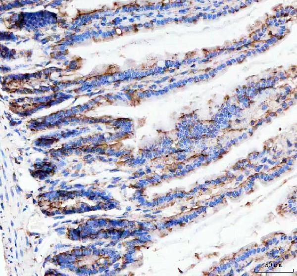
The IHC-tested TACR1 antibody has paraffin-section images from human lung cancer and mouse and rat colon (M01006 image captions). No IF data are supplied (M01006 catalog).
M01006 will render with an IHC figure from paraffin-embedded human lung cancer tissue (M01006 figure caption). Additional images show paraffin-embedded mouse and rat colon, and IHC is a listed application (M01006 image captions; M01006 catalog).
Which to pick: Choose M01006 for chromogenic IHC on paraffin sections: its figure documents EDTA retrieval at pH 8.0, a 1:50 primary dilution and peroxidase/DAB detection; the fixative is unreported (M01006 figure caption). For cross-species IHC, M01006 is a rabbit monoclonal with human, mouse and rat reactivity and corresponding paraffin-section images (M01006 catalog; M01006 image captions). There is no supported IF/ICC pick: M01006 has no listed IF application or IF image (M01006 catalog).