TAF12 / Transcription initiation factor TFIID subunit 12 · IHC design guide

Design Immunohistochemistry for TAF12

Plan chromogenic TAF12 IHC in paraffin sections using the observed nuclear and cytoplasmic tissue pattern (HPA tissue IHC). Compare positive glandular cells with unstained cell populations and assess unexpected signal against the nuclear molecular location (HPA tissue IHC; UniProt).

Evidence assembled Oct 2026 · For research use; verify linked source records and product datasheet before use
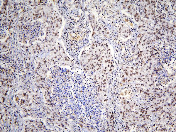
Immunohistochemistry protocol sheet for TAF12 (IHC for TAF12): expected localisation Nuclear and cytoplasmic tissue staining (HPA tissue IHC), antibody A06944-1, validated IHC image, and IHC protocol steps
Printable TAF12 IHC protocol sheet — expected localisation Nuclear and cytoplasmic tissue staining (HPA tissue IHC), antibody A06944-1, controls and protocol steps. Open the full TAF12 IHC guide →

TAF12 Immunohistochemistry Experimental Design Guide

Expected localisation, validated protocols, controls and antibodies — the at-a-glance facts below, then the full design guide.

Must know before staining
Expected localisation Nuclear and cytoplasmic tissue staining (HPA tissue IHC)
Staining pattern Nuclear and cytoplasmic staining across most tissues (HPA tissue IHC)
Antigen retrieval Citrate pH 6 HIER, heat-mediated (datasheet A06944-1)
Positive control ⓘ Adrenal gland+4 more · see all
Negative control ⓘ Adipose tissue+4 more · see all
Important caveats
Reasons your staining may differ from the expected pattern.
Fixation Keep formalin fixation consistent across sections. (standard IHC practice; not target-specific)
Caveat IHC–RNA agreement is moderate; verify unexpected staining (HPA tissue IHC)
Regulation Low tissue specificity; staining intensity varies (HPA tissue IHC)
Isoform / epitope 2 isoforms; antibody epitope coverage is unknown (UniProt)
Section 1

Recommended TAF12 IHC & IF Protocols

The catalog antibody’s IHC-P protocol is complemented by published TAF12 staining methods for human glioma (PMC9775265) and mouse choroid plexus carcinoma (PMC4458854).

Recommended immunohistochemistry (IHC-P) protocol parameters
SampleParaffin-embedded human lung cancer tissue; fixative not specified (datasheet A06944-1)
FixationImage fixative and duration unreported (datasheet A06944-1); verify before use.
Sectioning4–5 µm sections on charged slides (standard)
DeparaffinisationXylene, graded ethanol series to water (standard)
Antigen retrievalHeat retrieval: Citrate pH 6, 20 min (datasheet A06944-1)
Peroxidase block3% H2O2, 10 min, room temperature (standard)
Blocking10% goat serum (datasheet A06944-1)
Primary antibodyRabbit anti-TAF12, 1μg/ml (datasheet A06944-1)
Primary incubationOvernight at 4 °C (datasheet A06944-1)
DetectionStreptavidin-biotin complex (SABC), DAB chromogen (datasheet A06944-1)
CounterstainHematoxylin, blue, dehydrate and mount (standard)
Expected resultTAF12-positive staining in glandular cells of adrenal gland (HPA tissue IHC: High). HPA tissue profile: Cytoplasmic and nuclear expression in most tissues. No signal in the no-primary control.
💡Decision noteStart with heat-mediated citrate retrieval at pH 6 (datasheet A06944-1); neither emitted article specifies a retrieval method (PMC9775265; PMC4458854).
Section 2

What Is the Expected TAF12 Staining Pattern?

TAF12 is a nuclear transcription factor component with no transmembrane segment (UniProt Q16514). In paraffin-section IHC, expect nuclear staining, with possible cytoplasmic staining, across many tissues (HPA: cytoplasmic and nuclear expression in most tissues). Strong examples include glandular cells of colon and adrenal gland (HPA: High). HPA rates the tissue IHC pattern Approved, with medium consistency against RNA data and external verification pending (HPA: reliability description).

What am I looking at on my slide?
Distinct nuclear staining in colon glandular cells, with or without cytoplasmic staining.This fits the nuclear location assigned to TAF12 (UniProt Q16514) and the cytoplasmic and nuclear tissue pattern (HPA: profile). Colon glandular cells are a useful high-staining reference (HPA: High). Judge the cellular pattern alongside a matched negative control; HPA's Approved rating still has medium RNA concordance and awaits external verification (HPA: reliability description).
Signal outlines cell membranes or lies mainly outside cells, with little nuclear staining.That distribution conflicts with TAF12's nuclear location and lack of a transmembrane segment (UniProt Q16514). Treat it as suspect staining and inspect the negative control and tissue morphology before scoring it as TAF12. Cytoplasmic signal alone needs a more careful judgment because HPA reports cytoplasmic as well as nuclear tissue staining (HPA: profile).
Strong staining appears in adipocytes or esophageal squamous epithelial cells.Those cell types were not detected in HPA tissue IHC (HPA: adipocytes and esophageal squamous epithelial cells, Not detected). Unexpected staining can reflect cross-reactivity or endogenous chromogen-generating activity (general IHC practice). Compare an adjacent section with primary antibody omitted, then review whether the signal follows cell boundaries and nuclei or persists independently of the primary antibody.
A broad, even haze covers tissue, empty spaces, and many cell types.Diffuse background prevents a reliable cell-level call (general IHC practice). It does not resemble the reported cell-associated nuclear and cytoplasmic pattern (HPA: profile). Review the negative control, blocking and detection conditions, and antibody concentration before interpreting faint tissue signal. HPA's low tissue specificity for RNA does not make an indiscriminate slide-wide deposit a positive protein result (HPA: RNA specificity).
Colon glandular cells show no detectable staining.This conflicts with a reported high IHC level in that cell type (HPA: colon glandular cells, High), but one blank slide cannot establish absence of TAF12. Check that the glandular cells are present and interpretable, then review the catalog antibody's IHC-P conditions and detection controls (general IHC practice). HPA's tissue observations are references, not a guaranteed result for every specimen (HPA: Approved; external verification pending).
💡Expected TAF12 appearanceA convincing positive has nuclear staining in high-reference glandular cells such as colon, with possible cytoplasmic staining (UniProt Q16514; HPA: colon High; HPA: profile); membrane outlines, extracellular deposits, or strong signal in HPA-undetected adipocytes warrant investigation (UniProt Q16514 topology; HPA: adipocytes Not detected).
How each factor affects the staining
Subcellular locationTAF12 is assigned to the nucleus and has no transmembrane segment (UniProt Q16514). HPA nevertheless reports cytoplasmic and nuclear staining in most tissues (HPA: profile). Assess nuclear signal as the clearest location match, while recording cytoplasmic signal separately rather than automatically discarding it.
Cell and tissue selectionColon and adrenal glandular cells are reported High, whereas adipocytes and esophageal squamous epithelial cells are Not detected (HPA: tissue IHC). These provide contrasting cell-level references within the supplied observations. Low tissue specificity of RNA does not imply equal IHC intensity in every cell (HPA: RNA specificity; HPA: tissue IHC).
Strength of IHC evidenceThe listed antibody HPA008519 is Approved for IHC, and HPA describes medium consistency with RNA expression and pending external verification (HPA: antibody validation; HPA: reliability description). Use its tissue pattern as a reference for interpretation, and retain control-based checks when a specimen differs from it.
Isoforms and antibody recognitionUniProt lists two TAF12 isoforms, TAFII20 and TAFII15 (UniProt Q16514). The supplied evidence does not locate the IHC antibody's epitope or establish which isoforms it recognizes. A staining difference therefore cannot be assigned to isoform choice from this record alone.
Why is my staining missing, weak or wrong?
SituationLikely causeNext action
The positive-reference tissue is blank.The slide may have a workflow or detection failure; colon glandular cells are reported High in HPA tissue IHC (HPA: colon).Confirm the expected cells are present, inspect the detection control, and compare each step with the catalog antibody's IHC-P instructions (general IHC practice). Reassess the run before calling the sample biologically negative.
Membrane or extracellular signal dominates.This is inconsistent with nuclear TAF12 and its lack of a transmembrane segment (UniProt Q16514).Compare with a primary-omitted section and inspect morphology and deposits (general IHC practice). Record the pattern separately from nuclear staining; do not include it in a positive-cell score without corroboration.
Cells reported as undetected stain strongly.Adipocytes and esophageal squamous epithelial cells are Not detected in HPA tissue IHC (HPA: tissue IHC); cross-reactivity or endogenous detection activity is possible (general IHC practice).Run a primary-omitted control on the same tissue and review the detection system's endogenous-activity blocking step (general IHC practice). If staining persists without primary antibody, investigate that activity before interpreting the signal.
Nuclei are hard to distinguish from diffuse color.Widespread background can obscure the cell-associated pattern reported by HPA (HPA: profile; general IHC practice).Compare background in the negative control, verify blocking and washes, and review antibody and chromogen conditions (general IHC practice). Score only cells whose signal can be localized against the counterstain.
Staining is mainly cytoplasmic.HPA observes cytoplasmic and nuclear expression, while UniProt assigns TAF12 to the nucleus (HPA: profile; UniProt Q16514).Document nuclear and cytoplasmic staining separately. Check a high-reference glandular tissue and the negative control (HPA: colon High; general IHC practice). Treat a reproducible cytoplasmic pattern as an observation, without claiming its mechanism from these sources.
What should an IF/ICC signal look like?UniProt assigns TAF12 to the nucleus, but HPA supplies no ICC-IF images or main-location call for this target (UniProt Q16514; HPA: subcellular record).Expect a nuclear signal as a localization hypothesis (UniProt Q16514). Interpret any cytoplasmic signal cautiously in light of HPA tissue IHC, and use the separate IF/ICC guide for assay design (HPA: tissue IHC profile; HPA: subcellular record).

Sample controls for TAF12 IHC & IF

🧪Run adrenal gland first and require nuclear staining in its glandular cells (HPA: adrenal gland glandular cells High; UniProt Q16514: nucleus). Use adipose tissue adipocytes as the negative comparator (HPA: adipocytes Not detected); cells without nuclear staining on the adrenal slide should show only background chromogen, but they are not established TAF12-negative cells (UniProt Q16514: ubiquitous).
Positive control tissue: Adrenal gland (Glandular cells, HPA High)
Negative control tissue: Adipose tissue (HPA Not detected)
ICC-IF cell lines (HPA subcellular resource): HPA carries no ICC-IF cell line for TAF12; derive a cell-line control from the positive tissue's cell type (Glandular cells) and confirm it by RNA or western blot first.
Technical controls: Include no-primary (secondary-only), rabbit IgG isotype matched to the primary antibody’s clonality, and TAF12-knockout tissue as a biological negative (caption: rabbit anti-TAF12 A06944-1). On adrenal sections, quench endogenous peroxidase and check for endogenous biotin before interpreting signal from the SABC/DAB detection system (caption: SABC/DAB).
⚠️Feasibility: A target-specific fixation window or fixation effect is unreported, and the selected paraffin-section caption does not state its fixative (caption: fixative not stated). Heat retrieval in citrate buffer at pH 6 for 20 minutes is a documented starting condition; whether TAF12 staining depends on that retrieval is unreported (caption: citrate retrieval). Frozen sections or IF/ICC cannot be judged easier from these data (HPA: no ICC-IF image-bearing cell lines); endogenous biotin or peroxidase could complicate adrenal SABC/DAB interpretation (caption: SABC/DAB).

HPA tissue IHC evidence for TAF12

Comprehensive Human Protein Atlas IHC scoring per tissue (reliability: Approved — Medium consistency between antibody staining and RNA expression data. Pending external verification.). Rows are taken directly from the HPA tissue chart — click any row's HPA link to view the source.

Positive expression · recommended positive controls

TissueCell typeLevelEvidenceSource
Adrenal gland Glandular cells High Protein (IHC) HPA →
Appendix Glandular cells High Protein (IHC) HPA →
Breast Glandular cells High Protein (IHC) HPA →
Caudate Glial cells High Protein (IHC) HPA →
Cerebral cortex Neuronal cells High Protein (IHC) HPA →

Undetected expression · recommended negative controls

TissueCell typeLevelEvidenceSource
Adipose tissue Adipocytes Not detected Protein (IHC) HPA →
Esophagus Squamous epithelial cells Not detected Protein (IHC) HPA →
Ovary Ovarian stroma cells Not detected Protein (IHC) HPA →
Spleen Cells in red pulp Not detected Protein (IHC) HPA →
Vagina Squamous epithelial cells Not detected Protein (IHC) HPA →
Section 3

Advanced TAF12 IHC Tips

Use the catalog antibody’s paraffin-section IHC example as the starting point, then verify staining by compartment, cell type, and controls (datasheet A06944-1; UniProt Q16514; standard IHC practice).

What retrieval should I try first if TAF12 staining is weak?
Start with heat-mediated retrieval in citrate buffer at pH 6 for 20 minutes (datasheet A06944-1). Keep section thickness, heating method, and cooling conditions consistent while comparing retrieval runs, because these variables affect antigen exposure in paraffin sections (standard IHC practice). The catalog example detected TAF12 in a paraffin-embedded human lung cancer section after this retrieval, but its fixative was not reported (datasheet A06944-1). If staining remains weak, test a different retrieval buffer or heating duration on adjacent sections as a fallback, with a no-primary control alongside each condition (standard IHC practice). Judge improvement by interpretable nuclear staining and tissue preservation, since TAF12 is annotated as nuclear (UniProt Q16514).
Could fixation explain weak or uneven TAF12 staining?
The catalog caption identifies a paraffin-embedded section but does not state its fixative, so target-specific fixation sensitivity is unknown (datasheet A06944-1). Compare sections with documented fixation histories under the same citrate pH 6, 20-minute retrieval and detection conditions before assigning a weak result to fixation (datasheet A06944-1; standard IHC practice). Inspect whether patchy staining follows folds, torn areas, or poorly preserved morphology, and repeat with intact sections when it does (standard IHC practice). Record fixation duration for future samples and change only one processing variable at a time, since fixation and retrieval jointly affect antigen accessibility in paraffin IHC (standard IHC practice). Do not infer a TAF12-specific fixation effect from tissue-expression patterns or protein modifications (HPA tissue IHC; UniProt Q16514).
How should I assess cytoplasmic TAF12 staining when nuclear staining is weak?
Score nuclear and cytoplasmic signal separately: TAF12 is annotated as nuclear, while the tissue-IHC profile reports both cytoplasmic and nuclear expression in most tissues (UniProt Q16514; HPA tissue IHC). First confirm that the chromogen follows intact cells rather than section edges, pigment, or damaged tissue, using a matched no-primary section to assess background (standard IHC practice). Then compare staining across cell types on the same slide, because the HPA reports high staining in several glandular cell populations but undetected staining in selected others (HPA tissue IHC). If cytoplasmic signal dominates without convincing nuclei, repeat citrate pH 6 retrieval and antibody titration on adjacent sections before interpreting that pattern biologically (datasheet A06944-1; standard IHC practice).
Can this stain distinguish TAF12 isoforms or detect epitope masking?
The record lists 2 TAF12 isoforms, TAFII20 and TAFII15, but the supplied IHC caption does not map the antibody epitope or establish isoform discrimination (UniProt Q16514; datasheet A06944-1). Treat a positive section as antibody-reactive TAF12 staining, not evidence that one isoform is present or absent (standard IHC interpretation). TAF12 has a histone-fold region at residues 59–126 and annotated phosphorylation sites at 43, 51, and 59; their effects on this antibody’s IHC staining are unknown (UniProt Q16514; datasheet A06944-1). If an epitope question changes the conclusion, obtain antibody-region information and compare an independently validated epitope or orthogonal assay on matched material (standard IHC practice).
How should I design an IF follow-up to the paraffin IHC result?
Treat IF as a separate application requiring its own validation; the supplied catalog example demonstrates chromogenic IHC in a paraffin section, not IF performance (datasheet A06944-1). For multiplexing, pair TAF12 with an independently validated marker for the cell population being assessed, such as a glandular-cell marker when examining the high-staining glandular populations reported by HPA (HPA tissue IHC; standard IF practice). Choose spectrally separated fluorophores and consider a red or far-red TAF12 channel if the tissue shows strong shorter-wavelength autofluorescence (standard IF practice). Because TAF12 is nuclear and has no transmembrane segment, include a controlled permeabilisation step to give antibody access to the intracellular epitope, then check morphology and background (UniProt Q16514; standard IF practice).
How can I reduce diffuse or tissue-wide brown background?
Run a no-primary section through the same secondary, detection, and DAB steps to identify signal unrelated to primary-antibody binding (standard IHC practice). The catalog example used 10% goat serum blocking, 1 µg/ml primary antibody overnight at 4°C, and a biotin-based DAB detection system (datasheet A06944-1). For persistent diffuse stain, titrate the primary, improve washes, and assess endogenous peroxidase and biotin background with appropriate controls (standard IHC practice). Keep counterstain light enough to distinguish nuclear chromogen from hematoxylin, and compare tissue margins with central, well-preserved regions (standard IHC practice). Assess whether residual signal has a plausible cellular distribution before calling it TAF12, which is annotated as nuclear (UniProt Q16514).
What is a defensible way to quantify TAF12 IHC? ⚠ ANSWER MARKED FOR VERIFICATION
Define the tissue region and cell population before scoring, and exclude folds, necrosis, edges, and poorly preserved areas using the same rules for every section (standard IHC practice). Record nuclear and cytoplasmic staining separately because UniProt annotates nuclear TAF12, whereas HPA tissue IHC describes both compartments (UniProt Q16514; HPA tissue IHC). For a nuclear readout, report percent positive nuclei and an H-score from 0–300, or report positive-cell density per mm² when cell abundance itself matters (standard IHC practice). Normalize positive-cell counts to the number of eligible cells in the scored region, or density to measured tissue area, and keep acquisition and thresholds fixed (standard IHC practice). Include a matched control section to monitor staining-run variation (standard IHC practice).
When should I reject an apparent TAF12-positive result?
Interpret a positive result in intact cells and report nuclear and cytoplasmic compartments separately: TAF12 is annotated as nuclear, while HPA reports mixed tissue-IHC staining (UniProt Q16514; HPA tissue IHC). A signal confined to cut edges, necrotic areas, or morphologically damaged cells should prompt repeat staining on better-preserved sections (standard IHC practice). Check a no-primary control for endogenous enzyme or biotin-based detection background before attributing brown deposits to TAF12 (datasheet A06944-1; standard IHC practice). Compare the apparent positive cell type with the tissue context; HPA reports high glandular-cell staining in several tissues and undetected staining in some other cell populations (HPA tissue IHC). Treat disagreement as a reason to verify specificity, since HPA rates its tissue-IHC evidence Approved with medium RNA-staining consistency and pending external verification (HPA tissue IHC).
Boster reagents

Best TAF12 / Transcription initiation factor TFIID subunit 12 IHC Antibodies

A06944-1 has real IHC images from paraffin sections of human lung and mammary cancers and mouse and rat brain (A06944-1 IHC captions); no IF data are supplied (catalog).

Real IHC data IHC analysis of TAF12 using anti-TAF12 antibody (A06944-1). TAF12 was detected in paraffin-embedded section of human lung cancer tissue. Heat mediated antigen retrieval was performed in citrate buffer (pH6, epitope retrieval solution) for 20 mins. The tissue section was blocked with 10% goat serum. The tissue section was then incubated with 1μg/ml rabbit anti-TAF12 Antibody (A06944-1) overnight at 4°C. Biotinylated goat anti-rabbit IgG was used as secondary antibody and incubated for 30 minutes at 37°C. The tissue section was developed using Strepavidin-Biotin-Complex (SABC)(Catalog # SA1022) with DAB as the chromogen.
Anti-TAF12 Antibody ®
Cat # A06944-1

A06944-1 was demonstrated by IHC on paraffin sections of human lung and mammary cancers and mouse and rat brain (A06944-1 IHC captions). Its listed applications are IHC and WB, and its listed reactivity is human, mouse and rat (catalog: A06944-1).

Which to pick: Choose A06944-1 for tissue IHC on paraffin sections, supported by its own IHC captions; the fixative is unreported (A06944-1 IHC captions). For cross-species IHC, A06944-1 lists human, mouse and rat reactivity, with images from each species; clonality is unspecified (catalog: A06944-1; A06944-1 IHC captions). No SKU in this payload is listed for IF/ICC, so there is no IF/ICC recommendation (catalog: A06944-1 applications).

Each figure is that product's own IHC / IF validation image from its datasheet.

References

  1. UniProt Consortium. UniProt entry Q16514 (TAF12_HUMAN, Transcription initiation factor TFIID subunit 12).
  2. Human Protein Atlas. TAF12 tissue IHC expression (reliability: Approved).
  3. Human Protein Atlas. TAF12 subcellular location (ICC-IF): Highest expression in HEL: 119.2 nTPM.
  4. Human Protein Atlas. TAF12 antibody validation summary (1 antibodies).
  5. Urinary exosomal lnc-TAF12-2:1 promotes bladder cancer progression through the miR-7847-3p/ASB12 regulatory axis. Genes & diseases 2025 — PMC12036056.
  6. Elevated TAF12 Expression Predicts Poor Prognosis in Glioma Patients: Evidence from Bioinformatic and Immunohistochemical Analyses. Biomolecules 2022 — PMC9775265.
  7. Cross-Species Genomics Identifies TAF12, NFYC, and RAD54L as Choroid Plexus Carcinoma Oncogenes. Cancer cell 2015 — PMC4458854.
  8. PubMed PMID:8598932 — UniProt-cited evidence.
  9. PubMed PMID:8663456 — UniProt-cited evidence.
  10. PubMed PMID:8647459 — UniProt-cited evidence.