TAOK1 / Serine/threonine-protein kinase TAO1 · IHC design guide

Design Immunohistochemistry for TAOK1

Plan paraffin-section TAOK1 IHC around the reported cytoplasmic and membranous tissue pattern (HPA tissue IHC). The guide highlights high staining in neuronal cells and hepatocytes, alongside the reported low consistency between antibody staining and RNA expression (HPA tissue IHC).

Evidence assembled Oct 2026 · For research use; verify linked source records and product datasheet before use
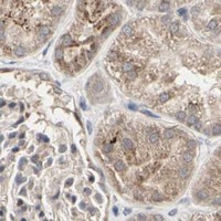
Immunohistochemistry protocol sheet for TAOK1 (IHC for TAOK1): expected localisation Cytoplasmic (UniProt); membranous tissue staining (HPA tissue IHC), antibody A07063, validated IHC image, and IHC protocol steps
Printable TAOK1 IHC protocol sheet — expected localisation Cytoplasmic (UniProt); membranous tissue staining (HPA tissue IHC), antibody A07063, controls and protocol steps. Open the full TAOK1 IHC guide →

TAOK1 Immunohistochemistry Experimental Design Guide

Expected localisation, validated protocols, controls and antibodies — the at-a-glance facts below, then the full design guide.

Must know before staining
Expected localisation Cytoplasmic (UniProt); membranous tissue staining (HPA tissue IHC)
Staining pattern General cytoplasmic and membranous staining (HPA tissue IHC)
Antigen retrieval Citrate pH 6.0 HIER, 95–98 °C, 20 min (rule: cytoplasmic / membrane antigen)
Positive control ⓘ Adipose tissue+4 more · see all
Negative control ⓘ None in HPA (detected in all 44 tissues); use no-primary + isotype controls
Important caveats
Reasons your staining may differ from the expected pattern.
Fixation Keep fixation consistent across sections. (standard IHC practice; not target-specific)
Caveat Staining and RNA expression show low consistency (HPA tissue IHC)
Regulation Stimulus-linked expression change is unreported (UniProt)
Isoform / epitope 3 isoforms; antibody epitope coverage is unspecified (UniProt; datasheet)
Section 1

Recommended TAOK1 IHC & IF Protocols

Compare the catalog antibody’s IHC-P protocol (datasheet) with published TAOK1 tissue protocols using chromogenic detection (PMC11855378; PMC11920640).

Recommended immunohistochemistry (IHC-P) protocol parameters
SampleParaffin-embedded human breast carcinoma tissue; fixative not specified (datasheet A07063)
FixationImage fixative and duration unreported (datasheet A07063); verify before use.
Sectioning4–5 µm sections on charged slides (standard)
DeparaffinisationXylene, graded ethanol series to water (standard)
Antigen retrievalHeat-induced epitope retrieval in citrate buffer, pH 6.0, 20 min at 95–98 °C (standard rule: cytoplasmic / membrane antigen)
Peroxidase block3% H2O2, 10 min, room temperature (standard)
Blocking10% normal serum of the secondary host, 30 min, room temperature (standard)
Primary antibodyRabbit anti-TAOK1, 1:100 - 1:300 (datasheet A07063)
Primary incubationOvernight at 4 °C (standard)
DetectionHRP-polymer secondary, DAB chromogen 5–10 min (standard)
CounterstainHematoxylin, blue, dehydrate and mount (standard)
Expected resultTAOK1-positive staining in adipocytes of adipose tissue (HPA tissue IHC: High). HPA tissue profile: General cytoplasmic and membranous expression. No signal in the no-primary control.
💡Decision noteStart with citrate pH 6.0 HIER at 95–98 °C for 20 min (page antigen-retrieval setting); neither cited excerpt specifies retrieval conditions (PMC11855378; PMC11920640).
Section 2

What Is the Expected TAOK1 Staining Pattern?

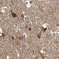
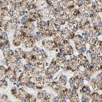
TAOK1 is cytoplasmic (UniProt Q7L7X3) and has no transmembrane segment (UniProt Q7L7X3 topology). In paraffin sections, expect cytoplasmic staining, sometimes with a membranous appearance, in cell types including cortical neurons and hepatocytes (HPA tissue IHC). HPA rates its tissue IHC pattern Approved but reports low consistency with RNA expression (HPA tissue IHC); treat the pattern as a guide to interpretation, not proof of specificity.

What am I looking at on my slide?
Cytoplasmic staining in cortical neurons or hepatocytes, with possible membrane-associated staining.This fits HPA's general cytoplasmic and membranous IHC profile; both cell types are reported High (HPA tissue IHC). Cytoplasmic localisation also agrees with UniProt Q7L7X3. Compare the stained cells with tissue morphology before scoring the result (general IHC practice).
Predominantly nuclear staining with little cytoplasmic signal.A nuclear-dominant pattern does not match the supplied cytoplasmic localisation (UniProt Q7L7X3) or HPA's tissue IHC profile (HPA tissue IHC). Treat it as suspect and check the counterstain, detection controls and antibody conditions before assigning it to TAOK1 (general IHC practice).
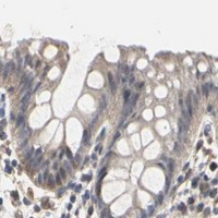
Conspicuous staining in a cell population expected to stain weakly.HPA reports Low staining in colon glandular cells, but provides no negative tissue group (HPA tissue IHC). Strong signal there deserves scrutiny for antibody cross-reactivity or endogenous detection activity; it is not, by itself, proof of either cause (general IHC practice).
Diffuse colour across cells and surrounding tissue, obscuring compartment boundaries.This is background rather than an interpretable TAOK1 distribution (general IHC practice). Assess a no-primary control and review blocking, washes and chromogen development; HPA's Approved rating and RNA-expression inconsistency do not identify the source of background (HPA tissue IHC).
No detectable signal in cortical neurons on an otherwise readable section.Cortical neurons are reported High by HPA, so absent staining challenges the run (HPA tissue IHC). Check the tissue and assay controls, retrieval and detection workflow (general IHC practice). A failed run cannot establish that the sampled cells lack TAOK1.
💡Expected TAOK1 appearanceA convincing positive is clear cytoplasmic staining, sometimes membranous, in a reported High population such as cortical neurons or hepatocytes (HPA tissue IHC); intense nuclear-only or structureless diffuse colour is suspect against the supplied localisation (UniProt Q7L7X3; HPA tissue IHC).
How each factor affects the staining
Compartment and topologyUniProt places TAOK1 in the cytoplasm and reports no transmembrane segment (UniProt Q7L7X3 topology). HPA describes cytoplasmic and membranous tissue staining (HPA tissue IHC). Interpret a membrane outline alongside cytoplasmic signal; do not infer that TAOK1 spans the membrane.
Choice of comparison tissueCortical neurons and hepatocytes are reported High, while colon glandular cells are reported Low (HPA tissue IHC). These are useful relative comparisons, but Low is not a negative control: HPA supplies no negative tissue entries (HPA tissue IHC).
Strength of the tissue evidenceHPA labels the tissue IHC profile Approved while noting low consistency between antibody staining and RNA expression (HPA tissue IHC). The listed High and Low levels describe observed staining; they should not be treated as independently confirmed protein abundance in every specimen.
Antibody validationHPA007669 has Approved IHC status, whereas the other listed antibodies have no IHC status in the supplied record (HPA antibodies). Antibody identities are not interchangeable evidence for a particular tissue result; check which antibody produced the image being compared (general IHC practice).
Isoforms and processingUniProt lists three TAOK1 isoforms, one 1–1001 chain, and no signal peptide or propeptide (UniProt Q7L7X3). The supplied record gives no antibody epitope or isoform-specific staining evidence, so an unusual pattern cannot be assigned to a particular isoform.
Why is my staining missing, weak or wrong?
SituationLikely causeNext action
Expected cytoplasmic signal is absent in a reported High tissue.An assay failure is possible; cortical neurons and hepatocytes are reported High, but HPA staining is not a guarantee for each specimen (HPA tissue IHC).Confirm tissue identity and section quality, inspect the positive control, then review the antibody's stated IHC conditions, retrieval and detection steps (general IHC practice). Do not infer a TAOK1-negative specimen until the run is valid.
Strong nuclear staining dominates the section.This conflicts with TAOK1's cytoplasmic annotation (UniProt Q7L7X3) and HPA's tissue pattern (HPA tissue IHC); the supplied evidence does not identify a specific mechanism.Compare with a no-primary control and a reported High cell population. Review counterstain and detection settings before scoring nuclear colour as specific (general IHC practice).
A reported Low cell population stains as strongly as a reported High one.HPA calls colon glandular cells Low and cortical neurons High, but supplies no negative tissue category (HPA tissue IHC). Cross-reactivity or endogenous detection activity are possibilities, not established diagnoses (general IHC practice).Compare matched sections and no-primary controls under the same detection conditions; score cell type, compartment and intensity together (general IHC practice).
Colour is diffuse or persists in the no-primary control.Background or endogenous detection activity can obscure cellular staining (general IHC practice). HPA's TAOK1 profile does not determine which workflow step caused it (HPA tissue IHC).Check blocking appropriate to the detection system, washing and chromogen development, then reassess whether cytoplasmic boundaries can be resolved (general IHC practice).
Membrane-like staining appears without an obvious cytoplasmic component.HPA describes membranous tissue staining, but UniProt reports a cytoplasmic protein without a transmembrane segment (HPA tissue IHC; UniProt Q7L7X3 topology). An isolated membrane outline therefore needs cautious interpretation.Inspect cell morphology and compare with cytoplasmic staining in a reported High population; check detection controls before calling the outline TAOK1-positive (general IHC practice).
How should an IF/ICC image be compared with the IHC result?HPA supports cytosolic ICC-IF localisation; plasma-membrane and primary-cilium assignments are uncertain, and vesicle localisation is Approved (HPA subcellular ICC-IF). These confidence labels differ from the tissue IHC assessment.Use the separate IF/ICC guide for that application. Here, use the ICC-IF record only as localisation context; do not treat an uncertain IF location as a required paraffin IHC pattern (HPA subcellular ICC-IF; HPA tissue IHC).

Sample controls for TAOK1 IHC & IF

🧪Run cerebral cortex first and score neuronal cells, which have high TAOK1 staining (HPA: High in cerebral-cortex neuronal cells). There is no supported negative tissue because TAOK1 is detected in all 44 scored tissues (HPA: no negative rows); use no-primary and isotype controls for the negative reference, and treat non-neuronal cells on the positive slide as internal negatives only if they show background-level chromogen, since their TAOK1-negative status is unreported (HPA: cerebral-cortex row).
Positive control tissue: Adipose tissue (Adipocytes, HPA High)
Negative control tissue: None in HPA: TAOK1 is detected in all 44 scored tissues. Use a no-primary (secondary-only) and an isotype control instead.
ICC-IF cell lines (HPA subcellular resource): HPA ICC-IF images show TAOK1 in Rh30, SiHa, SuSa, ASC52telo, RPTEC/TERT1, hTERT-RPE1 (serum starved), Sperm, KOLF2.1J, with annotated localisation: Plasma membrane (uncertain), Cytosol (supported) (HPA subcellular).
Technical controls: Include a no-primary, secondary-only control and a primary-antibody host-species-matched control: an isotype-matched antibody for a monoclonal primary or nonimmune IgG for a polyclonal primary (standard IHC practice). Confirm target-dependent staining with a TAOK1 knockout specimen if available or a peptide-block control (A07063 caption: peptide-blocked image); in cerebral cortex, quench endogenous peroxidase for chromogenic IHC and check for lipofuscin autofluorescence if assessing IF (standard IHC/IF practice).
⚠️Feasibility: A target-specific fixation window and the effect of fixation are unreported, and the paraffin-section example does not state its fixative (A07063 caption: fixative not stated). Whether antigen retrieval is required, or frozen sections or IF/ICC are easier than paraffin IHC, is unreported in the supplied application evidence (A07063 caption; HPA: tissue staining and ICC-IF localization). Cerebral-cortex lipofuscin can resemble chromogenic signal or fluoresce, so compare staining with the matched controls (standard IHC/IF practice).

HPA tissue IHC evidence for TAOK1

Comprehensive Human Protein Atlas IHC scoring per tissue (reliability: Approved — Low consistency between antibody staining and RNA expression data.). Rows are taken directly from the HPA tissue chart — click any row's HPA link to view the source.

Positive expression · recommended positive controls

TissueCell typeLevelEvidenceSource
Adipose tissue Adipocytes High Protein (IHC) HPA →
Caudate Neuronal cells High Protein (IHC) HPA →
Cerebellum Cells in molecular layer High Protein (IHC) HPA →
Cerebral cortex Neuronal cells High Protein (IHC) HPA →
Cervix Squamous epithelial cells High Protein (IHC) HPA →

Undetected expression · recommended negative controls

TissueCell typeLevelEvidenceSource
None in HPA: TAOK1 is detected in all 44 scored tissues. Use a no-primary (secondary-only) and an isotype control instead.
Section 3

Advanced TAOK1 IHC Tips

Troubleshoot TAOK1 chromogenic IHC in paraffin sections by checking retrieval, staining compartment, controls, and scoring consistency.

How should I retrieve TAOK1 in paraffin sections when staining is weak?
Start with citrate buffer at pH 6.0 for heat induced retrieval at 95–98 °C for 20 minutes (page retrieval rule). Allow sections to cool in the retrieval buffer before washing, and compare a retrieved section with an adjacent section processed without retrieval (standard IHC practice). If staining remains weak, test a second retrieval condition on serial sections while keeping antibody concentration and detection conditions fixed; this is an optimisation step, not a documented TAOK1 requirement (standard IHC practice). Score cytoplasmic staining first, while recording any membrane staining separately, because those patterns are reported for TAOK1 (UniProt Q7L7X3; HPA tissue IHC).
Does fixation explain variable TAOK1 staining between paraffin blocks?
TAOK1 specific sensitivity to fixation is unknown from the supplied evidence, and the selected A07063 paraffin section caption does not state a fixative (selected A07063 caption). Record the fixative, fixation duration, processing schedule, and block age for each specimen before comparing staining across blocks (standard IHC practice). When possible, stain sections of the same thickness from blocks processed together in one run, with the same retrieval and detection settings (standard IHC practice). If a block remains weak, compare its tissue preservation and staining with a concurrently processed control before assigning the difference to TAOK1 abundance (standard IHC practice).
Which staining compartments should I accept for TAOK1?
Prioritise cytoplasmic staining when reviewing TAOK1 sections, since UniProt places TAOK1 in the cytoplasm and HPA reports general cytoplasmic and membranous tissue expression (UniProt Q7L7X3; HPA tissue IHC). Record membrane staining as a separate pattern; the HPA subcellular record supports cytosol but marks plasma membrane localisation uncertain (HPA subcellular). Compare the pattern across at least 2 intact tissue regions and against the negative control, using identical microscope settings (standard IHC practice). Treat isolated nuclear staining cautiously because the supplied TAOK1 localisation records do not identify the nucleus as an expected site (UniProt Q7L7X3; HPA subcellular).
Can this stain distinguish TAOK1 isoforms or phosphorylation states?
Do not assign staining to a particular isoform without an epitope map: the record lists 3 TAOK1 isoforms, but the selected caption does not identify an isoform specific epitope (UniProt Q7L7X3; selected A07063 caption). Likewise, total TAOK1 staining cannot establish phosphorylation at the listed serine or threonine residues without a validated site specific reagent (UniProt Q7L7X3; standard IHC practice). The selected paraffin section image includes a synthesized peptide blocking comparison, which supports evaluating antibody dependent staining for that image (selected A07063 caption). For a disputed pattern, compare peptide blocked and unblocked sections under identical retrieval and detection conditions, then seek an independent specificity control (standard IHC practice).
How can I check the IHC pattern with multiplex immunofluorescence?
For a separate IF experiment, pair TAOK1 with a marker for the cell population under study; neuronal cells are one HPA reported high staining population (HPA tissue IHC). Select spectrally separated fluorophores, favouring a far red channel when tissue autofluorescence obscures shorter wavelength signal, and image a no primary control (standard IF practice). Because TAOK1 has no transmembrane segment and cytosol is its supported location, test mild permeabilisation when the mapped epitope is intracellular; establish epitope accessibility experimentally (UniProt Q7L7X3; HPA subcellular; standard IF practice). Compare at least 2 matched fields per condition without treating an IF pattern alone as validation of chromogenic IHC (standard IF practice).
What should I change if TAOK1 DAB staining is diffuse?
Run a no primary section to assess detection background, and inspect whether brown signal follows tissue edges, damaged areas, or deposits rather than intact cells (standard IHC practice). In a peroxidase and DAB workflow, apply a peroxidase block according to the detection system instructions before primary antibody incubation (standard IHC practice). If background persists, titrate the catalog antibody downward across 2–3 adjacent sections while holding retrieval, wash steps, and development time constant (standard IHC practice). Compare the result with the selected A07063 peptide blocked image as context, while recognising that its caption supplies neither a fixative nor a working dilution (selected A07063 caption).
How should I score TAOK1 across tissue samples? ⚠ ANSWER MARKED FOR VERIFICATION
Define the cell population and staining compartment before scoring; HPA reports both cytoplasmic and membranous TAOK1 tissue staining (HPA tissue IHC). For intact cells, record the percentage at each intensity grade 0–3 and calculate an H score from 0–300, separately for cytoplasmic and membrane signal if both are analysed (standard IHC practice). Normalise each score to the number of eligible cells scored, and report positive cell density per mm² only when tissue area is measured consistently (standard IHC practice). Use the same threshold, section selection rules, and staining run controls across samples, since HPA flags low consistency between staining and RNA data (standard IHC practice; HPA tissue IHC).
When is a positive TAOK1 stain convincing?
A convincing pattern has signal in intact cells in a reported compartment, with cytoplasm expected from UniProt and cytoplasmic or membranous tissue staining reported by HPA (UniProt Q7L7X3; HPA tissue IHC). Check whether comparable signal recurs away from section edges and necrotic regions, and whether a no primary section lacks the same deposit (standard IHC practice). High neuronal staining in several brain regions provides a reported cell context, but HPA labels its tissue IHC reliability Approved with low consistency against RNA expression (HPA tissue IHC). Treat nuclear only staining, edge limited colour, necrotic deposits, or signal retained in a no primary control as reasons to investigate artefact before reporting TAOK1 positivity (UniProt Q7L7X3; HPA subcellular; standard IHC practice).
Boster reagents

Best TAOK1 / Serine/threonine-protein kinase TAO1 IHC Antibodies

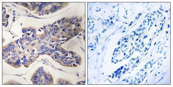
A07063 has an IHC image from a human paraffin-embedded breast carcinoma section (catalog image caption); IF is listed without an IF image (catalog applications and image records).

Real IHC data Immunohistochemistry analysis of paraffin-embedded human breast carcinoma tissue, using TAOK1 Antibody. The picture on the right is blocked with the synthesized peptide.
Anti-TAOK1/Psk2 Antibody
Cat # A07063

A07063 is listed for IHC and IF/ICC in human, mouse, and rat (catalog applications and reactivity). Its IHC image shows a human paraffin-embedded breast carcinoma section with a peptide-blocked comparison (catalog image caption).

Which to pick: For tissue IHC, choose A07063 for paraffin sections; its image documents that processing, but the fixative is unreported (catalog image caption). For IF/ICC or work across human, mouse, and rat, A07063 is the listed rabbit polyclonal option, with IF/ICC applications and those species in the catalog; no IF image is supplied (catalog host, dilution record, applications, reactivity, and image records).

Each figure is that product's own IHC / IF validation image from its datasheet.

References

  1. UniProt Consortium. UniProt entry Q7L7X3 (TAOK1_HUMAN, Serine/threonine-protein kinase TAO1).
  2. Human Protein Atlas. TAOK1 tissue IHC expression (reliability: Approved).
  3. Human Protein Atlas. TAOK1 subcellular location (ICC-IF): Mainly localized to the plasma membrane and cytosol. In addition localized to vesicles and primary cilium..
  4. Human Protein Atlas. TAOK1 antibody validation summary (3 antibodies).
  5. Deferasirox Targets TAOK1 to Induce p53-Mediated Apoptosis in Esophageal Squamous Cell Carcinoma. International journal of molecular sciences 2025 — PMC11855378.
  6. Multi-Omics Analysis Revealed That TAOK1 Can Be Used as a Prognostic Marker and Target in a Variety of Tumors, Especially in Cervical Cancer. OncoTargets and therapy 2025 — PMC11920640.
  7. The miR-183/Taok1 target pair is implicated in cochlear responses to acoustic trauma. PloS one 2013 — PMC3589350.
  8. PubMed PMID:13679851 — UniProt-cited evidence.
  9. PubMed PMID:10718198 — UniProt-cited evidence.
  10. PubMed PMID:14702039 — UniProt-cited evidence.