This website uses cookies to ensure you get the best experience on our website.


- Table of Contents
Plan TAOK2 chromogenic IHC in paraffin sections using the catalog antibody's 2–5 μg/ml range (datasheet A07466-1). Interpret the generally low cytoplasmic staining alongside medium signals in selected cell populations and the reported low antibody–RNA consistency (HPA tissue IHC).

Expected localisation, validated protocols, controls and antibodies — the at-a-glance facts below, then the full design guide.
| Expected localisation | Low cytoplasmic staining in most tissues (HPA tissue IHC) | |
| Staining pattern | Mostly low cytoplasmic staining; medium in colon glands (HPA tissue IHC) | |
| Antigen retrieval | EDTA pH 8.0 HIER, heat-mediated (datasheet A07466-1) | |
| Positive control | Cerebral cortex+4 more · see all | |
| Negative control | Adipose tissue+4 more · see all |
| Fixation | Matched tissue-IHC evidence does not establish the fixation claim. Validate the specimen-specific method before use. (selected-SKU IHC image A07466-1) | |
| Caveat | Antibody staining has low consistency with RNA expression (HPA tissue IHC) | |
| Regulation | Higher expression in testis and brain (UniProt) | |
| Isoform / epitope | 4 isoforms; C-terminal truncation may change epitope coverage (UniProt) |
The catalog antibody uses paraffin-section IHC with EDTA pH 8.0 retrieval (datasheet A07466-1); the published mouse-brain protocol uses frozen sections and chromogenic detection (PMC4011144).
| Sample | Paraffin-embedded human thyroid cancer tissue; fixative not specified (datasheet A07466-1) |
| Fixation | Image fixative and duration unreported (datasheet A07466-1); verify before use. |
| Sectioning | 4–5 µm sections on charged slides (standard) |
| Deparaffinisation | Xylene, graded ethanol series to water (standard) |
| Antigen retrieval | Heat retrieval: EDTA pH 8.0 (datasheet A07466-1); 20 min, 95–100 °C (standard) |
| Peroxidase block | 3% H2O2, 10 min, room temperature (standard) |
| Blocking | 10% goat serum (datasheet A07466-1) |
| Primary antibody | Rabbit anti-TAOK2, 2-5 μg/ml (datasheet A07466-1) |
| Primary incubation | Overnight at 4 °C (datasheet A07466-1) |
| Detection | HRP-conjugated secondary, DAB chromogen (datasheet A07466-1) |
| Counterstain | Hematoxylin, blue, dehydrate and mount (standard) |
| Expected result | TAOK2-positive staining in endothelial cells of cerebral cortex (HPA tissue IHC: Medium). HPA tissue profile: Low cytoplasmic expression in most tissues. No signal in the no-primary control. |
TAOK2 is expected to show low cytoplasmic staining in most tissues, with medium staining in selected endothelial, glandular, hepatocyte, and pancreatic endocrine cells (HPA tissue IHC; Approved, low consistency with RNA). Cytoplasmic vesicle membranes and the cytoskeleton are documented locations; TAOK2 has 5 transmembrane segments, and nuclear localization depends on isoform and processing (UniProt Q9UL54 topology/localization).
| Weak cytoplasmic staining across a section, with clearer signal in colon or duodenal glandular cells. | This fits the reported low cytoplasmic profile in most tissues and medium staining in these glandular cells (HPA tissue IHC). Compare the expected cells within the section; a uniformly dark slide would obscure this distinction. The HPA tissue assessment is Approved but has low consistency with RNA expression, so intensity alone does not establish specificity (HPA tissue IHC). |
| Medium cytoplasmic staining in cortical endothelial cells, hepatocytes, or pancreatic endocrine cells. | These are reported positive cell populations and useful places to assess whether staining follows tissue architecture (HPA tissue IHC). Interpret the named cell type rather than treating every cell in the organ as positive. Confirm its identity by morphology and counterstain as a general IHC practice. |
| Staining is confined to extracellular material or a glandular lumen, with no matching cellular pattern. | That distribution does not match the reported low cytoplasmic tissue pattern (HPA tissue IHC) or the documented vesicle, cytoskeletal, and conditional nuclear locations (UniProt Q9UL54 localization). Treat it as suspect; check section morphology and a no-primary control before scoring cellular TAOK2. |
| Strong staining appears in adipocytes or bone-marrow hematopoietic cells. | Those cell populations were not detected in the cited tissue survey (HPA tissue IHC). Cross-reactivity or endogenous detection activity is possible, especially if staining is widespread or also appears without primary antibody. Recheck cell identity and controls; a survey-level absence is not proof that every specimen must be negative. |
| A known-positive cell population has no visible signal, or the entire section has diffuse color. | Absent signal in colon glandular cells or hepatocytes conflicts with their reported medium staining; diffuse color prevents compartment scoring (HPA tissue IHC). Before interpreting biology, inspect tissue preservation, retrieval and detection controls as general IHC practice. The supplied sources do not establish TAOK2-specific fixation sensitivity. |
| Tissue and cell selection | Colon or duodenal glandular cells, hepatocytes, cortical endothelial cells, and pancreatic endocrine cells have reported medium staining; adipocytes and bone-marrow hematopoietic cells were not detected (HPA tissue IHC). Score the named cells, since an organ-wide label can conceal mixed populations. |
| Antibody evidence | The listed antibody HPA010650 is IHC Approved, and its ICC evidence is Supported (HPA antibody validation). The tissue assessment also reports low consistency between staining and RNA expression (HPA tissue IHC). Use independent controls when a result departs from the reported pattern; this record does not claim Enhanced IHC validation. |
| Isoform and epitope location | Full-length phosphorylated isoform 1 associates with perinuclear microtubule cables; a C-terminally truncated form can enter the nucleus, while full-length isoform 1 and isoform 2 are excluded (UniProt Q9UL54 localization). The antibody epitope is unspecified, so nuclear versus cytoplasmic recognition cannot be predicted from this record. |
| Membrane topology and retrieval | TAOK2 has 5 transmembrane segments near its C-terminal region (UniProt Q9UL54 topology). This does not identify the recognized epitope or an optimal antigen-retrieval condition. Retrieval comparisons are a general IHC optimization step; no TAOK2-specific fixation or retrieval effect is established here. |
| Situation | Likely cause | Next action |
|---|---|---|
| No signal in colon or duodenal glandular cells. | Possible assay failure; these cells show medium staining in the HPA survey (HPA tissue IHC). | Check section quality, retrieval, primary-antibody application, and detection controls as general IHC practice. Avoid assigning a TAOK2-specific fixation cause without evidence. |
| Every cell is uniformly brown, including cells expected to be negative. | Background or endogenous enzyme activity may obscure the reported cell pattern (HPA tissue IHC; general IHC practice). | Inspect a no-primary control; if enzyme detection is used, assess the relevant endogenous-activity block. Re-evaluate the primary concentration using the assay's documented conditions. |
| Color lies mainly in lumens, extracellular debris, or tissue edges. | The location conflicts with the reported cytoplasmic tissue profile (HPA tissue IHC). | Check morphology and no-primary staining, then score intact cells separately from deposits and edge effects (general IHC practice). |
| Only intense nuclear staining is seen throughout an IHC section. | Nuclear TAOK2 is possible for a truncated isoform, but universal nuclear dominance is not the reported tissue IHC pattern (UniProt Q9UL54 localization; HPA tissue IHC). | Check controls and cell identity; report nuclear staining separately. Do not infer which isoform was detected while the antibody epitope is unknown. |
| Pancreatic endocrine cells appear negative while surrounding tissue stains. | The result differs from the reported medium endocrine-cell signal (HPA tissue IHC); staining of other cells alone does not validate the expected pattern. | Confirm endocrine-cell identity by morphology or an appropriate adjacent marker, then review positive and no-primary controls (general IHC practice). |
| IF/ICC Q&A: Should nucleoplasmic signal be dismissed as an IHC false positive? | HPA reports supported nucleoplasmic and nucleolar IF locations, with uncertain cytosolic IF localization; UniProt describes conditional nuclear localization (HPA subcellular; UniProt Q9UL54). | No. Interpret IF/ICC on its own guide page and with its own controls. Do not transfer an IF compartment call directly to chromogenic tissue IHC (HPA subcellular; HPA tissue IHC). |
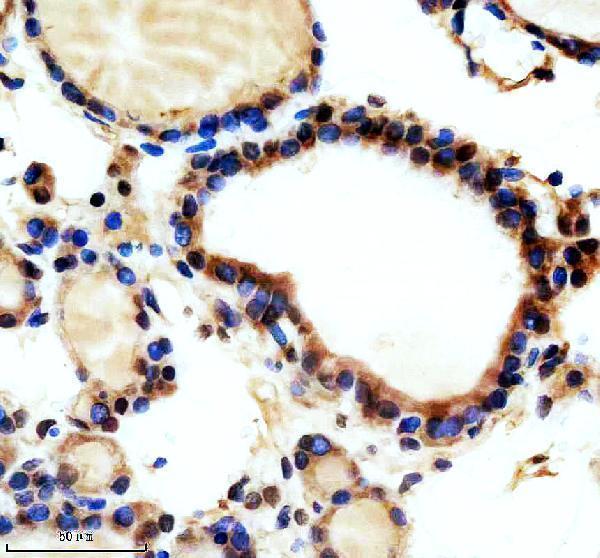
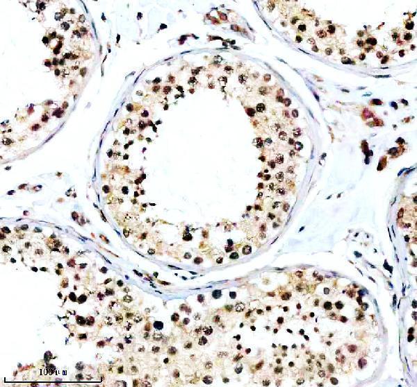
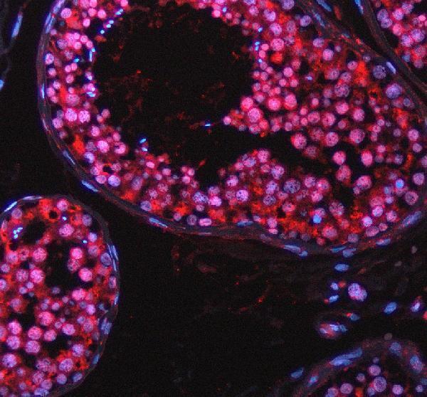
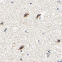


Comprehensive Human Protein Atlas IHC scoring per tissue (reliability: Approved — Low consistency between antibody staining and RNA expression data.). Rows are taken directly from the HPA tissue chart — click any row's HPA link to view the source.
| Tissue | Cell type | Level | Evidence | Source |
|---|---|---|---|---|
| Cerebral cortex | Endothelial cells | Medium | Protein (IHC) | HPA → |
| Colon | Glandular cells | Medium | Protein (IHC) | HPA → |
| Duodenum | Glandular cells | Medium | Protein (IHC) | HPA → |
| Liver | Hepatocytes | Medium | Protein (IHC) | HPA → |
| Pancreas | Pancreatic endocrine cells | Medium | Protein (IHC) | HPA → |
| Tissue | Cell type | Level | Evidence | Source |
|---|---|---|---|---|
| Adipose tissue | Adipocytes | Not detected | Protein (IHC) | HPA → |
| Bone marrow | Hematopoietic cells | Not detected | Protein (IHC) | HPA → |
| Caudate | Glial cells | Not detected | Protein (IHC) | HPA → |
| Cerebellum | Cells in granular layer | Not detected | Protein (IHC) | HPA → |
| Epididymis | Glandular cells | Not detected | Protein (IHC) | HPA → |
Troubleshoot TAOK2 staining in paraffin sections with the antibody’s documented retrieval conditions and compartment-aware controls.
A07466-1 has IHC images from human paraffin thyroid cancer and testis, plus an IF image from human paraffin testis (catalog image captions).
A07466-1 has IHC images from human paraffin thyroid cancer and testis (catalog IHC image captions). The same SKU has an IF image from human paraffin testis and lists Human, Mouse, and Rat reactivity (catalog IF image caption; catalog reactivity).
Which to pick: For tissue IHC, choose A07466-1: its own captions document human paraffin sections with EDTA pH 8 retrieval and 2 μg/ml primary antibody; the fixative is unreported (A07466-1 IHC image captions). For IF, A07466-1 has human paraffin testis data at 5 μg/ml; ICC validation is unreported (A07466-1 IF image caption; catalog applications). For cross-species work, A07466-1 lists Human, Mouse, and Rat reactivity, while the supplied IHC and IF images show human tissue (catalog reactivity; A07466-1 image captions).