TARBP2 / RISC-loading complex subunit TARBP2 · IHC design guide

Design Immunohistochemistry for TARBP2

Plan TARBP2 IHC-P around nuclear and cytoplasmic staining (HPA tissue IHC), using placenta decidual cells as a high-staining reference (HPA tissue IHC). Compare adipocytes, where staining was not detected (HPA tissue IHC), and score each cell type and compartment separately.

Evidence assembled Oct 2026 · For research use; verify linked source records and product datasheet before use
Immunohistochemistry protocol sheet for TARBP2 (IHC for TARBP2): expected localisation Nuclear and cytoplasmic tissue staining (HPA tissue IHC), antibody A01680-2, validated IHC image, and IHC protocol steps
Printable TARBP2 IHC protocol sheet — expected localisation Nuclear and cytoplasmic tissue staining (HPA tissue IHC), antibody A01680-2, controls and protocol steps. Open the full TARBP2 IHC guide →

TARBP2 Immunohistochemistry Experimental Design Guide

Expected localisation, validated protocols, controls and antibodies — the at-a-glance facts below, then the full design guide.

Must know before staining
Expected localisation Nuclear and cytoplasmic tissue staining (HPA tissue IHC)
Staining pattern Nuclear and cytoplasmic signal across cell types (HPA tissue IHC)
Antigen retrieval EDTA pH 8.0 HIER, heat-mediated (datasheet A01680-2)
Positive control ⓘ Caudate+4 more · see all
Negative control ⓘ Adipose tissue
Important caveats
Reasons your staining may differ from the expected pattern.
Fixation Keep fixation consistent across sections. (standard IHC practice; not target-specific)
Caveat Presumed off-target binding may complicate scoring (HPA tissue IHC)
Regulation Broad tissue expression (HPA tissue IHC)
Isoform / epitope 2 isoforms; epitope coverage is unspecified (UniProt)
Section 1

Recommended TARBP2 IHC & IF Protocols

The catalog antibody’s IHC-P protocol uses EDTA retrieval (datasheet A01680-2). The three published TARBP2 IHC protocols below provide tissue-specific conditions (PMC5147454; PMC6406945; PMC8745112).

Recommended immunohistochemistry (IHC-P) protocol parameters
SampleParaffin-embedded human lung cancer tissue; fixative not specified (datasheet A01680-2)
FixationImage fixative and duration unreported (datasheet A01680-2); verify before use.
Sectioning4–5 µm sections on charged slides (standard)
DeparaffinisationXylene, graded ethanol series to water (standard)
Antigen retrievalHeat retrieval: EDTA pH 8.0 (datasheet A01680-2); 20 min, 95–100 °C (standard)
Peroxidase block3% H2O2, 10 min, room temperature (standard)
Blocking10% goat serum (datasheet A01680-2)
Primary antibodyRabbit anti-TARBP2, 2-5 μg/ml (datasheet A01680-2)
Primary incubationOvernight at 4 °C (datasheet A01680-2)
DetectionHRP-conjugated secondary, DAB chromogen (datasheet A01680-2)
CounterstainHematoxylin, blue, dehydrate and mount (standard)
Expected resultTARBP2-positive staining in glial cells of caudate (HPA tissue IHC: High). HPA tissue profile: General nuclear and cytoplasmic expression. No signal in the no-primary control.
💡Decision noteStart with heat-mediated EDTA retrieval at pH 8.0 for the catalog antibody (datasheet A01680-2); use each paper’s stated retrieval when reproducing its protocol (PMC5147454; PMC6406945; PMC8745112).
Section 2

What Is the Expected TARBP2 Staining Pattern?

TARBP2 staining can be nuclear and cytoplasmic, with a perinuclear component (HPA tissue IHC: general nuclear and cytoplasmic expression; UniProt Q15633: cytoplasm, perinuclear region and nucleus). Assess defined cell populations, such as lung alveolar cells and pancreatic exocrine glandular cells, rather than whole organs (HPA: High in both). HPA rates its tissue IHC reliability Approved, with medium agreement between staining and RNA data and presumed off-target binding disregarded (HPA tissue IHC). TARBP2 has no transmembrane segment (UniProt Q15633 topology).

What am I looking at on my slide?
Nuclear and cytoplasmic staining in lung alveolar cells or pancreatic exocrine glandular cells (HPA: High in both).This fits the reported IHC pattern and provides a useful within-run positive comparison (HPA tissue IHC: general nuclear and cytoplasmic expression; HPA: High in both). Judge the named cells individually; a tissue-level label does not imply equal staining in every cell (HPA tissue IHC: cell-specific entries).
A sharply outlined plasma membrane or extracellular deposit dominates the signal.That compartment is inconsistent with the reported intracellular locations and lack of a transmembrane segment (UniProt Q15633: subcellular location and topology; HPA tissue IHC: nuclear and cytoplasmic expression). Check section artefact, reagent deposits and detection controls before scoring it as TARBP2 (general IHC practice).
Strong staining occurs chiefly outside the named positive cell population.Check cell identity before interpreting the signal: the HPA entries describe particular cells, including prostate glandular cells and placental decidual cells (HPA: High in both). Unexpected staining can reflect off-target binding or endogenous detection activity (HPA tissue IHC: presumed off-target binding observed; general IHC practice).
Diffuse color obscures cell boundaries or appears broadly across the section.That appearance makes compartment and cell-type scoring unreliable (general IHC practice). Uneven reagent coverage, excess background from detection reagents or inadequate blocking are workflow possibilities; they are not documented TARBP2-specific effects (general IHC practice).
No convincing signal appears in the expected positive cells.Check the run before calling the specimen negative: glial cells in caudate and cerebral cortex, and exocrine glandular cells in pancreas, are reported High (HPA tissue IHC). A failed positive control, weak detection or unsuitable antibody conditions can prevent an interpretable result (general IHC practice).
💡Expected TARBP2 appearanceA credible positive result shows nuclear and cytoplasmic staining in an HPA-reported High cell population, such as lung alveolar cells; dominant membrane or extracellular color warrants artefact review (HPA tissue IHC: general nuclear and cytoplasmic expression; HPA: High in lung alveolar cells; UniProt Q15633 topology).
How each factor affects the staining
Cell-specific tissue referenceUse reported cells as comparators: cervix and prostate glandular cells, hippocampal neuronal cells, and placental decidual cells are High (HPA tissue IHC). Adipocytes are Not detected, while liver cholangiocytes are Low; neither label describes every cell in its organ (HPA tissue IHC).
Compartment referenceTissue IHC is described as generally nuclear and cytoplasmic, and UniProt also lists a perinuclear location (HPA tissue IHC; UniProt Q15633). These references support intracellular scoring without requiring identical nuclear-to-cytoplasmic intensity in every section.
Antibody validationThe listed antibodies HPA051181, HPA061454 and CAB069406 each have Approved IHC status (HPA antibodies). The tissue profile has medium staining-to-RNA consistency and notes presumed off-target binding (HPA tissue IHC); an unusual pattern still needs control-based review (general IHC practice).
Isoforms and epitopeUniProt lists 2 TARBP2 isoforms, but the supplied records do not locate the antibodies' epitopes or show isoform-specific IHC staining (UniProt Q15633: isoforms; HPA antibodies: validation summaries). Do not assign a compartment or tissue difference to one isoform from these data.
IF/ICC cross-check: should the patterns match exactly?An exact match is not established: ICC-IF mainly places TARBP2 in the nucleoplasm, with nuclear bodies as an additional location, while tissue IHC reports general nuclear and cytoplasmic expression (HPA subcellular ICC-IF; HPA tissue IHC). Compare compartments while keeping the assay-specific observations distinct.
Chromogenic detection backgroundEndogenous enzyme activity and nonspecific reagent deposition can create color unrelated to primary-antibody binding (general IHC practice). Include detection controls when the signal is diffuse or appears in unexpected structures; the supplied HPA and UniProt records do not identify a TARBP2-specific detection artefact.
Why is my staining missing, weak or wrong?
SituationLikely causeNext action
Expected positive cells remain unstained.The IHC run may have failed, or the antibody and detection conditions may yield insufficient signal (general IHC practice).Check a same-run positive section containing a reported High cell population, such as pancreatic exocrine glandular cells (HPA: High). Review retrieval, antibody incubation and chromogen development as general IHC workflow checks; no TARBP2-specific retrieval requirement is supplied.
Adipocytes stain strongly.This conflicts with the HPA Not detected entry for adipocytes and may indicate background or off-target binding (HPA: adipocytes Not detected; HPA tissue IHC: presumed off-target binding observed).Confirm adipocyte identity, inspect a primary-antibody omission control, and compare the pattern with a reported High cell population in the same run (general IHC practice; HPA tissue IHC).
Color is widespread and cell boundaries are unclear.Nonspecific detection or uneven reagent handling may obscure localization (general IHC practice).Inspect omission and detection controls, reagent coverage, washing and blocking, then repeat the run if boundaries remain unreadable (general IHC practice). Do not score diffuse color as TARBP2 localization.
Only extracellular material or cell outlines stain.The pattern disagrees with the listed intracellular locations and no-transmembrane topology (UniProt Q15633; HPA tissue IHC).Check the counterstain to locate nuclei and cell borders, inspect for deposits, and compare the signal with a primary-antibody omission control (general IHC practice).
Nuclear staining is present but cytoplasmic staining is faint.The supplied tissue IHC profile is general, while ICC-IF reports mainly nucleoplasmic localization; neither source sets a required intensity ratio (HPA tissue IHC; HPA subcellular ICC-IF).Score the compartments separately in the named cells and compare same-run controls (general IHC practice). Avoid rejecting a result solely because the two compartments differ in intensity.
Signal differs between two antibodies.Approved IHC status does not guarantee identical staining; the HPA tissue profile reports medium agreement with RNA and presumed off-target binding (HPA antibodies; HPA tissue IHC).Compare the same cell type and controls for each antibody, then favor the pattern consistent with reported intracellular localization and cell-specific tissue entries (UniProt Q15633; HPA tissue IHC; general IHC practice).

Sample controls for TARBP2 IHC & IF

🧪Run lung first: alveolar cells should stain (HPA: Lung alveolar cells, High). Run adipose tissue as the negative control; adipocytes should lack specific staining (HPA: Adipose tissue adipocytes, Not detected), while unstained cells within the lung section should show counterstain without specific DAB signal (standard IHC practice).
Positive control tissue: Caudate (Glial cells, HPA High)
Negative control tissue: Adipose tissue (HPA Not detected)
ICC-IF cell lines (HPA subcellular resource): HPA ICC-IF images show TARBP2 in HeLa, MCF-7, U2OS, NIH 3T3, with annotated localisation: Nucleoplasm (supported) (HPA subcellular).
Technical controls: Include a no-primary, secondary-only control; a concentration-matched nonimmune rabbit IgG isotype control; and a TARBP2-knockout specimen if available (selected A01680-2 caption: rabbit primary; standard IHC practice). Quench endogenous peroxidase in lung sections and compare background with the no-primary slide (selected A01680-2 caption: lung tissue, HRP/DAB; standard IHC practice).
⚠️Feasibility: A TARBP2-specific fixation window or fixation effect is unreported, and the selected A01680-2 paraffin-section caption does not report a fixative (selected A01680-2 caption: fixative not stated). The caption uses heat retrieval in EDTA at pH 8.0, but does not establish that retrieval is required or that frozen sections or IF are easier (selected A01680-2 caption: EDTA pH 8.0). For IF/ICC, assess mainly nucleoplasmic signal with additional nuclear-body signal (HPA subcellular: nucleoplasm supported; nuclear bodies approved); for lung IHC, monitor endogenous peroxidase background with the no-primary control (selected A01680-2 caption: lung tissue, HRP/DAB; standard IHC practice).

HPA tissue IHC evidence for TARBP2

Comprehensive Human Protein Atlas IHC scoring per tissue (reliability: Approved — Medium consistency between antibody staining and RNA expression data. Presumed off target binding observed and disregarded.). Rows are taken directly from the HPA tissue chart — click any row's HPA link to view the source.

Positive expression · recommended positive controls

TissueCell typeLevelEvidenceSource
Caudate Glial cells High Protein (IHC) HPA →
Cerebral cortex Glial cells High Protein (IHC) HPA →
Cervix Glandular cells High Protein (IHC) HPA →
Hippocampus Neuronal cells High Protein (IHC) HPA →
Lung Alveolar cells High Protein (IHC) HPA →

Undetected expression · recommended negative controls

TissueCell typeLevelEvidenceSource
Adipose tissue Adipocytes Not detected Protein (IHC) HPA →
Section 3

Advanced TARBP2 IHC Tips

Use the catalog antibody’s paraffin-section conditions as a starting point, then assess compartment, cell type, and background before scoring TARBP2 staining.

How should I troubleshoot weak TARBP2 staining after antigen retrieval?
Start with heat-mediated retrieval in EDTA at pH 8.0 for paraffin sections (datasheet A01680-2). The catalog image used 2 μg/ml primary antibody overnight at 4°C, so check that concentration and incubation before attributing weak signal to retrieval (datasheet A01680-2). Keep heating, cooling, and section handling consistent across comparison slides because these steps affect staining intensity (standard IHC practice). If signal remains weak, test a second retrieval condition on adjacent sections as a fallback, recording its buffer and pH rather than treating it as validated for this antibody (standard IHC practice). Compare cellular staining with the matched no-primary control before selecting a condition (standard IHC practice).
Can fixation explain weak or uneven TARBP2 staining in paraffin sections?
Target-specific sensitivity to fixation is unknown because the selected paraffin-section caption does not state its fixative (datasheet A01680-2). Record the fixative, fixation duration, and tissue processing history for each specimen before comparing staining intensity (standard IHC practice). If an established control stains but a test section does not, repeat staining on a similarly processed section before changing the primary concentration (standard IHC practice). The demonstrated starting condition is 2 μg/ml antibody overnight at 4°C after EDTA retrieval at pH 8.0 (datasheet A01680-2). Avoid assigning a fixation mechanism to TARBP2 from its localisation or phosphorylation annotation alone (UniProt Q15633).
Should TARBP2 staining appear in the nucleus, cytoplasm, or both?
Assess nuclear and cytoplasmic staining separately: both are reported for TARBP2 in tissue IHC (HPA: general nuclear and cytoplasmic expression). UniProt also places TARBP2 in the cytoplasm, perinuclear region, and nucleus (UniProt Q15633: subcellular location). Predominantly nucleoplasmic signal is compatible with the reported subcellular profile, which also lists nuclear bodies (HPA: supported nucleoplasm; approved nuclear bodies). Compare each compartment with a no-primary section and score only staining that remains cellular and distinguishable from background (standard IHC practice). Do not require every cell to show both compartments; document the compartment and cell population in each specimen (standard IHC practice).
Could isoforms or epitope accessibility explain discordant TARBP2 staining?
TARBP2 has 2 annotated isoforms, but the supplied caption does not identify the catalog antibody’s epitope or isoform coverage (UniProt Q15633: isoforms; datasheet A01680-2). Its 366-residue reference sequence contains three double-stranded RNA-binding domains at residues 30–97, 159–227, and 293–361 (UniProt Q15633: domains). Phosphoserine at residue 152 is annotated, although no effect on this antibody’s staining is established here (UniProt Q15633: modified residues; datasheet A01680-2). For discordant sections, compare adjacent sections processed together and check whether the antibody’s documented immunogen covers the sequence of interest (standard IHC practice). Do not interpret a negative stain as isoform loss without epitope information and an independent measurement (standard IHC interpretation).
How can IF help assess TARBP2 localisation alongside a cell-type marker?
For the separate IF/ICC workflow, multiplex TARBP2 with a marker for the cell population being assessed; high tissue-IHC staining is reported in lung alveolar cells and several other specified populations (HPA: tissue IHC). Choose spectrally separated fluorophores and place the weaker signal in a channel with lower tissue autofluorescence, checking single-label and unstained controls (standard IF practice). TARBP2 has no transmembrane segment, so use permeabilisation appropriate to accessing intracellular epitopes, including nuclear epitopes when nuclear signal is being assessed (UniProt Q15633: topology and subcellular location; standard IF practice). Compare nuclear and cytoplasmic channels against the supported nucleoplasmic and reported cytoplasmic localisation (HPA: subcellular; UniProt Q15633: subcellular location). Keep IF/ICC fixation and permeabilisation conditions specific to that assay rather than inferring them from the paraffin-section caption (datasheet A01680-2).
How do I reduce diffuse brown background without losing TARBP2 signal?
First compare the stained section with a no-primary control and inspect whether brown signal follows tissue edges, damaged areas, or cellular compartments (standard IHC practice). The catalog paraffin-section example used 10% goat serum blocking, 2 μg/ml primary antibody overnight at 4°C, and DAB development (datasheet A01680-2). Include a peroxidase-blocking step and verify washing and chromogen development time because endogenous enzyme activity and residual reagents can contribute to background (standard chromogenic IHC practice). If background persists, titrate the primary antibody around the documented concentration while keeping retrieval and detection constant (standard IHC practice; datasheet A01680-2: 2 μg/ml). Reject widespread diffuse staining that cannot be separated from the no-primary signal (standard IHC interpretation).
What should I measure when comparing TARBP2 staining across sections? ⚠ ANSWER MARKED FOR VERIFICATION
Define the cell population and scoring compartment before measuring, because TARBP2 is reported in both nuclear and cytoplasmic compartments (HPA: tissue IHC). For chromogenic sections, record the percentage of positive cells and an intensity-based H-score separately for each compartment, using one prespecified threshold across matched slides (standard IHC quantification practice). If cell density varies, report positive cells per mm² of viable tissue as an additional measure (standard IHC quantification practice). Normalise counts to the number of eligible cells or viable tissue area, and compare sections with matched processing and imaging settings (standard IHC quantification practice). Exclude necrotic and edge regions before scoring, using the same exclusion rule for every section (standard IHC quantification practice).
When is a TARBP2-positive area more likely to be artefact?
A credible result has cellular nuclear or cytoplasmic staining consistent with reported TARBP2 localisation (HPA: tissue IHC; UniProt Q15633: subcellular location). Check the stained cell type: HPA reports high signal in lung alveolar cells, whereas adipocytes are listed as not detected (HPA: tissue IHC). Treat staining confined to section edges, necrotic regions, or a no-primary control as suspect, and check endogenous peroxidase activity in the chromogenic workflow (standard IHC interpretation). HPA rates its tissue staining Approved but notes medium RNA consistency and presumed off-target binding, so agreement with that profile alone does not establish specificity (HPA: reliability description). Resolve unexpected compartments or cell populations with an independent antibody or orthogonal expression evidence before drawing a biological conclusion (standard IHC validation practice).
Boster reagents

Best TARBP2 / RISC-loading complex subunit TARBP2 IHC Antibodies

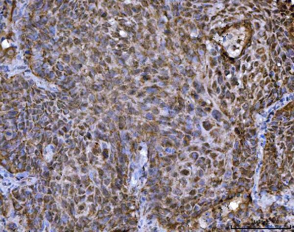
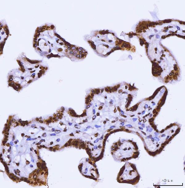
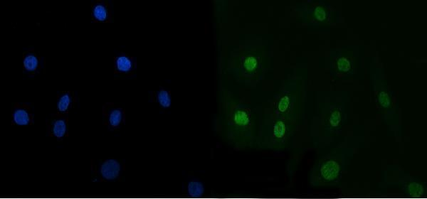
A01680-2 has pictured IHC in paraffin sections of human lung cancer, placenta, prostate cancer and rat testis, plus IF in A549 cells (catalog image captions).

Real IHC data IHC analysis of TRBP/TARBP2 using anti-TRBP/TARBP2 antibody (A01680-2). TRBP/TARBP2 was detected in a paraffin-embedded section of human lung cancer tissue. Heat mediated antigen retrieval was performed in EDTA buffer (pH 8.0, epitope retrieval solution). The tissue section was blocked with 10% goat serum. The tissue section was then incubated with 2 μg/ml rabbit anti-TRBP/TARBP2 Antibody (A01680-2) overnight at 4°C. Peroxidase Conjugated Goat Anti-rabbit IgG was used as secondary antibody and incubated for 30 minutes at 37°C. The tissue section was developed using HRP Conjugated Rabbit IgG Super Vision Assay Kit (Catalog # SV0002) with DAB as the chromogen.
Anti-TRBP/TARBP2 Antibody ®
Cat # A01680-2

A01680-2 has pictured paraffin-section IHC in human lung cancer, placenta and prostate cancer and rat testis, plus IF in A549 cells (catalog image captions). M01680 lists IHC and IF/ICC applications and human, mouse and rat reactivity, but provides no IHC or IF image captions (catalog applications, reactivity and image captions).

Which to pick: Choose A01680-2 for tissue IHC: its captions document paraffin sections, EDTA retrieval at pH 8.0 and 2 μg/ml primary antibody; the fixative is unreported (A01680-2 IHC image captions). For IF/ICC, A01680-2 has pictured A549 cell IF at 5 μg/ml, while M01680 lists IF/ICC without a pictured example (A01680-2 IF image caption; M01680 catalog applications and image captions). Consider rabbit monoclonal M01680 when mouse reactivity is required, but confirm performance in the intended sample: human, mouse and rat reactivity is listed without an IHC or IF image caption (M01680 catalog host, clone, reactivity and image captions).

Each figure is that product's own IHC / IF validation image from its datasheet.

References

  1. UniProt Consortium. UniProt entry Q15633 (TRBP2_HUMAN, RISC-loading complex subunit TARBP2).
  2. Human Protein Atlas. TARBP2 tissue IHC expression (reliability: Approved).
  3. Human Protein Atlas. TARBP2 subcellular location (ICC-IF): Mainly localized to the nucleoplasm. In addition localized to the nuclear bodies..
  4. Human Protein Atlas. TARBP2 antibody validation summary (3 antibodies).
  5. Clinicopathological Significance of TARBP2, APP, and ZNF395 in Breast Cancer. Breast cancer : basic and clinical research 2016 — PMC5147454.
  6. Clinical and functional impact of TARBP2 over-expression in adrenocortical carcinoma. Endocrine-related cancer 2013 — PMC3709642.
  7. TARBP2-Enhanced Resistance during Tamoxifen Treatment in Breast Cancer. Cancers 2019 — PMC6406945.
  8. TARBP2 Suppresses Ubiquitin-Proteasomal Degradation of HIF-1α in Breast Cancer. International journal of molecular sciences 2021 — PMC8745112.
  9. PubMed PMID:2011739 — UniProt-cited evidence.
  10. PubMed PMID:7774957 — UniProt-cited evidence.
  11. PubMed PMID:15489334 — UniProt-cited evidence.