This website uses cookies to ensure you get the best experience on our website.


- Table of Contents
Plan paraffin-section TBC1D5 IHC using the catalog antibody’s documented conditions (datasheet A09263). Compare cytoplasmic staining in glandular cells with appropriate controls, since tissue staining has uncertain reliability (HPA tissue IHC).

Expected localisation, validated protocols, controls and antibodies — the at-a-glance facts below, then the full design guide.
| Expected localisation | General cytoplasmic tissue staining (HPA tissue IHC) | |
| Staining pattern | Glandular cells show cytoplasmic staining (HPA tissue IHC) | |
| Antigen retrieval | EDTA pH 8.0 HIER, heat-mediated (datasheet A09263) | |
| Positive control | Adrenal gland+4 more · see all | |
| Negative control | Adipose tissue |
| Fixation | Keep fixation consistent across sections (standard IHC practice; not target-specific) | |
| Caveat | Tissue staining has uncertain reliability (HPA tissue IHC) | |
| Regulation | Starvation shifts compartmental signal (UniProt) | |
| Isoform / epitope | 2 isoforms; epitope coverage is unknown (UniProt) |
The catalog antibody has an IHC-P protocol (datasheet: A09263); the published protocol below describes chromogenic TBC1D5 staining in renal carcinoma samples (PMC10900628).
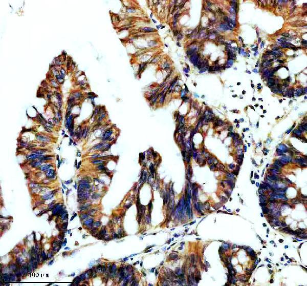
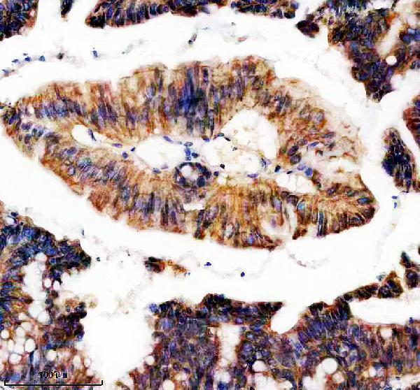
| Sample | Paraffin-embedded human colon cancer tissue; fixative not specified (datasheet A09263) |
| Fixation | Image fixative and duration unreported (datasheet A09263); verify before use. |
| Sectioning | 4–5 µm sections on charged slides (standard) |
| Deparaffinisation | Xylene, graded ethanol series to water (standard) |
| Antigen retrieval | Heat retrieval: EDTA pH 8.0 (datasheet A09263); 20 min, 95–100 °C (standard) |
| Peroxidase block | 3% H2O2, 10 min, room temperature (standard) |
| Blocking | 10% goat serum (datasheet A09263) |
| Primary antibody | Rabbit anti-TBC1D5, 1:50 recommended; image 1:200 (datasheet A09263) |
| Primary incubation | Overnight at 4 °C (datasheet A09263) |
| Detection | HRP-conjugated secondary, DAB chromogen (datasheet A09263) |
| Counterstain | Hematoxylin, blue, dehydrate and mount (standard) |
| Expected result | TBC1D5-positive staining in glandular cells of adrenal gland (HPA tissue IHC: High). HPA tissue profile: General cytoplasmic expression. No signal in the no-primary control. |
TBC1D5 is associated with endosome membranes and cytoplasmic vesicles, including autophagosomes; it has no transmembrane segment (UniProt Q92609 topology and subcellular annotation). In paraffin-section IHC, expect chiefly cytoplasmic staining in epithelial or glandular cells where HPA reports expression, including colon glandular cells (HPA: High). Treat this as a provisional pattern: HPA describes general cytoplasmic expression, but rates its tissue IHC reliability Uncertain (HPA: tissue IHC).
| Cytoplasmic staining in colon or duodenal glandular cells, with little nuclear signal. | This fits HPA’s High glandular-cell staining in both tissues and its general cytoplasmic profile (HPA: tissue IHC). Vesicle-scale puncta may be hard to resolve with chromogenic IHC; assess the predominant compartment before scoring intensity. |
| Predominantly nuclear, extracellular, or sharply surface-membrane staining. | This conflicts with the reported vesicle/endosome association and general cytoplasmic IHC profile (UniProt Q92609 subcellular annotation; HPA: tissue IHC). Treat an isolated wrong-compartment pattern as possible nonspecific staining; compare controls and antibody validation before interpreting it as TBC1D5. |
| Strong adipocyte staining, especially when adjacent expected-positive cells are weak. | HPA reports TBC1D5 as Not detected in adipocytes, although its tissue IHC reliability is Uncertain (HPA: adipose tissue; HPA: tissue IHC). Consider cross-reactivity or endogenous chromogen activity; compare a no-primary control and examine the cellular distribution. |
| Broad, hazy chromogen deposits across cells and surrounding tissue. | Diffuse background obscures the cytoplasmic pattern HPA describes (HPA: tissue IHC). In general IHC practice, incomplete blocking or washing, excessive antibody concentration, and detection background can contribute; use controls to locate the source before assigning a positive score. |
| No signal in a section selected because HPA reports High staining. | A negative result in colon glandular cells, for example, conflicts with HPA’s observed High level but does not prove biological absence or assay failure (HPA: colon glandular cells; HPA: IHC reliability Uncertain). Check tissue preservation, antigen retrieval, detection controls, and antibody performance. |
| Which compartment should guide IHC scoring? | Use HPA’s general cytoplasmic tissue pattern as the slide-level reference (HPA: tissue IHC). Endosome membrane and autophagosome association supports a vesicular interpretation (UniProt Q92609 subcellular annotation), but chromogenic resolution may not show individual vesicles. |
| Can cellular state change the pattern? | UniProt reports relocalization from endosomes to LC3-positive autophagosomes during starvation-induced autophagy (UniProt Q92609 subcellular annotation). A change in cytoplasmic puncta could therefore be biologically plausible, but the supplied HPA tissue images do not establish an IHC scoring rule for that state. |
| How strong is the tissue reference? | HPA reports High staining in several glandular-cell populations and low RNA tissue specificity, yet rates tissue IHC Uncertain because antibody staining and RNA data have low consistency (HPA: tissue IHC). Its two listed antibodies also have Uncertain IHC validation (HPA: HPA035125 and HPA035126). |
| Do isoforms or processing predict a different IHC pattern? | UniProt lists two isoforms, no signal peptide or propeptide, and a full-length chain of residues 1–795 (UniProt Q92609 isoforms and processing). The payload gives no antibody epitope or isoform-specific staining data, so it does not support separate IHC patterns. |
| What should IF/ICC show? | HPA places TBC1D5 mainly in vesicles, with additional Golgi localization, and marks the vesicle location Supported (HPA: subcellular ICC-IF). This answers the localization question for IF/ICC; the separate IF/ICC guide covers its experimental design. |
| Situation | Likely cause | Next action |
|---|---|---|
| A selected High-staining tissue has no visible cytoplasmic signal. | The HPA reference is uncertain; the run may also lack adequate antigen exposure or detection (HPA: tissue IHC reliability Uncertain). | Review morphology and positive detection controls, then optimize retrieval and antibody dilution as general IHC variables; use a second HPA High tissue to check whether the loss is section-specific (HPA: colon and duodenum glandular cells High). |
| Nuclei dominate the stain. | The distribution conflicts with HPA’s general cytoplasmic IHC pattern and UniProt’s vesicle/endosome localization (HPA: tissue IHC; UniProt Q92609 subcellular annotation). | Check a no-primary control and reassess antibody dilution, blocking, and detection background. Score TBC1D5 only after a credible cytoplasmic pattern is recovered. |
| Adipocytes stain strongly. | This differs from HPA’s Not detected adipocyte observation, although that IHC reference remains Uncertain (HPA: adipose tissue; HPA: tissue IHC). | Compare adjacent cell types and a no-primary control. Investigate endogenous detection activity or cross-reactivity before treating adipocyte staining as target expression. |
| Chromogen is diffuse across tissue or pools at section edges. | General IHC background or staining deposits can conceal the expected cytoplasmic distribution (HPA: general cytoplasmic expression). | Inspect no-primary and detection-only controls; adjust washing, blocking, or detection exposure as indicated. Interpret only well-preserved cells away from obvious deposits. |
| Two antibodies give different cell-type patterns. | HPA lists both antibodies as Uncertain for IHC and reports low consistency with RNA expression data (HPA: HPA035125, HPA035126; HPA: tissue IHC). | Record each antibody’s compartment and cell-type pattern separately. Favor a reproducible cytoplasmic result supported by controls; do not average discordant stains into a definitive tissue score. |
| Punctate staining differs between experimental conditions. | TBC1D5 can move from endosomes to LC3-positive autophagosomes during starvation-induced autophagy (UniProt Q92609 subcellular annotation). | Compare matched conditions and cytoplasmic morphology. Treat a shift as a hypothesis about vesicle localization, then seek an appropriate compartment marker before assigning it to autophagosomes. |


Comprehensive Human Protein Atlas IHC scoring per tissue (reliability: Uncertain — Low consistency between antibody staining and RNA expression data. Pending external verification.). Rows are taken directly from the HPA tissue chart — click any row's HPA link to view the source.
| Tissue | Cell type | Level | Evidence | Source |
|---|---|---|---|---|
| Adrenal gland | Glandular cells | High | Protein (IHC) | HPA → |
| Appendix | Glandular cells | High | Protein (IHC) | HPA → |
| Breast | Glandular cells | High | Protein (IHC) | HPA → |
| Cervix | Squamous epithelial cells | High | Protein (IHC) | HPA → |
| Colon | Glandular cells | High | Protein (IHC) | HPA → |
| Tissue | Cell type | Level | Evidence | Source |
|---|---|---|---|---|
| Adipose tissue | Adipocytes | Not detected | Protein (IHC) | HPA → |
Troubleshoot TBC1D5 staining in paraffin sections by checking retrieval, cellular pattern, controls and scoring before interpreting chromogenic signal.
A09263 has IHC images of human colon, human colon cancer, and rat testis paraffin sections; IF/ICC are listed applications, but no IF image is supplied (catalog: A09263 captions and applications).
A09263 will render with its human colon cancer paraffin-section IHC figure (catalog: A09263 figure caption). Additional captions show human colon and rat testis IHC; the catalog lists human, mouse, and rat reactivity and IF/ICC applications, without an IF image (catalog: A09263 captions, reactivity, and applications).
Which to pick: Choose A09263 for tissue IHC: its own caption documents heat retrieval in EDTA at pH 8.0 and antibody incubation at 1:200 overnight at 4°C in a paraffin section; the fixative is unreported (catalog: A09263 IHC caption). For IF/ICC or cross-species work, A09263 is the listed polyclonal option with human, mouse, and rat reactivity, though the supplied images document IHC in human and rat tissue only (catalog: A09263 dilution_raw, applications, reactivity, and captions).