TBR1 / T-box brain protein 1 · IHC design guide

Design Immunohistochemistry for TBR1

Plan TBR1 IHC in paraffin sections around nuclear staining in cerebral cortical neurons (HPA tissue IHC). Use the catalog antibody’s IHC dilution range of 1:200–1:1000 as a starting point (datasheet: M07503-2).

Evidence assembled Oct 2026 · For research use; verify linked source records and product datasheet before use
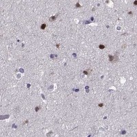
Immunohistochemistry protocol sheet for TBR1 (IHC for TBR1): expected localisation Nuclear staining in cortical neurons (HPA tissue IHC), antibody A07503, validated IHC image, and IHC protocol steps
Printable TBR1 IHC protocol sheet — expected localisation Nuclear staining in cortical neurons (HPA tissue IHC), antibody A07503, controls and protocol steps. Open the full TBR1 IHC guide →

TBR1 Immunohistochemistry Experimental Design Guide

Expected localisation, validated protocols, controls and antibodies — the at-a-glance facts below, then the full design guide.

Must know before staining
Expected localisation Nuclear staining in cortical neurons (HPA tissue IHC)
Staining pattern Neuronal nuclei stain in cerebral cortex (HPA tissue IHC)
Antigen retrieval Tris-EDTA pH 9.0 HIER, 95–98 °C, 20 min (rule: nuclear antigen)
Positive control ⓘ Cerebral cortex
Negative control ⓘ Adipose tissue+4 more · see all
Important caveats
Reasons your staining may differ from the expected pattern.
Fixation Keep fixation consistent across sections. (standard IHC practice; not target-specific)
Caveat Hippocampal neurons show low staining (HPA tissue IHC)
Regulation Brain-enriched expression (UniProt)
Isoform / epitope Two isoforms; epitope differences are unspecified (UniProt)
Section 1

Recommended TBR1 IHC & IF Protocols

The catalog antibody’s IHC-P protocol is followed by a published chromogenic TBR1 protocol for human prenatal brain (PMC9944618).

Recommended immunohistochemistry (IHC-P) protocol parameters
SampleParaffin-embedded rat brain tissue; fixative not specified (datasheet A07503)
FixationImage fixative and duration unreported (datasheet A07503); verify before use.
Sectioning4–5 µm sections on charged slides (standard)
DeparaffinisationXylene, graded ethanol series to water (standard)
Antigen retrievalHeat-induced epitope retrieval in Tris-EDTA buffer, pH 9.0, 20 min at 95–98 °C (standard rule: nuclear antigen)
Peroxidase block3% H2O2, 10 min, room temperature (standard)
Blocking10% normal serum of the secondary host, 30 min, room temperature (standard)
Primary antibodyRabbit anti-TBR1, 1:50-1:200 (datasheet A07503)
Primary incubationOvernight at 4 °C (standard)
DetectionHRP-polymer secondary, DAB chromogen 5–10 min (standard)
CounterstainHematoxylin, blue, dehydrate and mount (standard)
Expected resultTBR1-positive staining in neuronal cells of cerebral cortex (HPA tissue IHC: Medium). HPA tissue profile: Nuclear expression in neuronal cells in cerebral cortex. No signal in the no-primary control.
💡Decision noteStart with Tris-EDTA at pH 9.0, 95–98 °C for 20 min (page retrieval); the published protocol uses boiling citrate at pH 6.0 (PMC9944618).
Section 2

What Is the Expected TBR1 Staining Pattern?

TBR1 is a nuclear transcriptional regulator with no transmembrane segment (UniProt Q16650). In paraffin tissue IHC, expect staining in neuronal cell nuclei of the cerebral cortex; HPA reports medium staining there and rates its tissue profile Enhanced because staining is consistent with RNA expression (HPA tissue IHC). UniProt lists brain tissue specificity (UniProt Q16650).

What am I looking at on my slide?
Distinct nuclear staining in cerebral cortical neurons, with moderate overall intensity.This matches HPA’s medium staining in cortical neuronal cells and its nuclear profile (HPA tissue IHC). Judge the pattern by identifiable cells and compartment, not by uniform staining across the section (general IHC practice).
Predominantly cytoplasmic, membranous, or extracellular color in cortical cells.These compartments do not match the reported nuclear location or the absence of a transmembrane segment (UniProt Q16650; HPA tissue IHC). Treat the pattern as suspect and check the detection controls (general IHC practice).
Strong staining in adipocytes, adrenal glandular cells, or cerebellar granular-layer cells.HPA reports TBR1 as not detected in those specified cells (HPA tissue IHC). Check for nonspecific binding or endogenous chromogen-generating activity before assigning that color to TBR1 (general IHC practice).
Color spread across tissue, with cell borders or nuclei hard to distinguish.Diffuse background prevents a reliable nuclear call (general IHC practice). It cannot establish TBR1 expression in a cell type solely because that tissue contains positive cells elsewhere (HPA tissue IHC; general IHC practice).
No neuronal nuclear staining in a cerebral cortex section.This conflicts with HPA’s positive cortical-neuron observation (HPA tissue IHC). Check that neurons are present and that the IHC run worked before interpreting the absence as biological (general IHC practice).
💡Expected TBR1 appearanceCall a positive result when cerebral cortical neuronal nuclei show discernible, approximately medium chromogenic staining (HPA tissue IHC); widespread cytoplasmic color or staining in HPA-negative cell types is suspect (UniProt Q16650; HPA tissue IHC).
How each factor affects the staining
Tissue and cell selectionCerebral cortical neurons provide an observed positive reference; hippocampal neurons are reported at low level, while cerebellar granular-layer cells are not detected (HPA tissue IHC). Interpret weak hippocampal staining against that lower reference, not the cortical level.
Compartment and processingTBR1 is nuclear, has no transmembrane segment, and is listed as one 1–682 chain without signal peptide or propeptide processing (UniProt Q16650). These annotations support a nuclear interpretation but do not predict fixation sensitivity.
Isoforms and modified residuesUniProt lists two isoforms and four phosphorylated residues (UniProt Q16650). Without an antibody epitope or isoform-reactivity record in this payload, their effects on staining cannot be assigned.
Antibody evidenceHPA labels HPA078644 and HPA078657 Enhanced for IHC and HPA051256 Supported for IHC (HPA antibodies). Those labels apply to the listed antibodies; they do not establish the performance of an unspecified catalog antibody.
IF/ICC Q&A: What should a positive cell show?A nuclear signal is the expected compartment from UniProt and tissue IHC (UniProt Q16650; HPA tissue IHC). HPA supplies no ICC-IF images or subcellular location result, so an IF/ICC staining pattern is unconfirmed here (HPA subcellular).
Why is my staining missing, weak or wrong?
SituationLikely causeNext action
Cortical neuronal nuclei are blank.A failed staining run, absent neuronal profiles, or weak detection can resemble a negative result (general IHC practice); HPA reports cortical neurons as positive (HPA tissue IHC).Confirm neuronal morphology and section integrity, then compare a known-positive cortex processed in the same run; review primary-antibody, retrieval, and detection steps (general IHC practice).
Signal is faint in hippocampal neurons.HPA reports low staining there, compared with medium staining in cortical neurons (HPA tissue IHC).Use cortical neuronal nuclei as the stronger reference and compare against a control without primary antibody; avoid calling a faint hippocampal result a technical failure by intensity alone (HPA tissue IHC; general IHC practice).
Cytoplasm stains more strongly than nuclei.A dominant cytoplasmic pattern conflicts with the nuclear location reported for TBR1 (UniProt Q16650; HPA tissue IHC).Check nuclear counterstain alignment and the control without primary antibody; reassess antibody dilution and washing if off-target color persists (general IHC practice).
HPA-negative cell types show widespread color.Nonspecific antibody staining or endogenous detection activity may mimic a positive chromogenic result (general IHC practice); HPA reports several specified cell types as not detected (HPA tissue IHC).Compare the same tissue with a control lacking primary antibody; review blocking of endogenous enzyme activity and the detection reagents (general IHC practice).
The whole section has diffuse background.Excess reagent or insufficient washing can obscure cellular boundaries (general IHC practice).Check the control without primary antibody, then adjust antibody dilution, blocking, washing, or chromogen development within the assay workflow (general IHC practice).
An antibody gives a different pattern from an HPA image.HPA’s IHC validation labels are antibody specific; the payload does not establish equivalence with another antibody (HPA antibodies).Compare staining in cortical neuronal nuclei and HPA-negative cell types, then check the chosen antibody’s documented IHC validation and controls (HPA tissue IHC; HPA antibodies; general IHC practice).

Sample controls for TBR1 IHC & IF

🧪Run cerebral cortex first: neuronal cells should show nuclear staining at the HPA Medium level (HPA: cerebral cortex neuronal cells, Medium; UniProt Q16650: nucleus). Use adipose tissue as the negative tissue because adipocytes are not detected (HPA: adipose tissue adipocytes, Not detected); on the cortex slide, cells outside the stained neuronal population should lack convincing nuclear signal and serve as internal background comparators (HPA: cerebral cortex neuronal cells, Medium; UniProt Q16650: nucleus).
Positive control tissue: Cerebral cortex (Neuronal cells, HPA Medium)
Negative control tissue: Adipose tissue (HPA Not detected)
ICC-IF cell lines (HPA subcellular resource): HPA carries no ICC-IF cell line for TBR1; derive a cell-line control from the positive tissue's cell type (Neuronal cells) and confirm it by RNA or western blot first.
Technical controls: Include a no-primary, secondary-only control and an isotype control matched to the primary antibody’s host species, immunoglobulin class and concentration; use TBR1 knockout tissue or a validated immunizing-peptide block as a biological specificity control (standard IHC/IF practice). For chromogenic cortex IHC, quench endogenous peroxidase and check background on the no-primary slide; if using biotin-based detection, control endogenous biotin, and for IF assess tissue autofluorescence (standard IHC/IF practice).
⚠️Feasibility: No target-specific fixation window or retrieval dependency is reported in the supplied evidence; optimize antigen retrieval empirically for paraffin sections (A07503 caption; HPA: cerebral cortex neuronal cells, Medium). The selected A07503 caption shows paraffin-embedded rat brain with hematoxylin counterstain, but its fixative is unreported (A07503 caption). The supplied evidence does not establish whether frozen sections or IF are easier; cortical autofluorescence can complicate IF interpretation (standard IF practice).

HPA tissue IHC evidence for TBR1

Comprehensive Human Protein Atlas IHC scoring per tissue (reliability: Enhanced — High consistency between antibody staining and RNA expression data.). Rows are taken directly from the HPA tissue chart — click any row's HPA link to view the source.

Positive expression · recommended positive controls

TissueCell typeLevelEvidenceSource
Cerebral cortex Neuronal cells Medium Protein (IHC) HPA →

Undetected expression · recommended negative controls

TissueCell typeLevelEvidenceSource
Adipose tissue Adipocytes Not detected Protein (IHC) HPA →
Adrenal gland Glandular cells Not detected Protein (IHC) HPA →
Appendix Glandular cells Not detected Protein (IHC) HPA →
Bone marrow Hematopoietic cells Not detected Protein (IHC) HPA →
Breast Adipocytes Not detected Protein (IHC) HPA →
Section 3

Advanced TBR1 IHC Tips

Troubleshoot TBR1 staining in paraffin sections by assessing nuclear signal, tissue controls, processing conditions, and chromogenic detection.

How should I retrieve TBR1 in paraffin sections when nuclear staining is weak?
Start with Tris-EDTA at pH 9.0 for heat-induced retrieval at 95–98 °C for 20 min (page retrieval setting: nuclear antigen). Allow sections to cool consistently, then compare retrieved and unretrieved sections from the same run to judge whether retrieval improves nuclear contrast (standard IHC practice). TBR1 is nuclear (UniProt Q16650), so evaluate neuronal nuclei rather than treating diffuse cytoplasmic color as a successful rescue. If signal remains weak, vary heating time in a small controlled series while keeping antibody concentration and detection constant (standard IHC practice). Record tissue retention and background alongside nuclear signal, because excessive retrieval can impair morphology (standard IHC practice).
Could fixation explain weak or uneven TBR1 staining?
Target-specific fixation sensitivity for TBR1 is unknown from the supplied evidence; the catalog image identifies paraffin-embedded rat brain but does not state its fixative (A07503 tissue-IHC caption). For a controlled troubleshooting series, compare sections with documented fixation conditions while holding retrieval, antibody incubation, and chromogenic development constant (standard IHC practice). Check whether weak staining follows specimen thickness, delayed processing, or poorly preserved nuclear detail before changing antibody concentration (standard IHC practice). Score nuclear signal and morphology together, using the same neuronal region across sections (UniProt Q16650 localisation; HPA tissue IHC). Do not assign a TBR1-specific fixation effect without a direct comparison.
What staining pattern should count as correctly localised TBR1?
Expect predominantly nuclear staining because TBR1 is a nuclear protein (UniProt Q16650), and assess it in neuronal cells of the cerebral cortex, where tissue IHC reports nuclear expression (HPA tissue IHC). Use a hematoxylin counterstain to identify nuclei; the catalog image also uses hematoxylin in paraffin-embedded rat brain (A07503 tissue-IHC caption). Score a nucleus as positive only when chromogen is distinct from neighboring background and the cell remains morphologically interpretable (standard IHC practice). Widespread cytoplasmic color or staining restricted to section edges warrants a review of blocking, wash steps, and development time before biological interpretation (standard IHC practice).
How should isoforms and epitope accessibility affect troubleshooting?
TBR1 has 2 annotated isoforms (UniProt Q16650), but the supplied catalog caption does not identify the antibody epitope or establish isoform-specific recognition (A07503 tissue-IHC caption). Confirm the immunogen or epitope information for the antibody in use before claiming that a negative nucleus lacks every TBR1 isoform. TBR1 has no annotated transmembrane segment and is nuclear (UniProt Q16650); optimise access to the nuclear epitope through retrieval and section processing rather than assuming a membrane-facing target. Phosphorylated residues are annotated at positions 408, 410, 594, and 641 (UniProt Q16650), but their effect on this antibody's staining is unestablished.
How can I check a TBR1 result with multiplex immunofluorescence?
Use IF as an independent spatial check of the chromogenic result, pairing TBR1 with a validated neuronal marker because cortical neuronal nuclei are the expected positive population (HPA tissue IHC). Choose spectrally separated fluorophores after inspecting tissue autofluorescence and include single-stain controls to assess bleed-through (standard IF practice). TBR1 is nuclear and has no annotated transmembrane segment (UniProt Q16650), so use permeabilisation compatible with access to an intracellular nuclear epitope, then verify that nuclear morphology remains intact. The supplied catalog evidence is paraffin-section IHC, with no stated fixative or IF validation (A07503 tissue-IHC caption); optimise IF fixation and permeabilisation with IF controls rather than transferring IHC conditions.
What should I check when brown signal obscures TBR1-positive nuclei?
Compare a no-primary control with the stained section to separate detection-system color from antibody-dependent staining (standard IHC practice). Apply an appropriate endogenous peroxidase block before a peroxidase-based DAB workflow, and check whether prolonged DAB development raises color across the whole section (standard IHC practice). If diffuse staining persists, titrate the primary antibody and strengthen washing or blocking one variable at a time, recording nuclear contrast in the same region (standard IHC practice). TBR1 is nuclear (UniProt Q16650), while HPA reports nuclear staining in cerebral-cortex neuronal cells (HPA tissue IHC); diffuse extracellular or uniform cytoplasmic color should therefore trigger a background review.
How should I quantify TBR1 staining across paraffin sections? ⚠ ANSWER MARKED FOR VERIFICATION
Define the cortical region and analysis thresholds before scoring, then report the percentage of interpretable neuronal nuclei that are TBR1 positive (HPA tissue IHC; standard IHC practice). If intensity matters, use an H-score with fixed intensity bins and document the threshold and observer or image-analysis settings (standard IHC practice). For counts, report positive nuclei per mm² of sampled tissue and also the positive fraction among eligible neuronal nuclei, so sampling area and cell abundance remain visible (standard IHC practice). Exclude folds, damaged edges, and poorly preserved regions by a prespecified rule, and use identical counterstain and chromogen-development conditions across compared sections (standard IHC practice).
How can I distinguish genuine TBR1 staining from artefact?
A plausible positive result is discrete neuronal nuclear staining in cerebral cortex, consistent with TBR1's nuclear location and the reported tissue pattern (UniProt Q16650; HPA tissue IHC). Compare that pattern with a no-primary control and nearby intact tissue before accepting isolated brown deposits (standard IHC practice). Treat cytoplasmic staining, edge-only signal, and staining concentrated in necrotic or damaged areas as reasons to review preparation and detection rather than to call additional positive cells (standard IHC practice). Check whether residual endogenous enzyme activity explains color in the control when using peroxidase detection (standard IHC practice). HPA reports no detectable staining in several other sampled cell populations, but those observations do not establish universal absence (HPA tissue IHC).
Boster reagents

Best TBR1 / T-box brain protein 1 IHC Antibodies

Two anti-TBR1 antibodies have real brain IHC images (catalog image captions); both list Human, Mouse and Rat reactivity (catalog reactivity), and one also lists IF (M07503-2 applications).

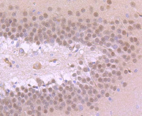
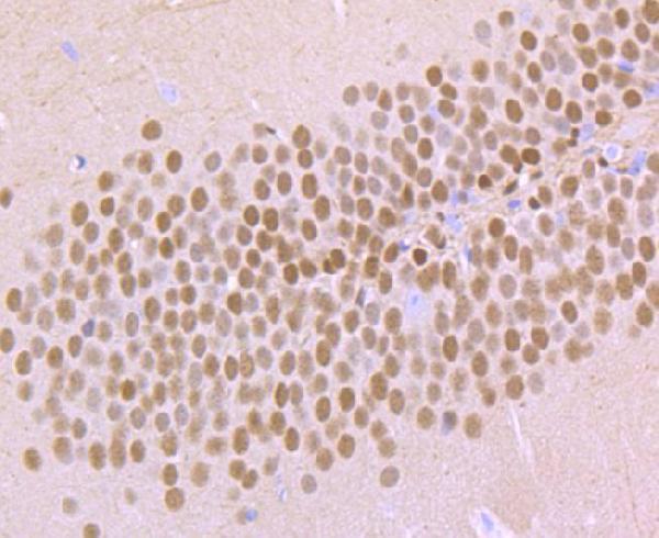
Real IHC data Immunohistochemical analysis of paraffin-embedded rat brain tissue using anti-TBR1 antibody. Counter stained with hematoxylin.
Anti-T-box brain protein 1 TBR1 Antibody
Cat # A07503
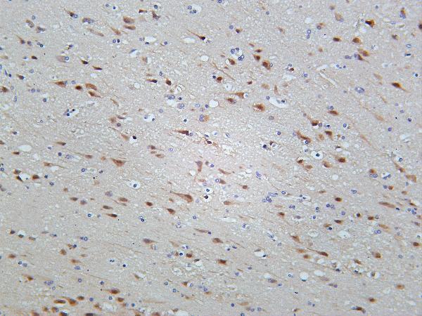
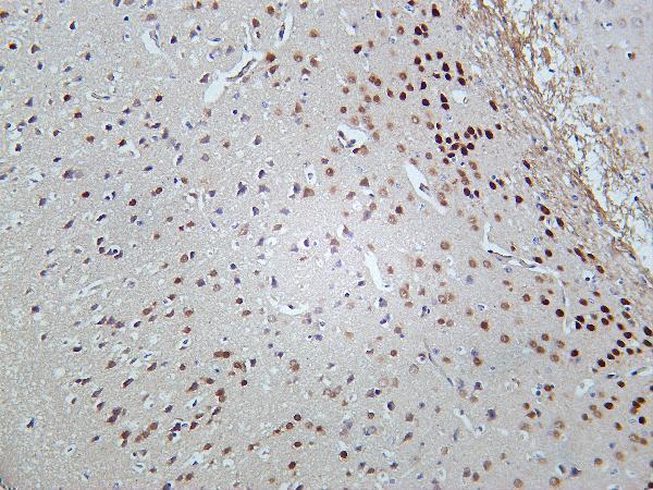
Real IHC data Human brain was stained with Anti-TBR1 rabbit antibody
Anti-TBR1 Rabbit Monoclonal Antibody
Cat # M07503-2

A07503 lists IHC and Human, Mouse and Rat reactivity, with images of paraffin-embedded rat and mouse brain sections counterstained with hematoxylin (A07503 applications, reactivity and image captions). M07503-2 lists IHC and IF and Human, Mouse and Rat reactivity, with IHC images of human and mouse brain (M07503-2 applications, reactivity and image captions).

Which to pick: For paraffin-section IHC, choose A07503 because its own images document that preparation in rat and mouse brain; its fixative is unreported (A07503 image captions). For IF, choose the monoclonal M07503-2 because IF is listed at 1:200–1:1000; ICC validation is unreported, and its brain IHC captions do not specify processing or fixative (M07503-2 catalog applications, dilution and image captions). Both list Human, Mouse and Rat reactivity for cross-species planning, while the depicted tissue evidence covers rat and mouse for A07503 and human and mouse for M07503-2 (catalog reactivity and image captions).

Each figure is that product's own IHC / IF validation image from its datasheet.

References

  1. UniProt Consortium. UniProt entry Q16650 (TBR1_HUMAN, T-box brain protein 1).
  2. Human Protein Atlas. TBR1 tissue IHC expression (reliability: Enhanced).
  3. Human Protein Atlas. TBR1 subcellular location (ICC-IF): Highest expression in HAP1: 0.2 nTPM.
  4. Human Protein Atlas. TBR1 antibody validation summary (3 antibodies).
  5. Laminar dynamics of deep projection neurons and mode of subplate formation are hallmarks of histogenetic subdivisions of the human cingulate cortex before onset of arealization. Brain structure & function 2023 — PMC9944618.
  6. Thalamo-cortical axons regulate the radial dispersion of neocortical GABAergic interneurons. eLife 2016 — PMC5167520.
  7. Developmental exposure to diesel exhaust upregulates transcription factor expression, decreases hippocampal neurogenesis, and alters cortical lamina organization: relevance to neurodevelopmental disorders. Journal of neurodevelopmental disorders 2020 — PMC7745370.
  8. Genetic interplay between the transcription factors Sp8 and Emx2 in the patterning of the forebrain. Neural development 2007 — PMC1868949.
  9. PubMed PMID:7619531 — UniProt-cited evidence.
  10. PubMed PMID:14702039 — UniProt-cited evidence.
  11. PubMed PMID:15815621 — UniProt-cited evidence.