TCOF1 / Treacle protein · IHC design guide

Design Immunohistochemistry for TCOF1

Plan chromogenic TCOF1 IHC in paraffin sections around nuclear and nucleolar staining (HPA tissue IHC). Use cerebellar GLUC cells as a high-staining reference (HPA tissue IHC), and titrate the IHC-validated antibody within 1:100–1:300 (datasheet: A02653).

Evidence assembled Oct 2026 · For research use; verify linked source records and product datasheet before use
Immunohistochemistry protocol sheet for TCOF1 (IHC for TCOF1): expected localisation Nucleolar and nuclear tissue staining (HPA tissue IHC), antibody A02653, validated IHC image, and IHC protocol steps
Printable TCOF1 IHC protocol sheet — expected localisation Nucleolar and nuclear tissue staining (HPA tissue IHC), antibody A02653, controls and protocol steps. Open the full TCOF1 IHC guide →

TCOF1 Immunohistochemistry Experimental Design Guide

Expected localisation, validated protocols, controls and antibodies — the at-a-glance facts below, then the full design guide.

Must know before staining
Expected localisation Nucleolar and nuclear tissue staining (HPA tissue IHC)
Staining pattern Many cell types show nuclear and nucleolar staining (HPA tissue IHC)
Antigen retrieval Tris-EDTA pH 9.0 HIER, 95–98 °C, 20 min (rule: nuclear antigen)
Positive control ⓘ Cerebellum+4 more · see all
Negative control ⓘ Soft tissue
Important caveats
Reasons your staining may differ from the expected pattern.
Fixation Keep fixation consistent across sections. (standard IHC practice; not target-specific)
Caveat Chondrocytes show no detectable staining (HPA tissue IHC)
Regulation Low tissue specificity (HPA tissue RNA)
Isoform / epitope 8 isoforms; check epitope coverage (UniProt)
Section 1

Recommended TCOF1 IHC & IF Protocols

The catalog antibody’s IHC-P protocol is accompanied by two published TCOF1 tissue IHC protocols (PMC13264060; PMC9850107).

Recommended immunohistochemistry (IHC-P) protocol parameters
SampleParaffin-embedded human lung carcinoma tissue; fixative not specified (datasheet A02653)
FixationImage fixative and duration unreported (datasheet A02653); verify before use.
Sectioning4–5 µm sections on charged slides (standard)
DeparaffinisationXylene, graded ethanol series to water (standard)
Antigen retrievalHeat-induced epitope retrieval in Tris-EDTA buffer, pH 9.0, 20 min at 95–98 °C (standard rule: nuclear antigen)
Peroxidase block3% H2O2, 10 min, room temperature (standard)
Blocking10% normal serum of the secondary host, 30 min, room temperature (standard)
Primary antibodyRabbit anti-TCOF1, 1:100 - 1:300 (datasheet A02653)
Primary incubationOvernight at 4 °C (standard)
DetectionHRP-polymer secondary, DAB chromogen 5–10 min (standard)
CounterstainHematoxylin, blue, dehydrate and mount (standard)
Expected resultTCOF1-positive staining in gLUC cells - nucleus of cerebellum (HPA tissue IHC: High). HPA tissue profile: Ubiquitous nucleolar and nuclear expression. No signal in the no-primary control.
💡Decision noteStart with Tris-EDTA pH 9.0 HIER at 95–98 °C for 20 min (page retrieval rule); TCOF1 is nucleolar (UniProt Q13428).
Section 2

What Is the Expected TCOF1 Staining Pattern?

TCOF1 should stain the nucleus, with a prominent nucleolar pattern (UniProt Q13428: nucleus, nucleolus; HPA: ubiquitous nucleolar and nuclear expression). Expect staining across many cell types, including high nuclear staining in cerebellar GLUC cells and medium staining in several glandular and epithelial cell populations (HPA tissue IHC). HPA rates the tissue staining “Enhanced” but reports only medium consistency with RNA expression; interpret intensity differences cautiously (HPA tissue IHC). TCOF1 has no transmembrane segment (UniProt Q13428 topology).

What am I looking at on my slide?
Distinct nuclear staining with visible nucleolar enrichment in cells of an HPA-positive tissue; cerebellar GLUC-cell nuclei may be especially strong (HPA tissue IHC).This matches the reported distribution (HPA tissue IHC; UniProt Q13428 localization). Compare each cell type with nearby morphology and score nuclear and nucleolar signal separately where the section permits; an overall brown nucleus can obscure nucleolar detail (general chromogenic IHC practice).
Predominantly membrane or cytoplasmic staining, without a convincing nuclear or nucleolar component.That compartment conflicts with TCOF1 localization (UniProt Q13428: nucleus, nucleolus; no transmembrane segment). Treat it as suspect until controls and staining conditions are checked; morphology alone cannot identify the cause of an off-target signal (general IHC interpretation).
Strong staining in soft-tissue chondrocytes, especially when expected nuclear staining elsewhere is weak.HPA lists chondrocytes as “Not detected” (HPA tissue IHC). Consider antibody cross-reactivity or endogenous detection activity, then compare a reagent-omission control and the cellular compartment (general IHC practice). One discordant field is insufficient to establish antibody specificity.
Diffuse staining across tissue, stroma, and nuclei, with little difference between positive and low-staining cell populations.The pattern is hard to reconcile with compartment-specific TCOF1 staining and HPA's reported variation among cell types (HPA tissue IHC). Background from detection reagents, incomplete blocking, or excess primary antibody is possible; assess controls before assigning a biological explanation (general IHC practice).
No nuclear signal in a section containing cerebellar GLUC cells or another HPA-positive population.Absence is discordant with HPA's high GLUC-cell nuclear staining or its listed medium-staining populations (HPA tissue IHC). First verify tissue and cell identification, antibody performance, detection reagents, and run conditions (general IHC practice); a failed stain does not establish TCOF1 absence.
💡Expected TCOF1 appearanceCall a result positive when staining is chiefly nuclear with nucleolar enrichment in the expected cells; high signal in cerebellar GLUC-cell nuclei is a useful reference (HPA tissue IHC; UniProt Q13428 localization). Dominant membrane or cytoplasmic staining, or strong chondrocyte staining, warrants scrutiny (UniProt Q13428 topology; HPA: chondrocytes not detected).
How each factor affects the staining
Cell type and tissue choiceHPA reports high nuclear staining in cerebellar GLUC cells, medium staining in selected adipose, glandular, respiratory epithelial, and glial cells, and low staining in several other populations (HPA tissue IHC). Choose an HPA-positive comparator and identify its cell type before judging a weak sample.
Interpretation of low or absent stainingHPA reports low staining in hippocampal neuronal cells, hepatocytes, and several glandular populations, while soft-tissue chondrocytes are not detected (HPA tissue IHC). These observations guide comparison; they do not make an entire tissue uniformly positive or negative.
Antibody evidenceHPA rates HPA038237 and HPA038238 “Enhanced” for IHC, and CAB033199 “Supported” (HPA antibody validation). Tissue IHC is also rated “Enhanced,” with medium agreement between staining and RNA data (HPA tissue IHC). These ratings support pattern review but do not validate every new specimen or run.
Protein forms and modificationUniProt lists eight TCOF1 isoforms and multiple modified residues, including phosphoserines and a phosphothreonine near positions 83–88 (UniProt Q13428). Without an epitope map, the supplied evidence cannot predict which forms a particular antibody detects or whether modification changes staining.
IF/ICC Q: What subnuclear pattern is reported?A: HPA places the main IF/ICC signal in the nucleoli fibrillar center and rates that localization “Enhanced” (HPA subcellular; HPA antibody validation). Use this as a localization comparison; chromogenic tissue IHC may not resolve the same fine structure (general microscopy practice).
Why is my staining missing, weak or wrong?
SituationLikely causeNext action
Positive comparator has no visible nuclear stain.A run or detection failure is possible when an HPA-positive cell population is unexpectedly blank (HPA tissue IHC; general IHC practice).Confirm that the expected cells are present; check the IHC-validated antibody's documented IHC-P conditions, detection reagents, and control slide, then repeat the run (general IHC practice).
Nuclear signal is faint in a sample expected to stain strongly.The sampled cell type may differ from the intended HPA comparator, or assay sensitivity may be inadequate (HPA tissue IHC; general IHC practice).Identify the cell population first. Compare it with an HPA-listed positive population and adjust only documented IHC-P conditions, such as primary dilution or retrieval, if applicable (general IHC practice).
Nuclei stain, but individual nucleoli are unclear.Section resolution, counterstain, or dense chromogen can limit subnuclear assessment (general microscopy practice); HPA reports nucleolar and nuclear tissue expression (HPA tissue IHC).Review lightly stained areas and the counterstain, and score nuclear signal without claiming fibrillar-center resolution on the chromogenic section (general IHC practice; HPA subcellular IF/ICC).
Brown signal appears mainly outside nuclei.This conflicts with the reported nuclear and nucleolar localization and lack of a transmembrane segment (UniProt Q13428); nonspecific staining or detection background is possible (general IHC practice).Check reagent-omission controls and cellular boundaries; compare with a validated positive compartment before interpreting the signal as TCOF1 (general IHC practice; HPA tissue IHC).
Chondrocytes stain strongly.HPA lists soft-tissue chondrocytes as “Not detected” (HPA tissue IHC); cross-reactivity or endogenous detection activity is possible (general IHC practice).Compare a reagent-omission control and an HPA-positive cell population on the same run; avoid calling the chondrocyte signal specific from intensity alone (general IHC practice).
Widespread diffuse background obscures the tissue pattern.Insufficient blocking, concentrated primary antibody, or detection-reagent background can reduce contrast (general IHC practice).Inspect omission controls and the documented IHC-P workflow; optimize blocking, washing, and primary dilution within the antibody's validated range before rescoring (general IHC practice).

Sample controls for TCOF1 IHC & IF

🧪Run cerebellum first and look for nuclear staining in GLUC cells (HPA: High in cerebellum GLUC-cell nuclei). Use soft tissue chondrocytes as the biological tissue negative (HPA: Not detected); on the cerebellum slide, cells without specific nucleolar staining should show counterstain only, but the supplied HPA rows do not identify a reliably negative cerebellar cell type (UniProt Q13428: nucleolar localization; HPA: cerebellum GLUC-cell nuclei).
Positive control tissue: Cerebellum (GLUC cells - nucleus, HPA High)
Negative control tissue: Soft tissue (HPA Not detected)
ICC-IF cell lines (HPA subcellular resource): HPA ICC-IF images show TCOF1 in A-431, U-251MG, U2OS, U2OS, siRNA 2 (10x), U2OS, siRNA 2 (40x), U2OS, scrambled (10x), U2OS, scrambled (40x), with annotated localisation: Nucleoli fibrillar center (enhanced) (HPA subcellular).
Technical controls: Include no-primary (secondary-only) and primary-host-species- and isotype-matched controls, plus a TCOF1 knockout sample or peptide-blocked primary as a target-specific control (standard IHC practice; selected A02653 tissue-IHC caption: peptide block). Quench endogenous peroxidase for chromogenic detection and distinguish any native pigment from the nuclear signal on the cerebellum slide (standard IHC practice; HPA: cerebellum GLUC-cell nuclei).
⚠️Feasibility: A target-specific fixation window, fixation effect, and antigen-retrieval dependency are unreported in the supplied evidence; the selected A02653 paraffin-section caption also leaves the fixative unreported (selected A02653 tissue-IHC caption). The available evidence does not establish that frozen sections or IF are easier than paraffin IHC for TCOF1 (selected A02653 tissue-IHC caption; HPA: ICC-IF nucleoli fibrillar center). In cerebellum, inspect apparent brown nuclear signal against pigment and the no-primary control before scoring it as TCOF1 (standard IHC practice; HPA: High in cerebellum GLUC-cell nuclei).

HPA tissue IHC evidence for TCOF1

Comprehensive Human Protein Atlas IHC scoring per tissue (reliability: Enhanced — Medium consistency between antibody staining and RNA expression data.). Rows are taken directly from the HPA tissue chart — click any row's HPA link to view the source.

Positive expression · recommended positive controls

TissueCell typeLevelEvidenceSource
Cerebellum GLUC cells - nucleus High Protein (IHC) HPA →
Adipose tissue Adipocytes Medium Protein (IHC) HPA →
Adrenal gland Glandular cells Medium Protein (IHC) HPA →
Appendix Glandular cells Medium Protein (IHC) HPA →
Breast Glandular cells Medium Protein (IHC) HPA →

Undetected expression · recommended negative controls

TissueCell typeLevelEvidenceSource
Soft tissue Chondrocytes Not detected Protein (IHC) HPA →
Section 3

Advanced TCOF1 IHC Tips

Troubleshoot TCOF1 chromogenic IHC in paraffin sections by checking retrieval, nuclear localisation, controls and scoring consistency.

What retrieval should I try when TCOF1 nuclear staining is weak?
Use Tris-EDTA at pH 9.0 for heat-induced epitope retrieval at 95–98 °C for 20 min (page IHC retrieval rule). Cool sections consistently before washing, then compare retrieved and untreated sections from the same block to distinguish inadequate retrieval from background caused by excessive heating (standard IHC practice). TCOF1 is a nuclear, mainly nucleolar protein, so assess improvement within nuclei rather than by an increase in diffuse cytoplasmic colour (UniProt Q13428; HPA subcellular). If staining remains weak, test a shorter or longer heating interval in parallel while holding antibody concentration, detection and imaging conditions constant (standard IHC practice).
Could fixation explain variable TCOF1 staining across paraffin sections?
The selected paraffin-section image does not state its fixative, and target-specific fixation sensitivity is unknown (A02653 tissue-IHC caption). Record the fixative and fixation duration for each specimen, then compare sections with matched processing before attributing a staining difference to TCOF1 abundance (standard IHC practice). For suspected overfixation, compare retrieval conditions on serial sections and judge whether nucleolar signal improves without widespread nuclear background or tissue damage (standard IHC practice; UniProt Q13428 localisation). Use an internal positive area and a no-primary control in each run; neither a published tissue pattern nor TCOF1 modifications establish a preferred fixative (HPA tissue IHC; UniProt Q13428 modified residues).
How should I judge diffuse staining when TCOF1 is expected in nucleoli?
Prioritise crisp intranuclear, especially nucleolar, staining: TCOF1 is annotated in the nucleus and nucleolus, with enhanced localisation to the nucleolar fibrillar centre (UniProt Q13428; HPA subcellular). Compare the chromogen with a nuclear counterstain at high magnification; a pale counterstain can make adjacent cytoplasmic deposit appear nuclear (standard IHC practice). If staining is broadly cytoplasmic or extracellular, check the no-primary control, antibody concentration and chromogen development time before scoring it as positive (standard IHC practice; UniProt Q13428 topology). TCOF1 has no transmembrane segment, so a dominant membrane outline lacks support from its annotated topology (UniProt Q13428 topology).
Could isoforms or epitope masking explain inconsistent TCOF1 IHC?
TCOF1 has 8 annotated isoforms and numerous modified residues, including phosphorylation near residues 83–88 (UniProt Q13428 isoforms and modified residues). Without the catalog antibody’s mapped epitope, differences between sections cannot be assigned to one isoform or modification (UniProt Q13428 isoforms; A02653 tissue-IHC caption). Compare serial sections under identical retrieval and detection conditions, then test any documented alternative antibody against the same blocks if its epitope is known (standard IHC practice). The selected image includes a peptide-blocked comparison, which supports an epitope-related staining check for that image but does not identify which isoform is detected (A02653 tissue-IHC caption).
How can I check TCOF1 localisation by IF alongside chromogenic IHC?
Treat IF as a separate assay and verify its fixation and antibody conditions independently; the selected paraffin IHC caption does not report a fixative (A02653 tissue-IHC caption). For epithelial regions, multiplex TCOF1 with an epithelial marker such as cytokeratin and a nuclear stain, then evaluate nucleolar signal within marker-positive cells (HPA tissue IHC: bronchus respiratory epithelial cells; HPA subcellular). Choose a far-red fluorophore if tissue autofluorescence is prominent, and inspect unstained and single-colour controls before interpreting overlap (standard IF practice). TCOF1 lacks a transmembrane segment and localises within nuclei, so permeabilise sufficiently for nuclear access while checking that nucleolar structure remains discernible (UniProt Q13428 topology and localisation; standard IF practice).
What should I check when brown signal obscures TCOF1-positive nuclei?
Run a no-primary control and inspect whether colour follows tissue edges, damaged regions or the expected nucleolar pattern (standard IHC practice; HPA subcellular). Apply a peroxidase block before chromogenic detection, and shorten DAB development if background rises without clearer nuclear detail; these are general IHC steps, not TCOF1-specific validation (standard IHC practice). Check blocking, washing and primary-antibody titration on adjacent sections while keeping retrieval fixed at Tris-EDTA pH 9.0, 95–98 °C for 20 min (standard IHC practice; page IHC retrieval rule). A broad brown haze should not be counted as TCOF1 solely because the tissue contains cells with reported nuclear expression (HPA tissue IHC).
How should I score TCOF1 IHC across samples? ⚠ ANSWER MARKED FOR VERIFICATION
Define positive staining by a reproducible nuclear or nucleolar threshold, using matched exposure and the same counterstain and detection settings across sections (UniProt Q13428 localisation; standard IHC practice). Report the percentage of positive nucleated cells and an H-score from 0–300 based on nuclear intensity; if counting spatial foci, report density per mm² of viable tissue (standard IHC scoring practice). Normalise cell counts to the number of eligible cells and area measurements to viable tissue area, excluding folds and necrosis (standard IHC practice). Score comparable cell populations separately because HPA reports differing staining levels among sampled cell types (HPA tissue IHC).
How do I distinguish genuine TCOF1 staining from artefact?
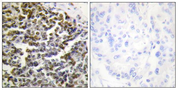
A convincing result shows nuclear, preferably nucleolar, staining in intact cells, consistent with TCOF1 localisation and the reported ubiquitous nuclear and nucleolar tissue profile (UniProt Q13428; HPA tissue IHC; HPA subcellular). Compare the sampled cell type with the relevant tissue annotation: HPA reports high nuclear staining in cerebellar GLUC cells, while chondrocytes in soft tissue were not detected (HPA tissue IHC). Treat dominant membrane staining, edge-enhanced colour and signal confined to necrosis as suspect, then compare no-primary and peroxidase-blocked controls for nonspecific or endogenous-enzyme signal (UniProt Q13428 topology; standard IHC practice). The selected lung carcinoma image includes peptide blocking, but its paraffin caption does not establish a fixative or validate every staining pattern (A02653 tissue-IHC caption).
Boster reagents

Best TCOF1 / Treacle protein IHC Antibodies

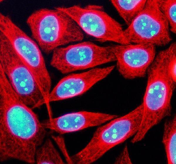
Two anti-TCOF1 antibodies have real IHC or IF images: paraffin-embedded human lung carcinoma tissue (A02653 IHC image caption) and SIHA cells (A02653-2 IF image caption).

Real IHC data Immunohistochemistry analysis of paraffin-embedded human lung carcinoma tissue, using TCOF1 Antibody. The picture on the right is blocked with the synthesized peptide.
Anti-TCOF1/Treacle Antibody
Cat # A02653
Real IF data IF analysis of TCOF1 using anti-TCOF1 antibody (A02653-2) and anti-Beta Tubulin antibody (M01857-3). TCOF1 was detected in an immunocytochemical section of SIHA cells. Enzyme antigen retrieval was performed using IHC enzyme antigen retrieval reagent (AR0022) for 15 mins. The cells were blocked with 10% goat serum. And then incubated with 5 μg/mL rabbit anti-TCOF1 Antibody (A02653-2) and mouse anti-Beta Tubulin antibody (M01857-3) overnight at 4°C. DyLight®488 Conjugated Goat Anti-Rabbit IgG (BA1127) and Cy3 Conjugated Goat Anti-Mouse IgG (BA1031) were used as secondary antibody at 1:500 dilution and incubated for 30 minutes at 37°C. The section was counterstained with DAPI. Visualize using a fluorescence microscope and filter sets appropriate for the label used.
Anti-TCOF1 Antibody ®
Cat # A02653-2

A02653 is listed for human and mouse IHC/IF and has an IHC image of paraffin-embedded human lung carcinoma tissue (A02653 catalog applications/reactivity; A02653 IHC image caption). A02653-2 is listed for human, mouse and rat reactivity and has an IF image of SIHA cells; IHC is not listed (A02653-2 catalog applications/reactivity; A02653-2 IF image caption).

Which to pick: Choose A02653 for tissue IHC: it is listed as a polyclonal IHC antibody, and its image shows paraffin-embedded tissue; the fixative is unreported (A02653 catalog dilution_raw; A02653 IHC image caption). For IF/ICC, both SKUs list those applications, while A02653-2 has the SIHA cell IF image (A02653 and A02653-2 catalog applications; A02653-2 IF image caption). For rat samples, A02653-2 lists rat reactivity, but its pictured IF result is from SIHA cells and it has no listed IHC application (A02653-2 catalog reactivity/applications; A02653-2 IF image caption).

Each figure is that product's own IHC / IF validation image from its datasheet.

References

  1. UniProt Consortium. UniProt entry Q13428 (TCOF_HUMAN, Treacle protein).
  2. Human Protein Atlas. TCOF1 tissue IHC expression (reliability: Enhanced).
  3. Human Protein Atlas. TCOF1 subcellular location (ICC-IF): Mainly localized to the nucleoli fibrillar center..
  4. Human Protein Atlas. TCOF1 antibody validation summary (3 antibodies).
  5. TCOF1 in extracellular vesicles predicts survival in patients with HCC treated with high-dose conformal radiotherapy. JHEP reports : innovation in hepatology 2026 — PMC13264060.
  6. TRPV1 Activation Promotes β-arrestin2 Interaction with the Ribosomal Biogenesis Machinery in the Nucleolus:Implications for p53 Regulation and Neurite Outgrowth. International journal of molecular sciences 2021 — PMC7956682.
  7. The oncogenic role of treacle ribosome biogenesis factor 1 (TCOF1) in human tumors: a pan-cancer analysis. Aging 2022 — PMC8833134.
  8. Construction and validation of a prognostic model for hepatocellular carcinoma: Inflammatory ferroptosis and mitochondrial metabolism indicate a poor prognosis. Frontiers in oncology 2022 — PMC9850107.
  9. PubMed PMID:8563749 — UniProt-cited evidence.
  10. PubMed PMID:9074926 — UniProt-cited evidence.
  11. PubMed PMID:9096354 — UniProt-cited evidence.