This website uses cookies to ensure you get the best experience on our website.


- Table of Contents
TCTN1 is a component of the ciliary transition-zone complex (UniProt). Use this IHC-P guide to plan staining controls and interpret the general cytoplasmic tissue pattern (HPA tissue IHC).

Expected localisation, validated protocols, controls and antibodies — the at-a-glance facts below, then the full design guide.
| Expected localisation | General cytoplasmic staining (HPA tissue IHC) | |
| Staining pattern | Ciliated cells: cytoplasmic staining at ciliary rootlets (HPA tissue IHC) | |
| Antigen retrieval | EDTA pH 8.0 HIER, heat-mediated (datasheet A09350-1) | |
| Positive control | Adrenal gland+4 more · see all | |
| Negative control | Adipose tissue+3 more · see all |
| Fixation | Matched tissue-IHC evidence does not establish the fixation claim. Validate the specimen-specific method before use. (selected-SKU IHC image A09350-1) | |
| Caveat | Antibody staining has low consistency with RNA expression (HPA tissue IHC) | |
| Regulation | Choroid plexus RNA is tissue enhanced (HPA tissue RNA) | |
| Isoform / epitope | 6 isoforms; signal peptide 1–22; no TM-defined epitope side (UniProt) |
The catalog antibody’s IHC-P protocol uses EDTA pH 8.0 retrieval (datasheet A09350-1). One published human GBM tissue-microarray protocol provides a comparison (PMC4198629).
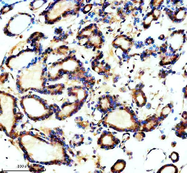
| Sample | Paraffin-embedded human thyroid cancer tissue; fixative not specified (datasheet A09350-1) |
| Fixation | Image fixative and duration unreported (datasheet A09350-1); verify before use. |
| Sectioning | 4–5 µm sections on charged slides (standard) |
| Deparaffinisation | Xylene, graded ethanol series to water (standard) |
| Antigen retrieval | Heat retrieval: EDTA pH 8.0 (datasheet A09350-1); 20 min, 95–100 °C (standard) |
| Peroxidase block | 3% H2O2, 10 min, room temperature (standard) |
| Blocking | 10% goat serum (datasheet A09350-1) |
| Primary antibody | Rabbit anti-TCTN1, 2-5 μg/ml (datasheet A09350-1) |
| Primary incubation | Overnight at 4 °C (datasheet A09350-1) |
| Detection | HRP-conjugated secondary, DAB chromogen (datasheet A09350-1) |
| Counterstain | Hematoxylin, blue, dehydrate and mount (standard) |
| Expected result | TCTN1-positive staining in glandular cells of adrenal gland (HPA tissue IHC: High). HPA tissue profile: General cytoplasmic expression. No signal in the no-primary control. |
TCTN1 localizes to the ciliary transition zone near the basal body and is also annotated in the cytoplasm (UniProt Q2MV58 localization). In tissue IHC, expect generally cytoplasmic staining, with high staining reported in bronchial and fallopian tube ciliated cells, kidney tubule cells, and selected neural cells (HPA tissue IHC). HPA rates its tissue staining Approved but reports low consistency with RNA expression (HPA tissue IHC). TCTN1 has no annotated transmembrane segment (UniProt Q2MV58 topology).
| Cytoplasmic staining in kidney tubule cells or adrenal glandular cells; staining at ciliary rootlets in bronchial or fallopian tube ciliated cells. | These match HPA-reported high tissue patterns (HPA tissue IHC). A sharply resolved rootlet signal supports the expected location, but chromogenic staining may not resolve the transition zone itself (UniProt Q2MV58 localization; general IHC practice). |
| Predominantly nuclear or cell-surface staining, without the expected cytoplasmic or ciliary-region pattern. | Review this as a possible artefact in tissue IHC (HPA tissue IHC; UniProt Q2MV58 localization). Nuclear signal alone is not decisive: nucleoplasm is an additional approved ICC-IF location, so assess the antibody, controls, and tissue context (HPA subcellular ICC-IF). |
| Strong staining in adipocytes, bone-marrow hematopoietic cells, ovarian stroma, or placental decidual cells. | These cell populations are reported as not detected (HPA tissue IHC). Check cell identification and controls; cross-reactivity or endogenous detection activity are possible explanations, not conclusions from staining alone (general IHC practice). |
| Uniform chromogen across unrelated cells, extracellular areas, or the whole section. | This distribution does not resemble HPA's general cytoplasmic profile or its cell-specific examples (HPA tissue IHC). Consider nonspecific detection, inadequate blocking or washing, and endogenous activity before assigning TCTN1 expression (general IHC practice). |
| No staining in a well-preserved bronchus or fallopian tube section containing identifiable ciliated cells. | Those ciliated cells are reported High at ciliary rootlets, so absence warrants a technical check (HPA tissue IHC). It does not alone establish biological absence; inspect section quality, retrieval, antibody conditions, and detection controls (general IHC practice). |
| Cell population and location | HPA reports High staining in ciliated cells, kidney tubules, adrenal glandular cells, selected neurons and glia, and lymph-node non-germinal-center cells; several other populations are Low or Not detected (HPA tissue IHC). Score the named cells rather than treating an entire tissue as uniformly positive. |
| Subcellular resolution | TCTN1 is annotated at the ciliary transition zone near the basal body (UniProt Q2MV58 localization). HPA tissue IHC specifically describes ciliary-rootlet staining in bronchus and fallopian tube (HPA tissue IHC). A broad cytoplasmic pattern can still fit its reported tissue profile. |
| Antibody validation and expression agreement | Two listed rabbit polyclonal antibodies, HPA039687 and HPA040036, have Approved IHC status (HPA antibodies). The overall tissue profile nevertheless has low staining–RNA consistency (HPA tissue IHC); treat unexpected tissue results as provisional until controls or another antibody support them. |
| Protein processing and variants | UniProt annotates a signal peptide at residues 1–22, a mature chain at 23–587, six isoforms, and three glycosylation sites (UniProt Q2MV58 processing, isoforms, glycosylation). Epitope information is absent, so these annotations cannot predict which variants this antibody stains or the effect of retrieval. |
| Secretion annotation and topology | UniProt lists TCTN1 as Secreted but cautions that full-length protein might not be secreted; it annotates no transmembrane segment (UniProt Q2MV58 localization, topology). Do not interpret an isolated extracellular or cell-surface deposit as a confirmed positive without supporting controls. |
| Situation | Likely cause | Next action |
|---|---|---|
| Expected ciliated-cell or kidney-tubule signal is absent. | The positive population may be missing from the section, or the IHC workflow may have failed (HPA tissue IHC; general IHC practice). | Confirm the relevant cells are present; check the positive control, retrieval conditions, antibody dilution, and detection reagents before scoring the section negative (general IHC practice). |
| Strong staining appears in an HPA Not detected population. | Cell misidentification, nonspecific antibody binding, or endogenous detection activity may account for the discrepancy (HPA tissue IHC; general IHC practice). | Recheck morphology and run omission and detection controls; compare the same cell population with an independently validated antibody when available (general IHC practice). |
| The whole section has diffuse chromogen. | Excess background can arise from detection chemistry, insufficient blocking or washing, or excessive antibody concentration (general IHC practice). | Inspect a no-primary control, blocking and washing steps, and the antibody titration; assess specific cells only after the diffuse signal is controlled (general IHC practice). |
| A nuclear-only pattern dominates the IHC slide. | The pattern differs from HPA's general cytoplasmic tissue profile, although nucleoplasm is an approved additional ICC-IF location (HPA tissue IHC; HPA subcellular ICC-IF). | Check tissue and detection controls, then compare cytoplasmic staining and the relevant cell types before assigning TCTN1 to nuclei in this section (general IHC practice). |
| Staining differs between antibodies or from RNA expectations. | HPA explicitly reports low consistency between tissue antibody staining and RNA expression, despite Approved IHC status for HPA039687 and HPA040036 (HPA tissue IHC; HPA antibodies). | Record the antibody and scored cell population; prioritize reproducible localization and controlled staining, and describe unresolved discordance rather than forcing a positive call (general IHC practice). |
| Can IF/ICC help assess a disputed compartment? | HPA approves actin filaments and primary cilium as main ICC-IF locations, with nucleoplasm as an additional location (HPA subcellular ICC-IF). | Use the separate IF/ICC guide for that application; compare its compartment evidence with IHC controls without assuming an IF pattern will be resolved in chromogenic paraffin sections (HPA subcellular ICC-IF; general IHC practice). |


Comprehensive Human Protein Atlas IHC scoring per tissue (reliability: Approved — Low consistency between antibody staining and RNA expression data.). Rows are taken directly from the HPA tissue chart — click any row's HPA link to view the source.
| Tissue | Cell type | Level | Evidence | Source |
|---|---|---|---|---|
| Adrenal gland | Glandular cells | High | Protein (IHC) | HPA → |
| Bronchus | Ciliated cells (ciliary rootlets) | High | Protein (IHC) | HPA → |
| Caudate | Glial cells | High | Protein (IHC) | HPA → |
| Cerebral cortex | Neuronal cells | High | Protein (IHC) | HPA → |
| Fallopian tube | Ciliated cells (ciliary rootlets) | High | Protein (IHC) | HPA → |
| Tissue | Cell type | Level | Evidence | Source |
|---|---|---|---|---|
| Adipose tissue | Adipocytes | Not detected | Protein (IHC) | HPA → |
| Bone marrow | Hematopoietic cells | Not detected | Protein (IHC) | HPA → |
| Ovary | Ovarian stroma cells | Not detected | Protein (IHC) | HPA → |
| Placenta | Decidual cells | Not detected | Protein (IHC) | HPA → |
Troubleshoot TCTN1 staining in paraffin sections by checking retrieval, compartment, controls and scoring before interpreting weak or diffuse signal.
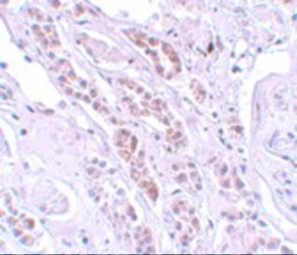
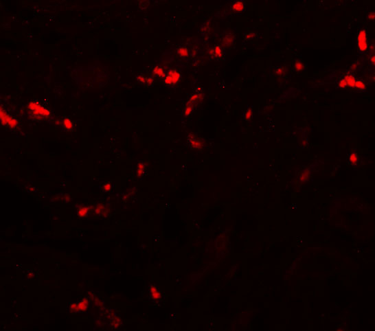
Two anti-TCTN1 antibodies have human tissue IHC images (catalog IHC captions); A09350 also has a human kidney-cell IF image (A09350 IF caption). Catalog reactivity spans human, mouse, and rat (catalog reactivity).
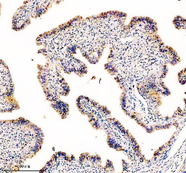
A09350-1 has paraffin-section IHC images from human thyroid cancer and fallopian tube tissue (A09350-1 IHC captions). A09350 has a human kidney IHC image and a human kidney-cell IF image (A09350 IHC and IF captions).
Which to pick: For tissue IHC, choose A09350-1 when you want a documented paraffin-section workflow; its IHC caption specifies EDTA retrieval and 2 μg/ml antibody, but does not report the fixative (A09350-1 IHC caption). For cell IF, choose A09350: IF is listed as an application and its image shows human kidney cells at 20 μg/mL; ICC validation is not separately reported (A09350 applications and IF caption). For cross-species planning, A09350-1 lists human, mouse, and rat reactivity, while A09350 lists human and mouse; the shown IHC evidence is human for both, and clonality is unreported (catalog reactivity and IHC captions; catalog clone fields).