TEK / Angiopoietin-1 receptor · IHC design guide

Design Immunohistochemistry for TEK

Plan chromogenic TEK IHC on paraffin sections using 0.5–1 µg/mL catalog antibody (datasheet A01274-1). Assess cytoplasmic staining in endothelial and other cell types (HPA tissue IHC), accounting for extracellular-domain shedding when interpreting epitope-dependent signal (UniProt).

Evidence assembled Oct 2026 · For research use; verify linked source records and product datasheet before use
Immunohistochemistry protocol sheet for TEK (IHC for TEK): expected localisation Cytoplasmic in several cell types, including endothelium (HPA tissue IHC), antibody A01274-1, validated IHC image, and IHC protocol steps
Printable TEK IHC protocol sheet — expected localisation Cytoplasmic in several cell types, including endothelium (HPA tissue IHC), antibody A01274-1, controls and protocol steps. Open the full TEK IHC guide →

TEK Immunohistochemistry Experimental Design Guide

Expected localisation, validated protocols, controls and antibodies — the at-a-glance facts below, then the full design guide.

Must know before staining
Expected localisation Cytoplasmic in several cell types, including endothelium (HPA tissue IHC)
Staining pattern Cytoplasmic staining in several cell types, including endothelium (HPA tissue IHC)
Antigen retrieval Citrate pH 6 HIER, heat-mediated (datasheet A01274-1)
Positive control ⓘ Testis
Negative control ⓘ Adipose tissue+4 more · see all
Important caveats
Reasons your staining may differ from the expected pattern.
Fixation Keep formalin fixation consistent across sections (standard IHC practice; not target-specific); Matched tissue-IHC evidence does not establish the fixation claim. Validate the specimen-specific method before use. (selected-SKU IHC image A01274-1)
Caveat Shedding can separate tissue staining from cell-bound TEK (UniProt)
Regulation ANGPT1 recruits TEK to cell contacts (UniProt)
Isoform / epitope 3 isoforms; shedding separates extracellular and cytoplasmic epitopes (UniProt)
Section 1

Recommended TEK IHC & IF Protocols

The catalog antibody protocol is accompanied by 2 published TEK IHC protocols (PMC3706492; PMC9097102).

Recommended immunohistochemistry (IHC-P) protocol parameters
SampleParaffin-embedded human lung cancer tissue; fixative not specified (datasheet A01274-1)
FixationImage fixative and duration unreported (datasheet A01274-1); verify before use.
Sectioning4–5 µm sections on charged slides (standard)
DeparaffinisationXylene, graded ethanol series to water (standard)
Antigen retrievalHeat retrieval: Citrate pH 6, 20 min (datasheet A01274-1)
Peroxidase block3% H2O2, 10 min, room temperature (standard)
Blocking10% goat serum (datasheet A01274-1)
Primary antibodyRabbit anti-TEK, 1μg/ml (datasheet A01274-1)
Primary incubationOvernight at 4 °C (datasheet A01274-1)
DetectionStreptavidin-biotin complex (SABC), DAB chromogen (datasheet A01274-1)
CounterstainHematoxylin, blue, dehydrate and mount (standard)
Expected resultTEK-positive staining in elongated or late spermatids of testis (HPA tissue IHC: High). HPA tissue profile: Cytoplasmic expression in several cell types, including endothelial cells. No signal in the no-primary control.
💡Decision noteStart with heat-mediated citrate retrieval at pH 6 (datasheet: A01274-1); one published TEK protocol also uses citrate pH 6 (PMC3706492).
Section 2

What Is the Expected TEK Staining Pattern?

TEK is a single-pass receptor with an extracellular region and cytoplasmic kinase region (UniProt Q02763 topology). Expect staining in endothelial cells, where TEK is predominantly expressed (UniProt Q02763 tissue specificity). HPA reports cytoplasmic staining in several cell types, including endothelial cells, and rates its tissue IHC profile Approved (HPA tissue IHC). Interpret the observed cytoplasmic pattern alongside the receptor’s membrane localization (UniProt Q02763; HPA subcellular).

What am I looking at on my slide?
Vessel-lining cells show discernible membrane-associated staining, with some cytoplasmic staining.This fits TEK’s membrane topology and endothelial expression (UniProt Q02763). Cytoplasmic signal alone does not disqualify a section: HPA reports cytoplasmic staining in several cell types, including endothelial cells (HPA tissue IHC). Judge the pattern against nearby background and the antibody’s control sections.
Strong, widespread nuclear staining dominates the section.A predominantly nuclear pattern does not match the supplied TEK locations: membrane, junctions, cytoskeleton and a shed extracellular form (UniProt Q02763), or HPA’s tissue cytoplasmic pattern (HPA tissue IHC). Treat it as suspect until controls and staining conditions exclude nonspecific detection.
Adipocytes or respiratory epithelial cells stain strongly while vascular profiles are unconvincing.HPA reports TEK as not detected in adipocytes of adipose tissue and respiratory epithelial cells of bronchus (HPA tissue IHC). Strong staining in those cell types warrants checks for cross-reactivity or endogenous detection activity; it does not by itself prove either cause.
Brown signal spreads across unrelated structures without clear cell boundaries.Diffuse chromogen deposition is difficult to score as TEK. Compare it with a primary-antibody omission control and inspect blocking, washing and detection steps (general IHC practice). HPA’s tissue profile describes cellular cytoplasmic expression, not indiscriminate staining throughout a section (HPA tissue IHC).
Late spermatids in testis show no convincing stain.HPA reports High staining in elongated or late spermatids of testis (HPA tissue IHC). Their absence in a matched positive-control section calls the run into question. Check the control’s staining and detection workflow before treating a negative study sample as evidence of absent TEK.
💡Expected TEK appearanceCall a section positive when defined endothelial cells show signal above local background, with membrane-associated or cytoplasmic distribution consistent with TEK and HPA tissue IHC (UniProt Q02763 topology; HPA tissue IHC); testis late spermatids are reported High (HPA tissue IHC), while dominant nuclear or diffuse cell-independent staining is suspect.
How each factor affects the staining
Receptor topology and epitope locationTEK spans the membrane at residues 749–769, with extracellular residues 23–748 and cytoplasmic residues 770–1124 (UniProt Q02763 topology). The antibody epitope is not supplied, so compartment differences cannot be assigned to a particular domain.
Processing and sheddingProteolysis produces a soluble extracellular domain detected in plasma (UniProt Q02763). Extracellular staining away from a cell boundary therefore needs careful interpretation; this record cannot establish whether a tissue deposit represents shed TEK, retained receptor, or background.
Isoforms and antibody scopeUniProt lists 3 TEK isoforms (UniProt Q02763). No epitope or isoform coverage is provided for the working antibody. Avoid inferring isoform-specific staining or using a negative section to rule out every isoform.
Tissue evidence and validationHPA rates the tissue IHC profile Approved and antibody CAB010359 IHC Approved (HPA tissue IHC; HPA antibodies). Those labels support use of the reported patterns but do not supply a TEK-specific retrieval setting, dilution, or evidence that a different antibody will reproduce them.
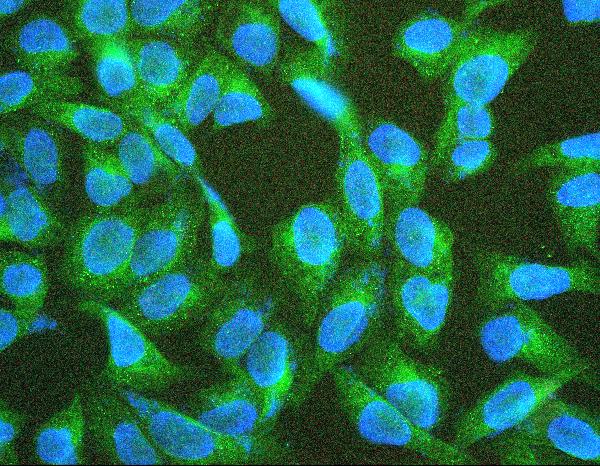
IF/ICC Q: Where should TEK appear?A: HPA supports plasma-membrane localization in ICC-IF; centrosome and basal-body locations are uncertain (HPA subcellular). UniProt also describes recruitment to endothelial cell contacts and focal adhesions (UniProt Q02763). Assess any puncta against those confidence levels; this guide supplies no IF/ICC protocol.
Why is my staining missing, weak or wrong?
SituationLikely causeNext action
The testis positive control is blank.A failed control run is plausible because HPA reports High staining in elongated or late spermatids (HPA tissue IHC); the section alone cannot identify the failed step.Confirm the expected cells are present, then review primary-antibody use, detection reagents and chromogen development against the control run (general IHC practice).
Vascular signal is weak but other cells stain.Endothelial staining is expected from TEK expression (UniProt Q02763), yet HPA describes cytoplasmic expression across several cell types (HPA tissue IHC); the supplied evidence sets no endothelial intensity threshold.Compare vessel-lining cells with adjacent background and a matched control. Score cell identity and compartment before increasing detection intensity (general IHC practice).
Nuclei are the strongest positive compartment.Nuclear localization is unsupported by the supplied TEK localization records (UniProt Q02763; HPA subcellular).Review the primary-antibody omission control and check for nonspecific secondary or chromogen signal; repeat scoring only after cell boundaries are clear (general IHC practice).
Adipocytes or bronchial respiratory epithelium stain strongly.HPA reports those cell types as Not detected (HPA tissue IHC). Cross-reactivity or endogenous detection activity is possible, but neither is established by appearance alone.Compare a primary-antibody omission control; assess endogenous peroxidase blocking for chromogenic detection and verify cell identity (general IHC practice).
The entire section has diffuse brown background.Diffuse signal obscures the cellular pattern HPA reports for TEK (HPA tissue IHC); residual detection activity or insufficient washing can contribute (general IHC practice).Inspect omission controls, blocking, wash steps and chromogen exposure, then reassess whether endothelial outlines remain distinguishable (general IHC practice).
A cytoplasmic signal seems inconsistent with a membrane receptor.TEK has a membrane-spanning segment (UniProt Q02763 topology), while HPA tissue IHC reports cytoplasmic expression, including in endothelial cells (HPA tissue IHC). The records do not resolve every cytoplasmic signal’s origin.Retain the finding as qualified when it is cell-specific and control-supported; do not reclassify all cytoplasmic staining as artifact solely from topology.

Sample controls for TEK IHC & IF

🧪Run testis first and score elongated or late spermatids for staining (HPA: High in elongated or late spermatids). Use adipose tissue adipocytes as the negative tissue (HPA: Not detected in adipocytes); on the testis slide, use morphologically distinct cells that remain at background as an internal negative reference, while assessing vascular endothelium separately (UniProt: TEK is predominantly expressed in endothelial cells).
Positive control tissue: Testis (Elongated or late spermatids, HPA High)
Negative control tissue: Adipose tissue (HPA Not detected)
ICC-IF cell lines (HPA subcellular resource): HPA ICC-IF images show TEK in AF22, BJ [Human fibroblast], U2OS, ASC52telo, RPTEC/TERT1, hTERT-RPE1 (serum starved), with annotated localisation: Plasma membrane (supported) (HPA subcellular).
Technical controls: Include a no-primary, secondary-only control and a concentration-matched rabbit isotype control for the rabbit primary antibody (selected-SKU tissue-IHC caption: rabbit anti-TEK), plus TEK-knockout tissue if available as a biological negative. For chromogenic detection, quench endogenous peroxidase and check endogenous biotin background because the caption uses a streptavidin–biotin complex with DAB (selected-SKU tissue-IHC caption).
⚠️Feasibility: A target-specific fixation window or fixation effect is unreported, and the selected-SKU tissue-IHC caption does not state the fixative (selected-SKU tissue-IHC caption). The reported paraffin-section procedure used citrate buffer at pH 6 with heat retrieval for 20 minutes; this supplies a starting condition, not evidence that retrieval is required for every specimen (selected-SKU tissue-IHC caption). The supplied evidence does not establish whether frozen sections or IF are easier, or identify a testis-specific artefact; assess background with the controls above.

HPA tissue IHC evidence for TEK

Comprehensive Human Protein Atlas IHC scoring per tissue (reliability: Approved — At least one protein variant secreted, tissue location of RNA and protein might differ and correlation is complex.). Rows are taken directly from the HPA tissue chart — click any row's HPA link to view the source.

Positive expression · recommended positive controls

TissueCell typeLevelEvidenceSource
Testis Elongated or late spermatids High Protein (IHC) HPA →

Undetected expression · recommended negative controls

TissueCell typeLevelEvidenceSource
Adipose tissue Adipocytes Not detected Protein (IHC) HPA →
Adrenal gland Glandular cells Not detected Protein (IHC) HPA →
Appendix Glandular cells Not detected Protein (IHC) HPA →
Bone marrow Hematopoietic cells Not detected Protein (IHC) HPA →
Breast Adipocytes Not detected Protein (IHC) HPA →
Section 3

Advanced TEK IHC Tips

Troubleshoot TEK staining in paraffin section IHC by checking retrieval, endothelial localisation, epitope accessibility and how signal is scored.

What retrieval should I use if TEK staining is weak in paraffin sections?
Start with heat mediated retrieval in citrate buffer at pH 6 for 20 minutes (datasheet A01274-1). That condition accompanies TEK staining in a paraffin section of human lung cancer tissue; the caption does not state the fixative (datasheet A01274-1). Keep section thickness, heating and cooling consistent across slides, then compare endothelial signal with a slide processed in the same run (standard IHC practice; UniProt Q02763 tissue specificity). If signal remains weak, optimise heating time on matched sections before considering a different retrieval buffer as a fallback (standard IHC practice). Excessive retrieval can impair morphology, so assess vessel structure alongside staining (standard IHC practice).
Could fixation explain inconsistent TEK staining between paraffin sections?
TEK specific sensitivity to fixation is unknown from the supplied evidence; the selected paraffin section caption does not report its fixative (datasheet A01274-1). Record each specimen’s fixative and fixation duration, and compare sections processed under matched conditions before attributing staining differences to TEK abundance (standard IHC practice). Use intact vessel morphology and a consistent endothelial staining reference to judge whether a run is technically interpretable (standard IHC practice; UniProt Q02763 tissue specificity). If signal varies, review retrieval, section age and detection conditions alongside fixation records (standard IHC practice). Do not infer a TEK specific fixation effect from tissue staining patterns (HPA tissue IHC).
Where should convincing TEK staining appear in a paraffin section?
Prioritise staining associated with endothelial cells lining vessels, because TEK is predominantly expressed in endothelial cells and their progenitors (UniProt Q02763 tissue specificity). Membrane and junctional signal is biologically plausible, including recruitment to cell contacts in quiescent endothelial cells (UniProt Q02763 subcellular location; HPA subcellular). Cytoplasmic staining can also be plausible: tissue IHC reports cytoplasmic expression in several cell types, including endothelial cells (HPA tissue IHC). Judge each pattern against vessel morphology, counterstain and a control section processed in the same run (standard IHC practice). Diffuse signal without a recognisable cellular pattern warrants checks of background and detection conditions before interpretation (standard IHC practice).
How does the antibody epitope affect interpretation of TEK staining?
Check the antibody’s documented immunogen or epitope before assigning a stained compartment to full length TEK (standard IHC practice). TEK has an extracellular region at residues 23–748, a transmembrane segment at 749–769 and a cytoplasmic region at 770–1124 (UniProt Q02763 topology). Proteolytic processing releases a soluble extracellular domain, so an extracellular epitope may detect material beyond membrane bound receptor (UniProt Q02763 processing and subcellular location). Three isoforms are listed, but the supplied evidence does not establish which ones the catalog antibody recognises (UniProt Q02763 isoforms; datasheet A01274-1). Interpret discordant patterns only after confirming epitope coverage and staining controls (standard IHC practice).
How should I assess TEK by multiplex IF alongside this IHC guide?
For multiplex IF, pair TEK with an independently validated endothelial marker and inspect whether signals occur in the same vessel lining cells (UniProt Q02763 tissue specificity; standard IF practice). Choose fluorophores after measuring tissue autofluorescence, favouring a spectrally separated channel with adequate contrast for the weaker signal (standard IF practice). An extracellular epitope may be accessible without permeabilisation, whereas an epitope in the cytoplasmic region at residues 770–1124 generally requires permeabilisation (UniProt Q02763 topology; standard IF practice). Confirm the antibody’s epitope before choosing that step, and include single stain and no primary controls for channel bleed through and background (standard IF practice). Assess membrane and junctional signal alongside cytoplasmic signal (HPA subcellular; HPA tissue IHC).
How can I reduce diffuse brown staining that obscures endothelial TEK?
First inspect a no primary control to separate detection background from antibody dependent staining (standard IHC practice). The selected paraffin section example used 10% goat serum block, 1 µg/mL primary antibody overnight at 4°C, a biotinylated secondary and DAB detection (datasheet A01274-1). Check endogenous peroxidase blocking, washing and reagent exposure when DAB appears outside identifiable cells (standard chromogenic IHC practice). Because the documented detection uses a biotin based complex, assess endogenous biotin background with an appropriate control if unexplained signal persists (datasheet A01274-1; standard IHC practice). Optimise antibody concentration against endothelial signal while preserving morphology and a clean no primary control (standard IHC practice; UniProt Q02763 tissue specificity).
How should I quantify TEK IHC across samples with different vascularity? ⚠ ANSWER MARKED FOR VERIFICATION
Define the analysis compartment before scoring: identify endothelial cells along vessels, then record the percentage positive and staining intensity using a prespecified H-score (UniProt Q02763 tissue specificity; standard IHC scoring practice). Report positive vessel density per mm² or positive endothelial cells as a fraction of all assessed endothelial cells when vascularity differs (standard IHC quantification practice). Keep threshold, region selection, counterstain and imaging settings consistent across slides (standard IHC quantification practice). Exclude folds, necrosis and tissue edges from the scored area, and document those exclusions (standard IHC practice). Report the endothelial denominator alongside the TEK score so altered vessel abundance is visible (standard IHC quantification practice).
How do I distinguish true TEK staining from artefact?
A convincing positive follows identifiable vessel lining endothelial cells and may show membrane, junctional or cytoplasmic staining (UniProt Q02763 tissue specificity and subcellular location; HPA tissue IHC). Check whether apparent signal is confined to section edges, folds or necrotic areas, which can produce misleading staining patterns (standard IHC practice). Diffuse brown deposits outside intact cells should prompt review of the no primary control and endogenous peroxidase blocking (standard chromogenic IHC practice). Do not treat all extravascular signal as artefact: TEK can yield a secreted extracellular domain, while tissue IHC also reports cytoplasmic staining in several cell types (UniProt Q02763 processing; HPA tissue IHC). Use morphology, controls and epitope information together before assigning a biological interpretation (standard IHC practice).
Boster reagents

Best TEK / Angiopoietin-1 receptor IHC Antibodies

Anti-TEK antibodies have IHC images from human, mouse and rat paraffin sections; one also has an IF/ICC image from U20S cells (catalog IHC and IF captions).

Real IHC data IHC analysis of TEK using anti-TEK antibody (A01274-1). TEK was detected in paraffin-embedded section of human lung cancer tissue. Heat mediated antigen retrieval was performed in citrate buffer (pH6, epitope retrieval solution) for 20 mins. The tissue section was blocked with 10% goat serum. The tissue section was then incubated with 1μg/ml rabbit anti-TEK Antibody (A01274-1) overnight at 4°C. Biotinylated goat anti-rabbit IgG was used as secondary antibody and incubated for 30 minutes at 37°C. The tissue section was developed using Strepavidin-Biotin-Complex (SABC)(Catalog # SA1022) with DAB as the chromogen.
Anti-TIE2/TEK Antibody ®
Cat # A01274-1
Real IHC data IHC analysis of TEK using anti-TEK antibody (A01274-2). TEK was detected in paraffin-embedded section of rat lung tissues. Heat mediated antigen retrieval was performed in citrate buffer (pH6, epitope retrieval solution) for 20 mins. The tissue section was blocked with 10% goat serum. The tissue section was then incubated with 1μg/ml rabbit anti-TEK Antibody (A01274-2) overnight at 4°C. Biotinylated goat anti-rabbit IgG was used as secondary antibody and incubated for 30 minutes at 37°C. The tissue section was developed using Strepavidin-Biotin-Complex (SABC)(Catalog # SA1022) with DAB as the chromogen.
Anti-TIE2/TEK Antibody ®
Cat # A01274-2

A01274-1 has IHC images from human lung cancer, mammary cancer and placenta sections, and mouse heart sections (A01274-1 IHC captions). A01274-2 has IHC images from rat lung and intestine and human tonsil and ovarian cancer sections, plus an IF/ICC image from U20S cells (A01274-2 IHC and IF captions).

Which to pick: For paraffin-section IHC, choose the SKU with an image closest to your sample: A01274-1 for its human or mouse examples, or A01274-2 for its human or rat examples (respective IHC captions); the fixative is unreported in those captions. For IF/ICC, choose A01274-2 because it has an IF/ICC application listing and cell image (A01274-2 catalog and IF caption). Both list human, mouse and rat reactivity for cross-species work; both are rabbit antibodies, and neither has a reported clone (catalog entries).

Each figure is that product's own IHC / IF validation image from its datasheet.

References

  1. UniProt Consortium. UniProt entry Q02763 (TIE2_HUMAN, Angiopoietin-1 receptor).
  2. Human Protein Atlas. TEK tissue IHC expression (reliability: Approved).
  3. Human Protein Atlas. TEK subcellular location (ICC-IF): Mainly localized to the plasma membrane. In addition localized to the centrosome and basal body..
  4. Human Protein Atlas. TEK antibody validation summary (2 antibodies).
  5. Expression of angiopoietin-TIE system components in angiosarcoma. Modern pathology : an official journal of the United States and Canadian Academy of Pathology, Inc 2013 — PMC3706492.
  6. Multi-omics analysis revealed TEK and AXIN2 are potential biomarkers in multifocal papillary thyroid cancer. Cancer cell international 2022 — PMC9097102.
  7. Iatrogenic Leptomeningeal Carcinomatosis Following Craniotomy for Resection of Metastatic Serous Ovarian Carcinoma: A Systematic Literature Review and Case Report. Frontiers in surgery 2022 — PMC9082594.
  8. Non-invasive Imaging and Modeling of Liver Regeneration After Partial Hepatectomy. Frontiers in physiology 2019 — PMC6652107.
  9. PubMed PMID:8382358 — UniProt-cited evidence.
  10. PubMed PMID:14702039 — UniProt-cited evidence.
  11. PubMed PMID:15164053 — UniProt-cited evidence.