This website uses cookies to ensure you get the best experience on our website.


- Table of Contents
Plan chromogenic TFCP2L1 IHC around the reported nucleolar tissue pattern (HPA tissue IHC). Use duodenal glandular cells as a positive reference and adipocytes as a negative reference, while interpreting staining cautiously because it shows low consistency with RNA expression (HPA tissue IHC).

Expected localisation, validated protocols, controls and antibodies — the at-a-glance facts below, then the full design guide.
| Expected localisation | Nucleolar staining reported in several tissues, including kidney (HPA tissue IHC) | |
| Staining pattern | Nucleolar signal in kidney tubule cells (HPA tissue IHC) | |
| Antigen retrieval | Tris-EDTA pH 9.0 HIER, 95–98 °C, 20 min (rule: nuclear antigen) | |
| Positive control | Duodenum+4 more · see all | |
| Negative control | Adipose tissue+4 more · see all |
| Fixation | Matched tissue-IHC evidence does not establish the fixation claim. Validate the specimen-specific method before use. (selected-SKU IHC image A08278) | |
| Caveat | Staining and RNA expression show low consistency (HPA tissue IHC) | |
| Regulation | Kidney and salivary RNA is tissue enhanced (HPA tissue RNA) | |
| Isoform / epitope | No isoforms or processing reported; one 1–479 chain (UniProt) |
Compare the catalog antibody’s IHC-P protocol with the published breast tissue FFPE protocol (PMC13562584).
| Sample | Tissue sections; selected-image fixative not specified (standard IHC workflow) |
| Fixation | Image fixative and duration unreported (datasheet A08278); verify before use. |
| Sectioning | 4–5 µm sections on charged slides (standard) |
| Deparaffinisation | Xylene, graded ethanol series to water (standard) |
| Antigen retrieval | Heat-induced epitope retrieval in Tris-EDTA buffer, pH 9.0, 20 min at 95–98 °C (standard rule: nuclear antigen) |
| Peroxidase block | 3% H2O2, 10 min, room temperature (standard) |
| Blocking | 10% normal serum of the secondary host, 30 min, room temperature (standard) |
| Primary antibody | Rabbit anti-TFCP2L1, 2.5 μg/mL (datasheet A08278) |
| Primary incubation | Overnight at 4 °C (standard) |
| Detection | HRP-polymer secondary, DAB chromogen 5–10 min (standard) |
| Counterstain | Hematoxylin, blue, dehydrate and mount (standard) |
| Expected result | TFCP2L1-positive staining in glandular cells of duodenum (HPA tissue IHC: High). HPA tissue profile: Nucleolar expression in several tissues, including kidney. No signal in the no-primary control. |
TFCP2L1 is a nuclear protein with no transmembrane segment (UniProt Q9NZI6). In paraffin-section IHC, expect staining mainly within nuclei of selected epithelial and glandular cells, including duodenal glandular cells and esophageal squamous epithelial cells (HPA tissue IHC). HPA describes a nucleolar pattern in several tissues, including kidney, but rates tissue IHC reliability uncertain because antibody staining has low consistency with RNA expression (HPA tissue IHC).
| Discrete nuclear or nucleolar staining in duodenal glandular cells or esophageal squamous epithelial cells. | Consistent with HPA's high staining observations in those cells and its nucleolar tissue profile (HPA tissue IHC). Assess the pattern in the named cells; HPA rates overall tissue IHC reliability uncertain (HPA tissue IHC). |
| Strong, widespread membrane staining or predominantly diffuse cytoplasmic staining. | Treat as a suspect compartment pattern: UniProt places TFCP2L1 in the nucleus and reports no transmembrane segment (UniProt Q9NZI6). Check antibody specificity and detection controls before scoring it as positive (standard IHC practice). |
| Staining concentrated in cells outside the expected cell population, such as adipocytes in adipose tissue. | HPA reports adipocytes as not detected (HPA tissue IHC). Consider cross-reactivity or endogenous detection activity; compare a no-primary control and the expected positive cell population (standard IHC practice). |
| Uniform haze or precipitate across tissue and empty areas of the section. | This distribution is difficult to assign to TFCP2L1-positive cells. Review blocking, washes, chromogen development and no-primary controls for nonspecific background (standard IHC practice); HPA's tissue profile is cell-selective (HPA tissue IHC). |
| No staining in duodenal glandular cells despite an intact, readable section. | HPA reports high staining there, so check the staining run, antigen retrieval and antibody conditions (HPA tissue IHC; standard IHC practice). A failed result does not establish target absence, especially given HPA's uncertain IHC reliability (HPA tissue IHC). |
| Compartment and image resolution | UniProt assigns TFCP2L1 to the nucleus; HPA describes nucleolar tissue staining (UniProt Q9NZI6; HPA tissue IHC). HPA ICC-IF refines its approved main location to the nucleoli fibrillar center, a detail that routine chromogenic IHC may not resolve (HPA subcellular; standard IHC practice). |
| Cell and tissue selection | HPA records high staining in duodenal glandular and esophageal or vaginal squamous epithelial cells, and medium staining in kidney tubule cells and salivary glandular cells (HPA tissue IHC). Compare named cell populations, not whole-section color (standard IHC practice). |
| Strength of tissue evidence | HPA labels tissue IHC uncertain because staining and RNA expression have low consistency; its RNA specificity is tissue enhanced in kidney and salivary gland (HPA tissue IHC). Treat staining levels as observed patterns rather than independently confirmed expression in every specimen. |
| Antibody validation by application | HPA029708 is rated uncertain for IHC and approved for ICC (HPA antibodies). HPA ICC-IF reports mitochondrial localization as uncertain (HPA subcellular). Neither ICC rating nor that additional location validates mitochondrial staining in paraffin-section IHC. |
| Protein topology and processing | UniProt lists one chain spanning residues 1–479, no signal peptide or propeptide, no transmembrane segment, and no annotated glycosylation sites (UniProt Q9NZI6). Those entries provide no basis to expect a surface or secreted IHC pattern. |
| Retrieval and detection controls | Antigen retrieval and detection conditions can affect paraffin-section staining in general (standard IHC practice). The supplied UniProt and HPA records give no TFCP2L1-specific fixation sensitivity or retrieval setting; assess each run against tissue and no-primary controls. |
| Situation | Likely cause | Next action |
|---|---|---|
| Expected positive cells show little or no nuclear signal. | The run may have inadequate retrieval or detection, or the tissue result may differ from HPA's uncertain IHC profile (standard IHC practice; HPA tissue IHC). | Check section integrity and run controls, then review retrieval and antibody conditions using duodenal glandular cells as an observed high reference (standard IHC practice; HPA tissue IHC). |
| Membrane staining dominates the section. | This conflicts with nuclear localization and the absence of a transmembrane segment (UniProt Q9NZI6); nonspecific antibody or detection staining is possible (standard IHC practice). | Inspect a no-primary control and the nuclear pattern in the expected cells before assigning a positive score (standard IHC practice; HPA tissue IHC). |
| Adipocytes stain as strongly as the proposed positive cells. | HPA reports adipocytes in adipose tissue as not detected; cross-reactivity or endogenous detection activity is possible (HPA tissue IHC; standard IHC practice). | Compare no-primary staining, review blocking and chromogen conditions, and score the named cell populations separately (standard IHC practice). |
| The entire slide has diffuse brown background. | Nonspecific binding, endogenous detection activity or excessive chromogen development can obscure cellular localization (standard IHC practice). | Check the no-primary control; review blocking, wash steps and development time, then reassess nuclear signal above background (standard IHC practice; UniProt Q9NZI6). |
| Kidney tubule cells stain, but the pattern is hard to classify. | HPA reports medium kidney tubule staining and a nucleolar tissue profile, while overall tissue IHC reliability remains uncertain (HPA tissue IHC). | Score tubule cells and subcellular distribution separately; compare run controls and avoid treating an unresolved nucleolar detail as a failed chromogenic stain (standard IHC practice; HPA tissue IHC). |
| An ICC-IF image suggests mitochondrial signal that the IHC slide lacks. | HPA calls mitochondria an uncertain additional ICC-IF location; its approved main ICC-IF location is the nucleoli fibrillar center (HPA subcellular). | Interpret paraffin-section IHC using its nuclear or nucleolar tissue pattern and IHC controls; do not require mitochondrial signal for a positive call (HPA tissue IHC; HPA subcellular; standard IHC practice). |
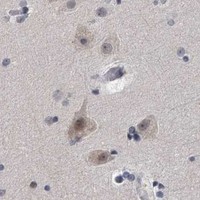
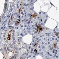
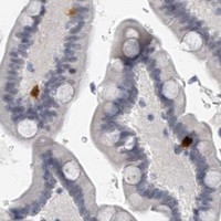
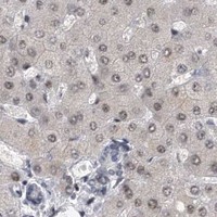
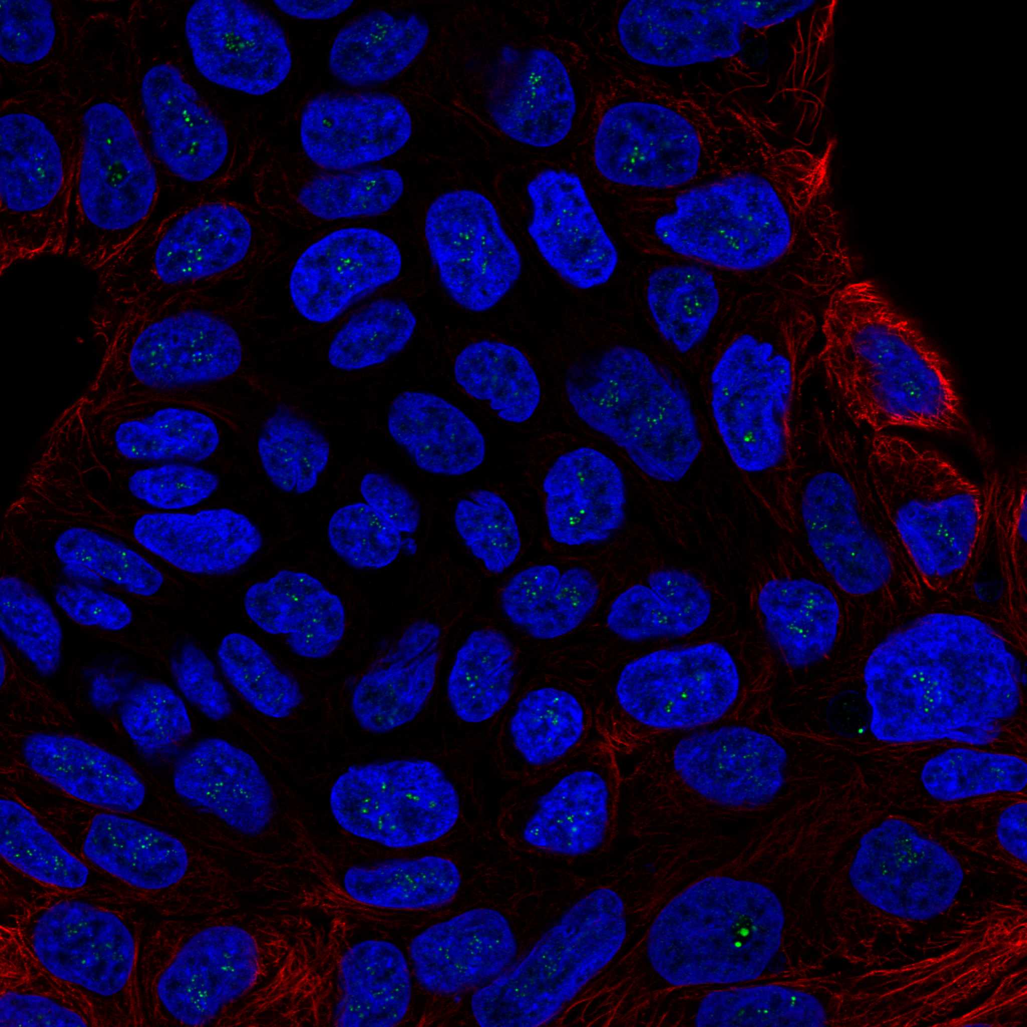
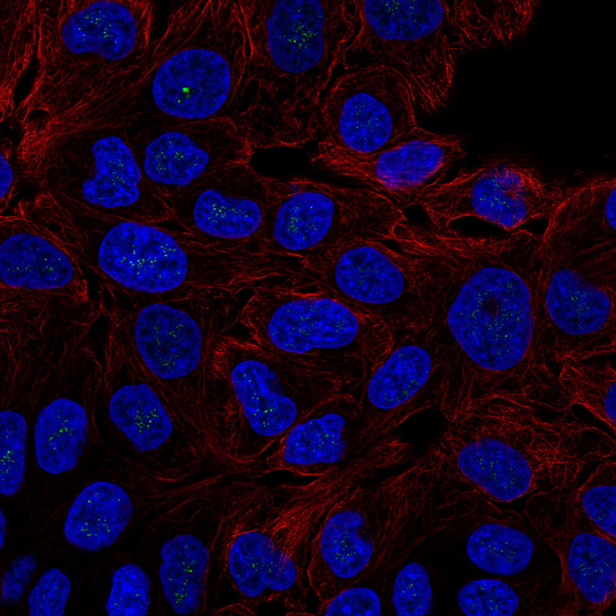
Comprehensive Human Protein Atlas IHC scoring per tissue (reliability: Uncertain — Low consistency between antibody staining and RNA expression data.). Rows are taken directly from the HPA tissue chart — click any row's HPA link to view the source.
| Tissue | Cell type | Level | Evidence | Source |
|---|---|---|---|---|
| Duodenum | Glandular cells | High | Protein (IHC) | HPA → |
| Esophagus | Squamous epithelial cells | High | Protein (IHC) | HPA → |
| Vagina | Squamous epithelial cells | High | Protein (IHC) | HPA → |
| Colon | Glandular cells | Medium | Protein (IHC) | HPA → |
| Kidney | Cells in tubules | Medium | Protein (IHC) | HPA → |
| Tissue | Cell type | Level | Evidence | Source |
|---|---|---|---|---|
| Adipose tissue | Adipocytes | Not detected | Protein (IHC) | HPA → |
| Bone marrow | Hematopoietic cells | Not detected | Protein (IHC) | HPA → |
| Breast | Adipocytes | Not detected | Protein (IHC) | HPA → |
| Bronchus | Respiratory epithelial cells | Not detected | Protein (IHC) | HPA → |
| Cervix | Glandular cells | Not detected | Protein (IHC) | HPA → |
Use nuclear staining, tissue context, and matched controls to troubleshoot TFCP2L1 chromogenic IHC (UniProt Q9NZI6: nucleus; HPA tissue IHC: uncertain reliability).
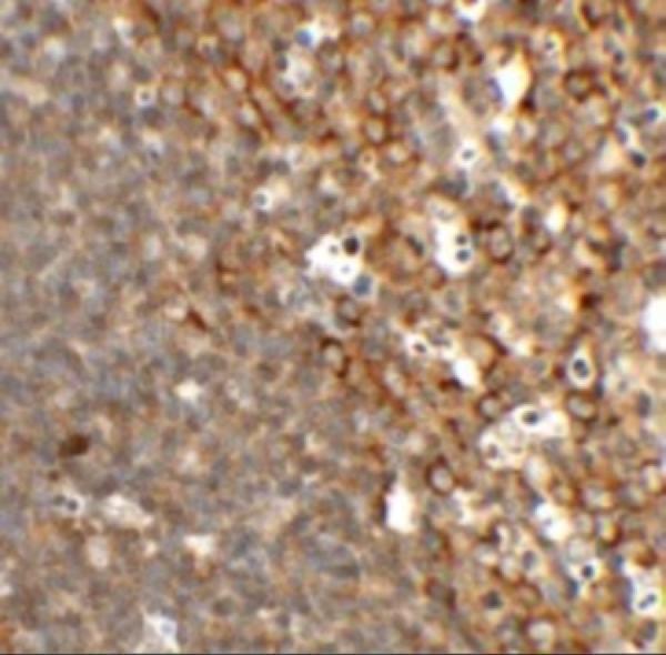
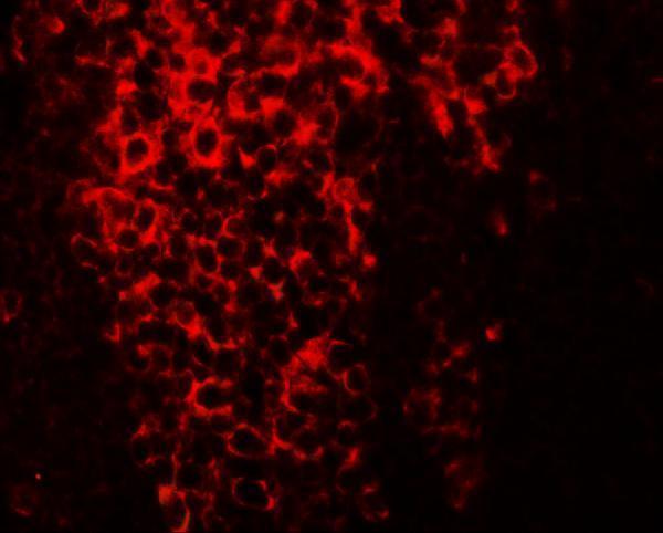
A08278 has rat-colon IHC and IF images (A08278 image captions) and lists human, mouse, and rat reactivity (catalog: A08278 reactivity).
A08278 lists IHC-P and shows rat-colon IHC at 2.5 μg/mL (catalog: A08278 applications; A08278 IHC caption). It also lists IF and shows rat-colon IF at 20 μg/mL (catalog: A08278 applications; A08278 IF caption).
Which to pick: For paraffin-section tissue IHC, choose A08278: it lists IHC-P and has its own rat-colon IHC image at 2.5 μg/mL; the fixative is unreported (catalog: A08278 applications; A08278 IHC caption). For IF, A08278 has a rat-colon image at 20 μg/mL; ICC-specific validation is unreported (A08278 IF caption; catalog: A08278 applications). For cross-species work, A08278 lists human, mouse, and rat reactivity, though its shown IHC and IF images are from rat colon; clonality is unreported (catalog: A08278 reactivity and clone; A08278 image captions).