THY1 / Thy-1 membrane glycoprotein · IHC design guide

Design Immunohistochemistry for THY1

Plan THY1 chromogenic IHC on paraffin sections using reported tissue staining to choose controls (HPA tissue IHC). Interpret the predominantly cytoplasmic CNS and peripheral-nerve staining alongside THY1's annotated cell-membrane location (HPA tissue IHC; UniProt).

Evidence assembled Oct 2026 · For research use; verify linked source records and product datasheet before use
Immunohistochemistry protocol sheet for THY1 (IHC for THY1): expected localisation Cytoplasmic tissue staining; molecularly at the cell membrane (HPA tissue IHC; UniProt), antibody M01818, validated IHC image, and IHC protocol steps
Printable THY1 IHC protocol sheet — expected localisation Cytoplasmic tissue staining; molecularly at the cell membrane (HPA tissue IHC; UniProt), antibody M01818, controls and protocol steps. Open the full THY1 IHC guide →

THY1 Immunohistochemistry Experimental Design Guide

Expected localisation, validated protocols, controls and antibodies — the at-a-glance facts below, then the full design guide.

Must know before staining
Expected localisation Cytoplasmic tissue staining; molecularly at the cell membrane (HPA tissue IHC; UniProt)
Staining pattern Cytoplasmic staining is most abundant in CNS and peripheral nerves (HPA tissue IHC)
Antigen retrieval Citrate pH 6.0 HIER, 95–98 °C, 20 min (rule: cytoplasmic / membrane antigen)
Positive control ⓘ Caudate+3 more · see all
Negative control ⓘ Adipose tissue+4 more · see all
Important caveats
Reasons your staining may differ from the expected pattern.
Fixation Keep paraffin-section fixation consistent (standard IHC practice; not target-specific); Matched tissue-IHC evidence does not establish the fixation claim. Validate the specimen-specific method before use. (selected-SKU IHC image M01818)
Caveat Cytoplasmic IHC staining may differ from membrane localisation (HPA tissue IHC; UniProt)
Regulation Expression regulation is unannotated (UniProt)
Isoform / epitope 0 isoforms; mature chain 20–130 has no cytoplasmic tail (UniProt)
Section 1

Recommended THY1 IHC & IF Protocols

The catalog antibody’s IHC-P protocol is accompanied by three published THY1 IHC protocols with usable preparation or staining details (PMC5228576; PMC5531053; PMC8770266).

Recommended immunohistochemistry (IHC-P) protocol parameters
SampleParaffin-embedded human tonsil tissue; fixative not specified (datasheet M01818)
FixationImage fixative and duration unreported (datasheet M01818); verify before use.
Sectioning4–5 µm sections on charged slides (standard)
DeparaffinisationXylene, graded ethanol series to water (standard)
Antigen retrievalHeat-induced epitope retrieval in citrate buffer, pH 6.0, 20 min at 95–98 °C (standard rule: cytoplasmic / membrane antigen)
Peroxidase block3% H2O2, 10 min, room temperature (standard)
Blocking10% normal serum of the secondary host, 30 min, room temperature (standard)
Primary antibodyRabbit monoclonal (clone CEB-20) anti-THY1, 1:50 (datasheet M01818)
Primary incubationOvernight at 4 °C (standard)
DetectionHRP-polymer secondary, DAB chromogen 5–10 min (standard)
CounterstainHematoxylin, blue, dehydrate and mount (standard)
Expected resultTHY1-positive staining in neuronal cells of caudate (HPA tissue IHC: Medium). HPA tissue profile: Cytoplasmic expression most abundant in CNS and peripheral nerves. No signal in the no-primary control.
💡Decision noteStart with citrate pH 6.0 retrieval at 95–98 °C for 20 min (page antigen-retrieval specification); use the reported unmasking buffer when reproducing PMC5531053.
Section 2

What Is the Expected THY1 Staining Pattern?

THY1 is GPI-anchored and lacks a transmembrane segment (UniProt P04216). CNS neuronal and neuropil IHC staining has Enhanced reliability (HPA).

What am I looking at on my slide?
Medium neuronal or neuropil staining in CNS (HPA tissue IHC).Fits the reported cytoplasmic CNS pattern (HPA tissue IHC).
Medium proximal-tubule microvillus staining in kidney (HPA tissue IHC).Fits the reported kidney pattern (HPA tissue IHC).
Nuclear-only chromogen in a tissue section.Possible IHC artifact; tissue IHC reports mainly cytoplasm (HPA tissue IHC).
Adipocytes stain in adipose tissue.Off-pattern; assess cross-reactivity or endogenous activity (HPA tissue IHC; general IHC practice).
Diffuse brown staining or no signal in caudate neurons.Suspect background or a failed run; caudate neurons stain Medium (HPA tissue IHC; general IHC practice).
💡Expected THY1 appearanceMedium CNS neuronal cytoplasm/neuropil is expected (HPA IHC); diffuse brown suggests background (general IHC practice).
How each factor affects the staining
Membrane attachment (UniProt P04216).THY1 is GPI-anchored and lacks a transmembrane segment (UniProt P04216).
Protein processing (UniProt P04216).The mature chain spans residues 20–130; antibody epitope is unspecified (UniProt P04216).
IHC antibody validation (HPA antibodies).HPA003733 is Enhanced; CAB068243 and CAB068244 are Supported (HPA antibodies).
IF/ICC Q: Where is THY1 seen?A: Mainly nucleoplasm, also plasma membrane (HPA ICC-IF); this is a separate assay.
Why is my staining missing, weak or wrong?
SituationLikely causeNext action
No caudate neuronal signal (HPA: Medium).Retrieval or detection failure is possible (general IHC practice).Check the positive run control, retrieval and detection steps (general IHC practice).
Diffuse brown background across the section.Inadequate blocking or washing is possible (general IHC practice).Review blocking, washes and chromogen development (general IHC practice).
Nuclear-only IHC staining.Possible artifact; tissue IHC reports mainly cytoplasm (HPA tissue IHC).Compare with an HPA positive tissue pattern and run controls (HPA tissue IHC; general IHC practice).
Adipocytes stain in adipose tissue (HPA: Not detected).Cross-reactivity or endogenous detection activity is possible (general IHC practice).Check omission and detection controls (general IHC practice).
Neuropil signal is obscured by heavy counterstain (HPA tissue IHC).Excess counterstain can hide weak detail (general IHC practice).Reduce counterstain and compare with the positive control (general IHC practice).

Sample controls for THY1 IHC & IF

🧪Run caudate first and score neuronal cells for staining (HPA: Medium in caudate neuronal cells). Use adipose tissue adipocytes as the negative comparator (HPA: Not detected in adipocytes); on the caudate slide, record morphologically identified cells without specific staining as internal negatives, without assuming that every non-neuronal cell lacks THY1 (HPA: caudate row specifies neuronal cells only).
Positive control tissue: Caudate (Neuronal cells, HPA Medium)
Negative control tissue: Adipose tissue (HPA Not detected)
ICC-IF cell lines (HPA subcellular resource): HPA ICC-IF images show THY1 in PC-3, U-251MG, with annotated localisation: Nucleoplasm (approved) (HPA subcellular).
Technical controls: Include a no-primary, secondary-only slide and an isotype control matched to the primary antibody’s host species and immunoglobulin class or subclass; compare both with the stained section under identical detection conditions (standard IHC practice). A THY1 knockout specimen is a stronger biological negative where available (standard IHC practice); quench endogenous peroxidase for chromogenic detection and assess brain pigment before scoring caudate staining (standard IHC practice).
⚠️Feasibility: No target-specific fixation window or fixation effect is reported in the supplied evidence, and the fixative is unreported in the selected M01818 paraffin-section caption (M01818 caption). Antigen retrieval dependence is unreported, so optimize retrieval on the caudate positive control alongside the negative comparator (standard IHC practice; HPA: Medium in caudate neuronal cells; HPA: Not detected in adipocytes). THY1 is annotated at the cell membrane with a GPI anchor (UniProt P04216 topology and keywords), but the supplied evidence does not establish that frozen sections or IF are easier; brain pigment can complicate interpretation, especially through autofluorescence in IF (standard IHC/IF practice).

HPA tissue IHC evidence for THY1

Comprehensive Human Protein Atlas IHC scoring per tissue (reliability: Enhanced — Medium consistency between antibody staining and RNA expression data. External characterization data supports antibody staining.). Rows are taken directly from the HPA tissue chart — click any row's HPA link to view the source.

Positive expression · recommended positive controls

TissueCell typeLevelEvidenceSource
Caudate Neuronal cells Medium Protein (IHC) HPA →
Cerebral cortex Neuropil Medium Protein (IHC) HPA →
Hippocampus Neuronal cells Medium Protein (IHC) HPA →
Kidney Proximal tubules (microvilli) Medium Protein (IHC) HPA →

Undetected expression · recommended negative controls

TissueCell typeLevelEvidenceSource
Adipose tissue Adipocytes Not detected Protein (IHC) HPA →
Adrenal gland Glandular cells Not detected Protein (IHC) HPA →
Appendix Glandular cells Not detected Protein (IHC) HPA →
Bone marrow Hematopoietic cells Not detected Protein (IHC) HPA →
Breast Adipocytes Not detected Protein (IHC) HPA →
Section 3

Advanced THY1 IHC Tips

Troubleshoot THY1 staining in paraffin sections by checking retrieval, tissue morphology, cellular location, and matched controls before scoring chromogenic signal.

How should I optimize retrieval when THY1 staining is weak?
Start with citrate buffer at pH 6.0 for heat-induced retrieval at 95–98 °C for 20 minutes (page retrieval rule). Cool sections consistently, then compare a short series of retrieval times on adjacent sections while keeping antibody concentration and detection unchanged (standard IHC practice). THY1 has an extracellular Ig-like domain, glycosylation sites, and a GPI anchor, so assess both staining and tissue preservation rather than assuming stronger heating improves specificity (UniProt P04216 topology and processing; standard IHC practice). If staining remains weak, test another retrieval buffer as a documented fallback alongside the citrate condition and a no-primary control (standard IHC practice).
Could fixation explain inconsistent THY1 staining between paraffin blocks?
Record the fixative, fixation duration, processing schedule, and section age for each block before comparing THY1 staining (standard IHC practice). Target-specific fixation sensitivity is unknown here: the catalog antibody’s paraffin-section caption does not state a fixative, and neither the tissue profile nor THY1 topology establishes a fixation effect (M01818 caption; HPA tissue IHC; UniProt P04216 topology). Compare matched blocks using the same pH 6.0 citrate retrieval and identical detection conditions, then assess morphology alongside signal (page retrieval rule; standard IHC practice). If one block underperforms, report the processing difference as a possible confounder rather than a demonstrated THY1-specific failure (standard IHC practice).
Where should convincing THY1 chromogenic staining appear?
Evaluate staining at cell boundaries and along recognizable processes because THY1 is a cell-membrane, GPI-anchored protein without a transmembrane segment (UniProt P04216 topology and keywords). Tissue IHC can also show broader apparent cytoplasmic staining, especially in CNS and peripheral nerves, so interpret the pattern within preserved anatomy (HPA tissue IHC). HPA reports medium staining in caudate neuronal cells, cerebral-cortex neuropil, hippocampal neuronal cells, and kidney proximal-tubule microvilli (HPA tissue IHC). Compare these compartment patterns with a no-primary section and neighboring negative structures; diffuse precipitate without cellular boundaries needs further specificity checks (standard IHC practice).
How do THY1 processing and glycosylation affect epitope interpretation?
THY1 is synthesized as a 161-amino-acid precursor, with a signal peptide at residues 1–19 and a mature chain at 20–130 (UniProt P04216 processing). Its Ig-like V-type domain spans 20–126, and reported glycosylation sites are 42, 79, 119, and 139 (UniProt P04216 domains and glycosylation). Check the antibody’s stated immunogen or epitope before attributing a staining difference to glycosylation or processing; no epitope mapping is supplied here (standard IHC practice; supplied product evidence). No isoforms are listed, so do not explain discordant tissue patterns by invoking an undocumented THY1 isoform (UniProt P04216 isoforms).
How can I check a THY1 finding by multiplex immunofluorescence?
Pair THY1 with a validated marker of the cell population being examined, then verify overlap within individual cells rather than relying on adjacent fluorescence (standard IF practice). For neuronal regions or kidney microvilli, choose the companion marker according to the structure under study; those THY1-positive patterns are reported in tissue IHC (HPA tissue IHC). Select fluorophores after inspecting unstained tissue autofluorescence, and put the weaker signal in a cleaner spectral channel (standard IF practice). Because THY1 is GPI-anchored at the cell surface, begin with minimal permeabilisation for an accessible external epitope; evaluate permeabilisation separately if the antibody epitope requires intracellular access (UniProt P04216 topology; standard IF practice).
What should I adjust when THY1 DAB staining is widespread?
Inspect a no-primary section first to distinguish detection-system signal from staining associated with the primary antibody (standard IHC practice). For chromogenic detection, block endogenous peroxidase before applying a peroxidase-based detection system, and inspect whether DAB develops in the control (standard IHC practice). Titrate the primary antibody and detection time on adjacent sections while preserving the specified pH 6.0 citrate retrieval as the starting condition (page retrieval rule; standard IHC practice). Check whether color follows tissue folds, damaged edges, or necrotic areas; such distribution warrants a repeat on intact sections before calling THY1 positive (standard IHC practice).
How should I score THY1 across heterogeneous tissue sections? ⚠ ANSWER MARKED FOR VERIFICATION
Define the anatomical compartment and eligible cell population before scoring, because THY1 tissue staining varies by cell type and location (HPA tissue IHC; standard IHC practice). For cell-associated staining, record percent-positive cells and intensity or calculate an H-score; for fine processes, measure positive area or density per mm² within a defined region (standard IHC practice). Normalize to the number of eligible cells or analyzed tissue area, and exclude folds, necrosis, and missing tissue using the same rules for every slide (standard IHC practice). Keep retrieval, imaging, threshold, and DAB development consistent across the comparison set (standard IHC practice).
How do I distinguish true THY1 staining from artefact?
Favor reproducible staining that follows identifiable cell surfaces or processes and fits the tissue compartment under examination (UniProt P04216 topology; standard IHC practice). Medium THY1 staining has been reported in neuronal cells, neuropil, and proximal-tubule microvilli, while adipocytes and several listed glandular or epithelial populations were not detected in HPA tissue IHC (HPA tissue IHC). Investigate isolated nuclear color, staining restricted to cut edges or necrosis, and DAB signal in a no-primary control before assigning positivity (UniProt P04216 subcellular location; standard IHC practice). HPA also reports an approved nucleoplasmic IF location, so treat compartment disagreement as a reason for orthogonal validation rather than dismissing either observation outright (HPA subcellular; standard IHC practice).
Boster reagents

Best THY1 / Thy-1 membrane glycoprotein IHC Antibodies

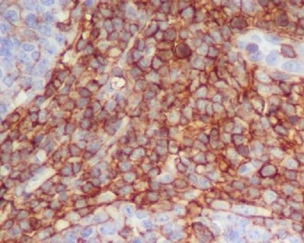
The anti-THY1 catalog antibodies show IHC in human paraffin-embedded tonsil and kidney sections (catalog IHC captions); one also shows IF in U87-MG cells (M01818-1 IF caption).

Real IHC data Immunohistochemical analysis of paraffin-embedded human tonsil, using CD90 / Thy1 Antibody.
Anti-CD90 / Thy1 Rabbit Monoclonal Antibody
Cat # M01818
Real IHC data Immunohistochemical analysis of paraffin-embedded human kidney, using CD90 Antibody.
Anti-THY1/Cd90 Rabbit Monoclonal Antibody
Cat # M01818-1

M01818 lists IHC and human and rat reactivity, with an IHC image from paraffin-embedded human tonsil (catalog applications, reactivity and IHC caption). M01818-1 lists human IHC and IF/ICC, with an IHC image from paraffin-embedded human kidney and an IF image from U87-MG cells (catalog applications, reactivity and image captions).

Which to pick: For tissue IHC, choose the rabbit monoclonal M01818 for human tonsil or M01818-1 for human kidney; each has its own paraffin-section IHC image, and neither caption reports the fixative (catalog clones and IHC captions). For IF/ICC, choose M01818-1, which lists both applications and shows IF in U87-MG cells (catalog applications and IF caption). For cross-species work, M01818 lists human and rat reactivity, while A01818-1 lists human, mouse and rat reactivity but specifies human for paraffin IHC; confirm IHC performance in other species before use (catalog reactivity; A01818-1 dilution data).

Each figure is that product's own IHC / IF validation image from its datasheet.

References

  1. UniProt Consortium. UniProt entry P04216 (THY1_HUMAN, Thy-1 membrane glycoprotein).
  2. Human Protein Atlas. THY1 tissue IHC expression (reliability: Enhanced).
  3. Human Protein Atlas. THY1 subcellular location (ICC-IF): Mainly localized to the nucleoplasm. In addition localized to the plasma membrane..
  4. Human Protein Atlas. THY1 antibody validation summary (3 antibodies).
  5. Overexpression of Thy1 and ITGA6 is associated with invasion, metastasis and poor prognosis in human gallbladder carcinoma. Oncology letters 2016 — PMC5228576.
  6. Thy1 (CD90) Expression Is Elevated in Radiation-Induced Periprosthetic Capsular Contracture: Implication for Novel Therapeutics. Plastic and reconstructive surgery 2017 — PMC5531053.
  7. Detection of pancreatic ductal adenocarcinoma in mice by ultrasound imaging of thymocyte differentiation antigen 1. Gastroenterology 2013 — PMC3783557.
  8. Identification of Key Pathways and Genes Related to Immunotherapy Resistance of LUAD Based on WGCNA Analysis. Frontiers in oncology 2021 — PMC8770266.
  9. PubMed PMID:2864690 — UniProt-cited evidence.
  10. PubMed PMID:10944468 — UniProt-cited evidence.
  11. PubMed PMID:17974005 — UniProt-cited evidence.