This website uses cookies to ensure you get the best experience on our website.


- Table of Contents
Plan chromogenic TICAM1 IHC in paraffin sections using granular cytoplasmic staining as the tissue reference (HPA tissue IHC). Bronchial respiratory epithelial cells show high staining, while liver cholangiocytes are reported as undetected (HPA tissue IHC).

Expected localisation, validated protocols, controls and antibodies — the at-a-glance facts below, then the full design guide.
| Expected localisation | Granular cytoplasm in most tissues (HPA tissue IHC) | |
| Staining pattern | Granular cytoplasmic staining in many tissue cell types (HPA tissue IHC) | |
| Antigen retrieval | Citrate pH 6.0 HIER, 95–98 °C, 20 min (rule: cytoplasmic / membrane antigen) | |
| Positive control | Adrenal gland+4 more · see all | |
| Negative control | Liver+1 more · see all |
| Fixation | Keep fixation consistent across paraffin sections (standard IHC practice; not target-specific) | |
| Caveat | Staining has medium consistency with RNA expression (HPA tissue IHC) | |
| Regulation | No expression regulation specified (UniProt) | |
| Isoform / epitope | No isoforms listed; one 1–712 chain (UniProt) |
The catalog antibody’s IHC-P protocol is paired with one published TICAM1 protocol for endometrial tissue (PMC13257043).
| Sample | Tissue sections; selected-image fixative not specified (standard IHC workflow) |
| Fixation | Image fixative and duration unreported (datasheet PA1902); verify before use. |
| Sectioning | 4–5 µm sections on charged slides (standard) |
| Deparaffinisation | Xylene, graded ethanol series to water (standard) |
| Antigen retrieval | Heat-induced epitope retrieval in citrate buffer, pH 6.0, 20 min at 95–98 °C (standard rule: cytoplasmic / membrane antigen) |
| Peroxidase block | 3% H2O2, 10 min, room temperature (standard) |
| Blocking | 10% normal serum of the secondary host, 30 min, room temperature (standard) |
| Primary antibody | Rabbit anti-TICAM1, 0.5-1μg/ml (datasheet PA1902) |
| Primary incubation | Overnight at 4 °C (standard) |
| Detection | HRP-polymer secondary, DAB chromogen 5–10 min (standard) |
| Counterstain | Hematoxylin, blue, dehydrate and mount (standard) |
| Expected result | TICAM1-positive staining in glandular cells of adrenal gland (HPA tissue IHC: High). HPA tissue profile: Granular cytoplasmic expression in most tissues. No signal in the no-primary control. |
In paraffin-section IHC, expect granular cytoplasmic TICAM1 staining in many tissues, including glandular and epithelial cells (HPA: tissue IHC). TICAM1 is reported in the cytosol, autophagosomes and mitochondria, and has no transmembrane segment (UniProt Q8IUC6). The HPA tissue pattern is Approved, with medium consistency between staining and RNA data (HPA: tissue IHC reliability).
| Granular cytoplasmic staining in colon glandular cells or bronchial respiratory epithelial cells. | This matches the reported tissue pattern and high staining in those cell types (HPA: tissue IHC). Compare signal within the relevant cells; tissue-wide colour alone cannot establish the expected pattern (general IHC practice). |
| Predominantly nuclear or crisp surface staining in a paraffin section. | The reported tissue IHC pattern is granular cytoplasmic (HPA: tissue IHC). HPA's nucleoplasmic, nucleolar and surface ICC-IF locations are uncertain (HPA: subcellular); check the IHC pattern before assigning the signal to TICAM1. |
| Strong signal in liver cholangiocytes or skeletal-muscle myocytes. | Those cells were reported as not detected (HPA: tissue IHC). Consider cross-reactivity or endogenous detection activity, particularly if the signal persists in a control lacking primary antibody (general IHC practice). |
| Diffuse colour across tissue and empty spaces, obscuring cell boundaries. | This is inconsistent with granular cytoplasmic staining (HPA: tissue IHC). Assess background with a control lacking primary antibody, then review blocking, washing and detection conditions (general IHC practice). |
| No signal in colon glandular cells or bronchial respiratory epithelial cells. | Both are reported high-staining cell types (HPA: tissue IHC). Check section integrity and the IHC detection workflow before interpreting absence of signal as biological loss (general IHC practice). |
| IHC evidence and antibody choice | The tissue profile is Approved with medium staining–RNA consistency (HPA: tissue IHC reliability). HPA055421 is IHC Approved; HPA042460 has no listed IHC status (HPA: antibodies). |
| Cell type within a tissue | Liver cholangiocytes are not detected by HPA IHC, although UniProt reports higher TICAM1 expression in liver overall (HPA: tissue IHC; UniProt Q8IUC6 tissue specificity). Interpret the specified cell population. |
| Subcellular distribution | UniProt lists cytosol, autophagosomes and mitochondria, with no transmembrane segment (UniProt Q8IUC6). HPA describes the tissue IHC signal as granular cytoplasmic; it does not resolve individual organelles in that profile (HPA: tissue IHC). |
| IF/ICC: should nuclear or membrane signal count as an IHC positive? | HPA labels its nucleoplasmic, nucleolar and plasma-membrane ICC-IF locations uncertain (HPA: subcellular). Use the granular cytoplasmic tissue profile to judge paraffin-section IHC (HPA: tissue IHC). |
| Antigen retrieval and fixation | No target-specific retrieval setting or fixation sensitivity is supplied by UniProt or HPA (UniProt Q8IUC6; HPA: tissue IHC). Record the conditions used and assess controls when comparing sections (general IHC practice). |
| Situation | Likely cause | Next action |
|---|---|---|
| No stain in a reported high-staining cell type. | A failed IHC workflow is possible; the expected cell-type signal is high (HPA: tissue IHC). | Check the positive-control section, antibody and detection steps, then review the conditions used for antigen retrieval (general IHC practice). |
| Only nuclei appear positive. | Nuclear signal differs from the tissue IHC profile; nuclear ICC-IF locations are uncertain (HPA: tissue IHC; HPA: subcellular). | Reassess compartment boundaries with the counterstain and a control lacking primary antibody (general IHC practice). |
| Cholangiocytes or myocytes stain strongly. | HPA reports these cell types as not detected (HPA: tissue IHC). Cross-reactivity or detection background is possible (general IHC practice). | Compare with a control lacking primary antibody and with a reported high-staining cell type (general IHC practice; HPA: tissue IHC). |
| Diffuse signal masks the granular pattern. | Excess background may obscure the reported cytoplasmic granules (HPA: tissue IHC; general IHC practice). | Review blocking, washing, antibody concentration and detection exposure; compare the control lacking primary antibody (general IHC practice). |
| A liver section seems to conflict with UniProt's liver expression statement. | UniProt describes liver overall; HPA specifically reports cholangiocytes as not detected (UniProt Q8IUC6; HPA: tissue IHC). | Score identified cell populations separately, and document which cells carry any signal (general IHC practice). |
| An antibody gives an unexpected compartment pattern. | HPA lists HPA055421 as IHC Approved, while HPA042460 has no listed IHC status (HPA: antibodies). | Confirm the antibody identifier, compare the staining with the granular cytoplasmic profile, and inspect controls (HPA: antibodies; HPA: tissue IHC; general IHC practice). |

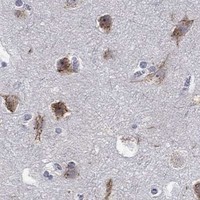
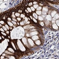
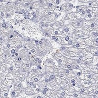
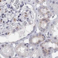


Comprehensive Human Protein Atlas IHC scoring per tissue (reliability: Approved — Medium consistency between antibody staining and RNA expression data.). Rows are taken directly from the HPA tissue chart — click any row's HPA link to view the source.
| Tissue | Cell type | Level | Evidence | Source |
|---|---|---|---|---|
| Adrenal gland | Glandular cells | High | Protein (IHC) | HPA → |
| Appendix | Glandular cells | High | Protein (IHC) | HPA → |
| Bronchus | Respiratory epithelial cells | High | Protein (IHC) | HPA → |
| Cerebral cortex | Neuronal cells | High | Protein (IHC) | HPA → |
| Cervix | Squamous epithelial cells | High | Protein (IHC) | HPA → |
Troubleshoot TICAM1 staining by tracking section processing, cellular compartment, background controls and cell-specific scoring.
The catalog contains one human-reactive IHC antibody with a real paraffin-section image from human mammary cancer tissue (PA1902 image caption; catalog applications and reactivity). No IF image is supplied (catalog image list).
PA1902 will render with an IHC(P) image of human mammary cancer tissue (PA1902 image caption). Its listed applications are IHC and WB, and its listed reactivity is Human (PA1902 catalog).
Which to pick: Choose PA1902 for human paraffin-section IHC: its own image caption documents IHC(P) on human mammary cancer tissue, but does not report the fixative (PA1902 image caption). No SKU here is listed for IF/ICC or for another species; PA1902 is rabbit-hosted, has no reported clone, and lists Human reactivity with IHC and WB applications (PA1902 catalog).