TIMD4 / T-cell immunoglobulin and mucin domain-containing protein 4 · IHC design guide

Design Immunohistochemistry for TIMD4

Plan TIMD4 chromogenic IHC on paraffin sections with the catalog antibody at 2 μg/mL (datasheet: IHC-P). Interpret cytoplasmic staining in testis and immune subsets of lymphoid tissues and liver in light of reported off-target binding (HPA tissue IHC).

Evidence assembled Oct 2026 · For research use; verify linked source records and product datasheet before use
Immunohistochemistry protocol sheet for TIMD4 (IHC for TIMD4): expected localisation Observed cytoplasmic (HPA tissue IHC); membrane expected (UniProt), antibody A06637-1, validated IHC image, and IHC protocol steps
Printable TIMD4 IHC protocol sheet — expected localisation Observed cytoplasmic (HPA tissue IHC); membrane expected (UniProt), antibody A06637-1, controls and protocol steps. Open the full TIMD4 IHC guide →

TIMD4 Immunohistochemistry Experimental Design Guide

Expected localisation, validated protocols, controls and antibodies — the at-a-glance facts below, then the full design guide.

Must know before staining
Expected localisation Observed cytoplasmic (HPA tissue IHC); membrane expected (UniProt)
Staining pattern Cytoplasmic in testis and immune subsets in lymphoid tissues and liver (HPA tissue IHC)
Antigen retrieval Citrate pH 6.0 HIER, 95–98 °C, 20 min (rule: cytoplasmic / membrane antigen)
Positive control ⓘ Bone marrow+4 more · see all
Negative control ⓘ Adipose tissue+4 more · see all
Important caveats
Reasons your staining may differ from the expected pattern.
Fixation Keep fixation consistent across sections. (standard IHC practice; not target-specific)
Caveat Presumed off-target binding in unannotated structures (HPA tissue IHC)
Regulation No specific expression regulator reported (UniProt)
Isoform / epitope 2 isoforms; check extracellular versus cytoplasmic epitope coverage (UniProt)
Section 1

Recommended TIMD4 IHC & IF Protocols

Compare the catalog antibody’s IHC-P protocol with two published TIMD4 IHC protocols (PMC11532026; PMC8013729).

Recommended immunohistochemistry (IHC-P) protocol parameters
SampleTissue sections; selected-image fixative not specified (standard IHC workflow)
FixationImage fixative and duration unreported (datasheet A06637-1); verify before use.
Sectioning4–5 µm sections on charged slides (standard)
DeparaffinisationXylene, graded ethanol series to water (standard)
Antigen retrievalHeat-induced epitope retrieval in citrate buffer, pH 6.0, 20 min at 95–98 °C (standard rule: cytoplasmic / membrane antigen)
Peroxidase block3% H2O2, 10 min, room temperature (standard)
Blocking10% normal serum of the secondary host, 30 min, room temperature (standard)
Primary antibodyRabbit anti-TIMD4, 2 μg/mL (datasheet A06637-1)
Primary incubationOvernight at 4 °C (standard)
DetectionHRP-polymer secondary, DAB chromogen 5–10 min (standard)
CounterstainHematoxylin, blue, dehydrate and mount (standard)
Expected resultTIMD4-positive staining in hematopoietic cells of bone marrow (HPA tissue IHC: Medium). HPA tissue profile: Cytoplasmic expression in testis and subsets of immune cells in lymphoid tissues and liver. No signal in the no-primary control.
💡Decision noteStart with the page’s citrate pH 6.0 retrieval at 95–98 °C for 20 min; consider the published EDTA or Tris-EDTA conditions if staining needs optimization (PMC11532026; PMC8013729).
Section 2

What Is the Expected TIMD4 Staining Pattern?

TIMD4 is a cell-surface protein with an extracellular region at residues 25–314 and a transmembrane segment at 315–335 (UniProt Q96H15 topology). It is expressed by macrophages and dendritic cells (UniProt Q96H15 tissue specificity). In tissue IHC, expect staining in selected immune-cell populations and testis; HPA describes its observed pattern as cytoplasmic and rates the tissue staining profile Approved, while noting presumed off-target binding (HPA tissue IHC).

What am I looking at on my slide?
Selected cells stain in spleen red pulp, lymph-node or tonsil germinal centers, or bone marrow.These are documented medium-staining sites (HPA tissue IHC). Interpret signal by cell population and tissue context; HPA does not identify every stained cell as a macrophage. UniProt reports macrophage and dendritic-cell expression (UniProt Q96H15 tissue specificity).
Elongated or late spermatids show staining in testis.This is a documented medium IHC pattern, despite TIMD4’s immune-cell association (HPA tissue IHC; UniProt Q96H15 tissue specificity). Do not reject testis staining solely because it occurs outside lymphoid tissue.
Strong nuclear staining dominates, or signal forms a uniform rim around most cells.Neither pattern matches the reported cytoplasmic staining in selected cells on tissue sections (HPA tissue IHC). Reassess specificity and slide controls. Plasma-membrane localization in ICC-IF does not establish that every cell should show a distinct membrane rim in chromogenic IHC (HPA subcellular ICC-IF).
Broad staining appears in respiratory or squamous epithelium.HPA reports TIMD4 as not detected in bronchial respiratory epithelial cells and cervical squamous epithelial cells (HPA tissue IHC). Consider cross-reactivity or endogenous detection activity; HPA specifically notes presumed off-target binding in its approved tissue profile (HPA tissue IHC).
A known-positive section is blank, while other tissue also lacks signal.Spleen red-pulp cells, germinal-center cells, bone-marrow hematopoietic cells, and late spermatids offer documented medium-staining reference populations (HPA tissue IHC). A blank reference section makes a negative test section inconclusive until the staining run and detection controls are checked (general IHC practice).
💡Expected TIMD4 appearanceCall a section positive when staining is concentrated in the documented cell populations, typically at medium intensity and described as cytoplasmic in tissue IHC; broad epithelial or dominant nuclear staining warrants review as a possible false-positive pattern (HPA tissue IHC).
How each factor affects the staining
Tissue and cell selectionChoose a documented medium-staining population for a positive reference, such as spleen red-pulp cells or testis late spermatids (HPA tissue IHC). Placental trophoblastic cells are reported at low staining, so a weak result there is less decisive (HPA tissue IHC).
Compartment and antibody epitopeTIMD4 has an extracellular region at 25–314, a membrane span at 315–335, and a cytoplasmic region at 336–378 (UniProt Q96H15 topology). No antibody epitope is supplied; topology alone cannot predict how sharply a chromogenic section will outline the membrane.
Isoforms and processingUniProt lists 2 isoforms, a signal peptide at 1–24, and a mature chain beginning at 25 (UniProt Q96H15 isoforms and processing). Without an antibody epitope, do not assume equal recognition of both isoforms or infer a distinct tissue staining pattern from processing alone.
Validation and reported limitsTwo listed antibodies have Approved IHC status (HPA antibody validation: HPA015625 and CAB026027). The tissue profile is also Approved, with presumed off-target binding observed and disregarded (HPA tissue IHC). Treat unexpected cell staining as a finding to verify, not as established TIMD4 expression.
IF/ICC: where should signal appear?HPA reports approved plasma-membrane localization in ICC-IF, consistent with TIMD4 membrane topology (HPA subcellular ICC-IF; UniProt Q96H15 topology). This answers the IF/ICC localization question; the separate tissue IHC description remains cytoplasmic staining in selected cells (HPA tissue IHC).
Why is my staining missing, weak or wrong?
SituationLikely causeNext action
No staining in a test sectionThe selected cells may have little detectable TIMD4; HPA reports only low staining in placental trophoblastic cells (HPA tissue IHC).Run a documented medium-staining reference population alongside the test section (HPA tissue IHC). If that reference is also blank, check the IHC detection run and controls (general IHC practice).
Positive reference is blankA run or reagent failure is possible when a documented positive population shows no signal (HPA tissue IHC; general IHC practice).Check antibody and detection-reagent steps against the chosen IHC method, then repeat with the same positive reference and a negative control (general IHC practice). No target-specific fixation sensitivity is reported in the supplied evidence.
Diffuse color obscures cell boundariesBackground from detection chemistry or insufficient washing can obscure a cell-restricted pattern (general IHC practice).Review the negative control, blocking, washing, and chromogen development for that run (general IHC practice). Score TIMD4 only where staining can be assigned to cells matching the documented tissue distribution (HPA tissue IHC).
Many bronchial or cervical epithelial cells stainThose cell types are reported as not detected; broad staining could reflect cross-reactivity or endogenous detection activity (HPA tissue IHC; general IHC practice).Compare with a no-primary control and a documented positive tissue; review endogenous-activity blocking for the detection chemistry used (general IHC practice; HPA tissue IHC).
A membrane rim is absent in chromogenic IHCHPA describes cytoplasmic tissue staining, although ICC-IF localizes TIMD4 to the plasma membrane (HPA tissue IHC; HPA subcellular ICC-IF).Judge the tissue section by its documented stained cell populations and controls (HPA tissue IHC; general IHC practice). Do not require a crisp membrane rim as the sole positive criterion.
An unexpected isolated structure stainsHPA notes presumed off-target binding and staining in cells or structures outside its annotations (HPA tissue IHC reliability description).Record its location, compare the negative control and documented positive pattern, and seek independent antibody or orthogonal support before assigning it to TIMD4 (general IHC practice; HPA tissue IHC).

Sample controls for TIMD4 IHC & IF

🧪Run spleen first and score cells in the red pulp, where TIMD4 staining is Medium (HPA: spleen, cells in red pulp, Medium; selected-SKU tissue-IHC caption: human spleen). Use adipose tissue as the negative tissue and score adipocytes, where staining is Not detected (HPA: adipose tissue, adipocytes, Not detected). On the spleen slide, use cells outside the annotated red-pulp population as background comparators; HPA does not establish those cells as TIMD4-negative (HPA: spleen, cells in red pulp, Medium).
Positive control tissue: Bone marrow (Hematopoietic cells, HPA Medium)
Negative control tissue: Adipose tissue (HPA Not detected)
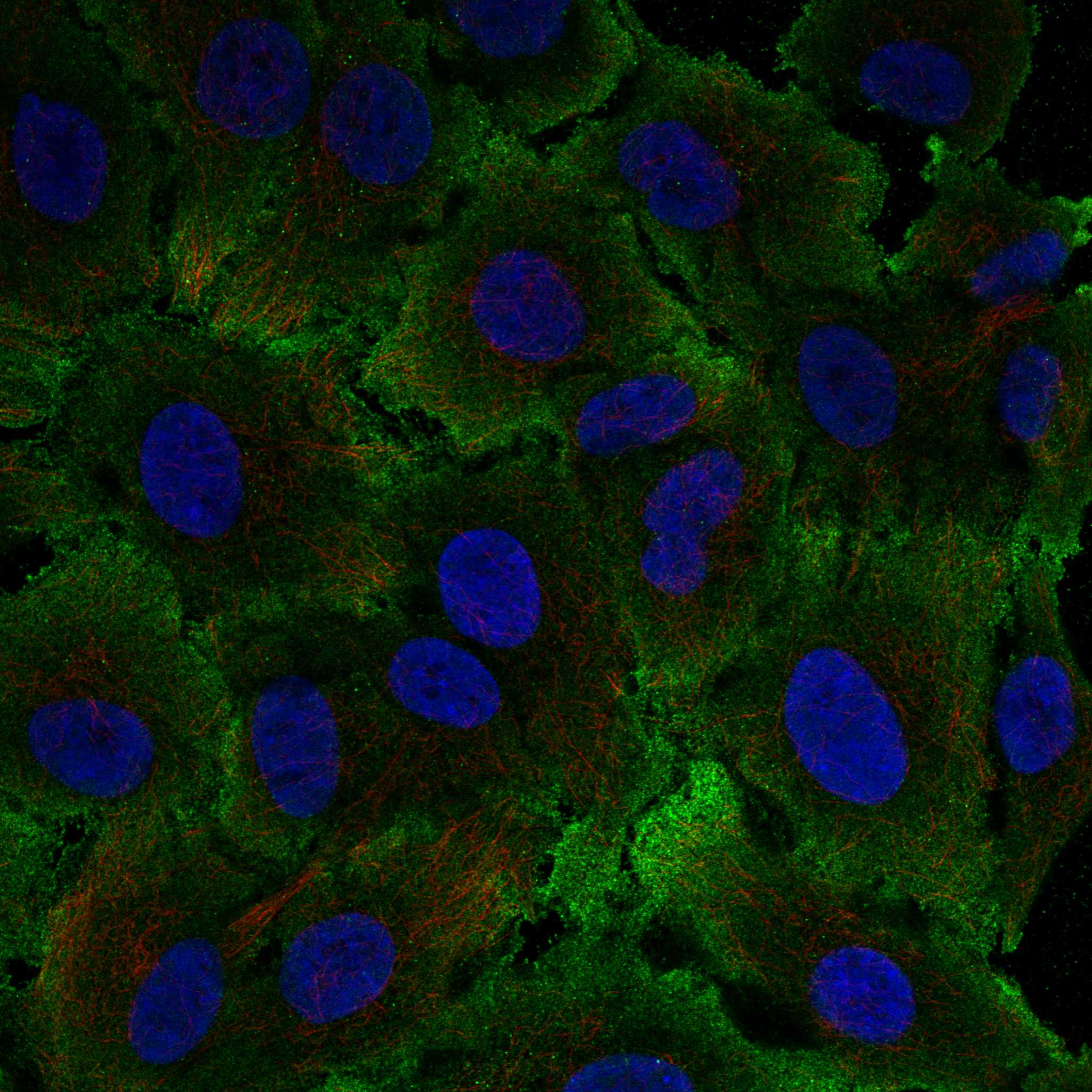
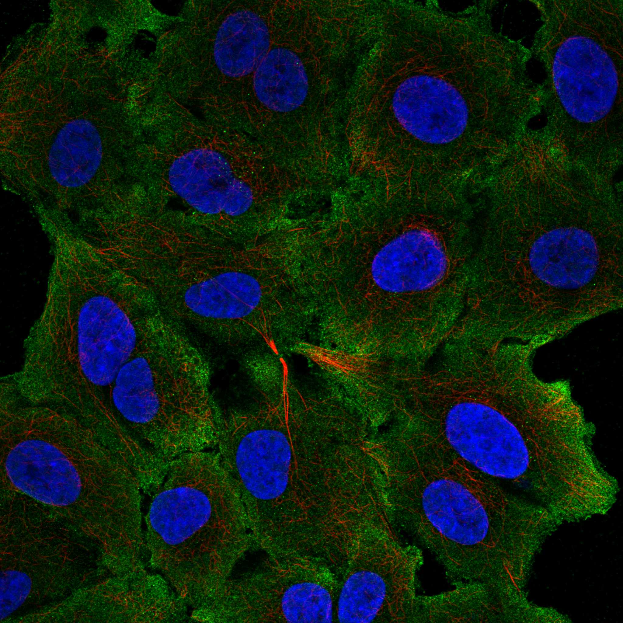
ICC-IF cell lines (HPA subcellular resource): HPA ICC-IF images show TIMD4 in A-549, CACO-2, U2OS, with annotated localisation: Plasma membrane (approved) (HPA subcellular).
Technical controls: Include a no-primary, secondary-only control; an isotype control matched to the primary antibody’s host, immunoglobulin class, and monoclonal or polyclonal format; and TIMD4 knockout material or peptide competition if the immunizing peptide is available (standard IHC controls). For chromogenic spleen IHC, block endogenous peroxidase and check red-pulp background, including signal from blood-rich areas (standard IHC practice).
⚠️Feasibility: A TIMD4-specific fixation window, fixation effect, and antigen-retrieval dependency are unreported in the supplied evidence; the selected A06637-1 spleen-IHC caption reports 2.5 μg/ml but leaves the fixative unreported (selected-SKU tissue-IHC caption). The evidence does not establish whether frozen sections or IF are easier than paraffin IHC; IF can help assess the expected plasma-membrane localization (HPA: approved plasma membrane location; UniProt Q96H15 topology). In spleen, blood-rich red pulp can produce endogenous peroxidase background in chromogenic detection (standard IHC practice).

HPA tissue IHC evidence for TIMD4

Comprehensive Human Protein Atlas IHC scoring per tissue (reliability: Approved — Presumed off target binding observed and disregarded. Antibody staining in cells/structures not annotated, view images.). Rows are taken directly from the HPA tissue chart — click any row's HPA link to view the source.

Positive expression · recommended positive controls

TissueCell typeLevelEvidenceSource
Bone marrow Hematopoietic cells Medium Protein (IHC) HPA →
Lymph node Germinal center cells Medium Protein (IHC) HPA →
Spleen Cells in red pulp Medium Protein (IHC) HPA →
Testis Elongated or late spermatids Medium Protein (IHC) HPA →
Tonsil Germinal center cells Medium Protein (IHC) HPA →

Undetected expression · recommended negative controls

TissueCell typeLevelEvidenceSource
Adipose tissue Adipocytes Not detected Protein (IHC) HPA →
Adrenal gland Glandular cells Not detected Protein (IHC) HPA →
Breast Adipocytes Not detected Protein (IHC) HPA →
Bronchus Respiratory epithelial cells Not detected Protein (IHC) HPA →
Caudate Glial cells Not detected Protein (IHC) HPA →
Section 3

Advanced TIMD4 IHC Tips

Troubleshoot TIMD4 staining in chromogenic paraffin-section IHC using compartment, cell-type and control evidence; the IF entry addresses the separate application.

How should I adjust retrieval when TIMD4 staining is weak or uneven?
Start with citrate pH 6.0 heat-induced epitope retrieval at 95–98 °C for 20 min (page retrieval rule). If staining is weak, compare a second retrieval condition on adjacent sections while keeping antibody concentration, detection and imaging constant (standard IHC practice). Check whether any gain is confined to expected immune cells, since TIMD4 is expressed by macrophages and dendritic cells (UniProt Q96H15 tissue specificity). Excessive retrieval can damage morphology or increase nonspecific staining, so inspect cell boundaries alongside signal (standard IHC practice). Record retrieval conditions with every scored section to avoid mistaking a processing difference for a biological difference (standard IHC practice).
Could fixation explain weak TIMD4 staining in my paraffin sections?
TIMD4-specific fixation sensitivity is unknown from the supplied evidence; the selected tissue-IHC caption does not state a fixative (A06637-1 caption). Record the fixative, fixation duration and processing history for each section before comparing staining (standard IHC practice). If samples differ, run matched sections through the same citrate pH 6.0, 95–98 °C, 20 min retrieval and detection workflow (page retrieval rule; standard IHC practice). Compare signal within similar cell populations and assess morphology, because processing differences can affect epitope detection and tissue preservation (standard IHC practice). Do not assign a TIMD4-specific fixation effect without a controlled comparison (supplied fixation evidence; standard IHC practice).
Is cytoplasmic TIMD4 staining compatible with its expected localisation?
TIMD4 has an extracellular region at residues 25–314, a transmembrane segment at 315–335 and a cytoplasmic tail at 336–378 (UniProt Q96H15 topology). Plasma-membrane localisation is supported by the subcellular record, while tissue IHC reports cytoplasmic staining in testis and subsets of immune cells (HPA subcellular; HPA tissue IHC). In chromogenic sections, assess whether cytoplasmic signal occurs in plausible cells and accompanies discernible cell-associated staining (standard IHC practice; UniProt Q96H15 tissue specificity). Compare that pattern with adjacent negative controls and tissue morphology before calling it TIMD4 (standard IHC practice). Diffuse staining across unrelated cell populations warrants a background or specificity check (standard IHC practice).
How do epitope location and isoforms affect TIMD4 IHC interpretation?
TIMD4 has 2 listed isoforms, but the supplied evidence does not map the catalog antibody’s epitope to either one (UniProt Q96H15 isoforms; supplied antibody evidence). Its extracellular region spans residues 25–314, and one glycosylation site is listed at residue 291 (UniProt Q96H15 topology; UniProt Q96H15 glycosylation). Check the antibody’s documented immunogen before inferring whether staining represents an extracellular region, the cytoplasmic tail or both isoforms (standard antibody-validation practice). Compare the same antibody across controls processed together, because retrieval may alter access to a given epitope (standard IHC practice). Do not assign a stained cell to a specific TIMD4 isoform from chromogenic signal alone (UniProt Q96H15 isoforms; standard IHC interpretation).
How can IF help resolve ambiguous TIMD4 staining in tissue?
For the separate IF application, pair TIMD4 with an independently validated macrophage or dendritic-cell marker to test cell identity (UniProt Q96H15 tissue specificity; standard multiplex IF practice). Choose fluorophores after measuring tissue autofluorescence in an unstained control, and separate their emission channels sufficiently for the imaging system (standard IF practice). The extracellular domain spans residues 25–314, whereas the cytoplasmic tail spans 336–378; choose permeabilisation according to the antibody’s documented epitope side (UniProt Q96H15 topology; standard IF practice). If that epitope is unknown, compare permeabilised and nonpermeabilised preparations rather than assuming surface access (supplied antibody evidence; standard IF practice). Use single-stain controls to check bleed-through before interpreting colocalisation (standard multiplex IF practice).
What should I check when TIMD4 IHC looks diffusely positive?
Inspect a no-primary control and the distribution of chromogen before changing the TIMD4 antibody condition (standard IHC practice). In chromogenic IHC, incomplete endogenous peroxidase blocking, nonspecific antibody binding and precipitated chromogen can produce signal unrelated to the target (standard IHC practice). Confirm peroxidase blocking and appropriate protein blocking, then compare matched sections using the same development time (standard IHC practice). TIMD4 expression is reported in macrophages and dendritic cells, while HPA reports staining in subsets of immune cells and testis (UniProt Q96H15 tissue specificity; HPA tissue IHC). Broad staining that ignores cell boundaries or appears in the no-primary control should be treated as background (standard IHC practice).
How should I score TIMD4 staining across heterogeneous tissue regions? ⚠ ANSWER MARKED FOR VERIFICATION
Define the cell population and anatomical region before scoring, then use identical retrieval, detection and image settings across specimens (standard IHC quantification practice). For cell-associated chromogenic signal, report the percentage of positive cells and, if intensity is reproducibly separable, an H-score using intensity grades 0–3 (standard IHC quantification practice). For spatial comparisons, report TIMD4-positive cells per mm² and normalise to the counted tissue area or eligible cell population (standard IHC quantification practice). Keep macrophage-rich regions distinct from other compartments because TIMD4 is expressed by macrophages and dendritic cells (UniProt Q96H15 tissue specificity). State the positivity threshold and exclude folds, edges and necrotic areas consistently (standard IHC quantification practice).
When is a TIMD4-positive IHC pattern convincing?
A convincing call combines staining in plausible cells with suitable controls and preserved morphology (standard IHC interpretation). TIMD4 is a membrane protein expressed by macrophages and dendritic cells, and HPA reports plasma-membrane localisation (UniProt Q96H15 topology and tissue specificity; HPA subcellular). HPA tissue IHC also reports cytoplasmic staining in testis and subsets of immune cells, so compartment alone cannot establish specificity (HPA tissue IHC; standard IHC interpretation). Compare suspect signal with no-primary controls and inspect section edges, necrotic areas and endogenous enzyme staining before scoring (standard IHC practice). The HPA tissue-IHC annotation notes presumed off-target binding, making cell identity and independent controls especially useful (HPA tissue IHC reliability description).
Boster reagents

Best TIMD4 / T-cell immunoglobulin and mucin domain-containing protein 4 IHC Antibodies

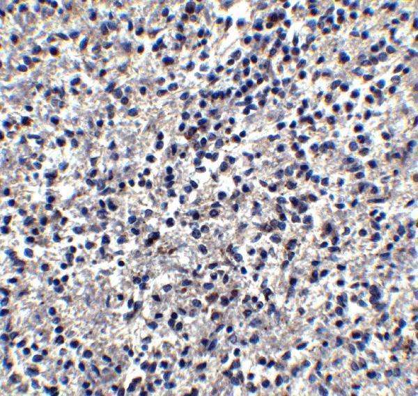
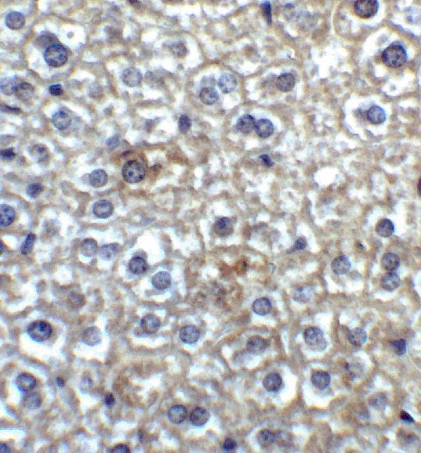
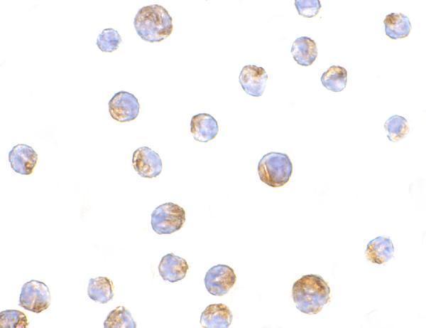
The anti-TIMD4 antibody has IHC images from human spleen and mouse liver, plus IF images from Jurkat cells and human spleen (catalog image captions).

Real IHC data Immunohistochemistry of TIM-4 in human spleen tissue with TIM-4 antibody at 2.5 μg/ml.
Anti-TIM-4 TIMD4 Antibody
Cat # A06637-1

A06637-1 lists IHC-P and human and mouse reactivity (catalog: applications; reactivity); its IHC images show human spleen at 2.5 μg/mL and mouse liver at 2 μg/mL (catalog image captions). The same SKU lists IF and ICC (catalog: applications), with images of Jurkat cells for both and human spleen for IF (catalog image captions).

Which to pick: Choose A06637-1 for tissue IHC-P based on its human spleen and mouse liver IHC captions (catalog image captions); the fixative is unreported (catalog image captions). For IF/ICC, choose A06637-1 based on its application list and Jurkat cell and human spleen images (catalog: applications; image captions). It is also the listed human–mouse choice (catalog: reactivity); the host is rabbit and clonality is unreported (catalog: host; clone).

Each figure is that product's own IHC / IF validation image from its datasheet.

References

  1. UniProt Consortium. UniProt entry Q96H15 (TIMD4_HUMAN, T-cell immunoglobulin and mucin domain-containing protein 4).
  2. Human Protein Atlas. TIMD4 tissue IHC expression (reliability: Approved).
  3. Human Protein Atlas. TIMD4 subcellular location (ICC-IF): Localized to the plasma membrane..
  4. Human Protein Atlas. TIMD4 antibody validation summary (2 antibodies).
  5. Identification and validation of efferocytosis-related biomarkers for the diagnosis of metabolic dysfunction-associated steatohepatitis based on bioinformatics analysis and machine learning. Frontiers in immunology 2024 — PMC11532026.
  6. Up-regulation of blood-circulating TIM-4-expressing monocytes and potential diagnostic biomarker sTIM-4 in primary liver cancer. Scientific reports 2025 — PMC12350753.
  7. Characterization of Subpopulations of Chicken Mononuclear Phagocytes That Express TIM4 and CSF1R. Journal of immunology (Baltimore, Md. : 1950) 2019 — PMC6436730.
  8. Single-Cell RNA-seq Reveals Characteristics of Malignant Cells and Immune Microenvironment in Subcutaneous Panniculitis-Like T-Cell Lymphoma. Frontiers in oncology 2021 — PMC8013729.
  9. PubMed PMID:14702039 — UniProt-cited evidence.
  10. PubMed PMID:15372022 — UniProt-cited evidence.
  11. PubMed PMID:15489334 — UniProt-cited evidence.