TIMELESS / Protein timeless homolog · IHC design guide

Design Immunohistochemistry for TIMELESS

Plan chromogenic IHC for TIMELESS in paraffin sections using the IHC-validated antibody at 1:50 (datasheet M00831). Use the nuclear tissue profile and high staining in testis spermatogonia to guide controls and scoring (HPA tissue IHC).

Evidence assembled Oct 2026 · For research use; verify linked source records and product datasheet before use
Immunohistochemistry protocol sheet for TIMELESS (IHC for TIMELESS): expected localisation Nuclear staining in several tissues (HPA tissue IHC), antibody M00831, validated IHC image, and IHC protocol steps
Printable TIMELESS IHC protocol sheet — expected localisation Nuclear staining in several tissues (HPA tissue IHC), antibody M00831, controls and protocol steps. Open the full TIMELESS IHC guide →

TIMELESS Immunohistochemistry Experimental Design Guide

Expected localisation, validated protocols, controls and antibodies — the at-a-glance facts below, then the full design guide.

Must know before staining
Expected localisation Nuclear staining in several tissues (HPA tissue IHC)
Staining pattern Nuclear staining; high in spermatogonia (HPA tissue IHC)
Antigen retrieval EDTA pH 8.0 HIER, heat-mediated (datasheet M00831)
Positive control ⓘ Appendix+4 more · see all
Negative control ⓘ Adipose tissue+4 more · see all
Important caveats
Reasons your staining may differ from the expected pattern.
Fixation Keep fixation conditions consistent across sections. (standard IHC practice; not target-specific)
Caveat Ciliary and microvillar staining may complicate nuclear scoring (HPA tissue IHC)
Regulation DNA breaks recruit TIMELESS to damage sites (UniProt)
Isoform / epitope 2 isoforms; epitope coverage is unspecified (UniProt)
Section 1

Recommended TIMELESS IHC & IF Protocols

The catalog antibody protocol is accompanied by a published TIMELESS IHC protocol for human breast cancer sections (PMC10280842).

Recommended immunohistochemistry (IHC-P) protocol parameters
SampleParaffin-embedded human breast cancer tissue; fixative not specified (datasheet M00831)
FixationImage fixative and duration unreported (datasheet M00831); verify before use.
Sectioning4–5 µm sections on charged slides (standard)
DeparaffinisationXylene, graded ethanol series to water (standard)
Antigen retrievalHeat retrieval: EDTA pH 8.0 (datasheet M00831); 20 min, 95–100 °C (standard)
Peroxidase block3% H2O2, 10 min, room temperature (standard)
Blocking10% goat serum (datasheet M00831)
Primary antibodyRabbit monoclonal (clone 22T48) anti-TIMELESS, 1:50 (datasheet M00831)
Primary incubationOvernight at 4 °C (datasheet M00831)
DetectionHRP-conjugated secondary, DAB chromogen (datasheet M00831)
CounterstainHematoxylin, blue, dehydrate and mount (standard)
Expected resultTIMELESS-positive staining in endocrine cells of appendix (HPA tissue IHC: High). HPA tissue profile: Nuclear expression in several tissues. No signal in the no-primary control.
💡Decision noteStart with heat-mediated EDTA retrieval at pH 8.0 (datasheet: M00831); the published breast cancer excerpt does not specify retrieval (PMC10280842).
Section 2

What Is the Expected TIMELESS Staining Pattern?

TIMELESS is a nuclear and chromosome-associated protein with no transmembrane segment (UniProt Q9UNS1 topology and subcellular location). In paraffin-section IHC, expect nuclear staining in selected cells, including testicular spermatogonia and appendix endocrine cells, where HPA reports high staining (HPA tissue IHC). HPA describes nuclear expression in several tissues and rates its tissue IHC evidence Enhanced, with medium consistency between staining and RNA expression (HPA tissue IHC).

What am I looking at on my slide?
Distinct nuclear stain in testicular spermatogonia or appendix endocrine cells, with a readable counterstain (HPA: High in both cell types).This fits the expected compartment and two observed high-staining populations (UniProt Q9UNS1 subcellular location; HPA tissue IHC). Score the named cells and their nuclear signal; do not treat every cell in the section as an expected positive. HPA also reports low or undetected staining in other specified cell populations (HPA tissue IHC).
Widespread cytoplasmic stain with little nuclear signal, outside the reported ciliary and microvillar patterns (HPA tissue IHC).This does not fit the predominant nuclear expectation (UniProt Q9UNS1 subcellular location; HPA tissue IHC). Check section morphology and detection background before interpreting it as TIMELESS. Compartment alone is not an absolute exclusion: HPA reports staining at fallopian-tube cilia tips, bronchial ciliary rootlets and colon enterocyte microvilli (HPA tissue IHC).
Strong stain in a cell population HPA reports as undetected, such as adipocytes or pancreatic exocrine glandular cells (HPA: Not detected).An isolated result conflicts with the reported cell-specific pattern and calls for checks for cross-reactivity or endogenous detection activity (HPA tissue IHC; general IHC practice). Do not use a tissue-wide label as a substitute for identifying cells: HPA reports undetected pancreatic exocrine cells, while UniProt reports TIMELESS expression in pancreas without specifying those cells (UniProt Q9UNS1 tissue specificity).
Diffuse colour across nuclei, cytoplasm and empty areas, without a clear cell-specific pattern (general IHC practice).A diffuse deposit limits interpretation of the nuclear pattern; nonspecific binding or detection background is possible (general IHC practice). Compare a section processed without primary antibody and inspect tissue edges and damaged regions. An apparently positive area is persuasive only when its cells and compartment match the expected pattern (HPA tissue IHC; UniProt Q9UNS1 subcellular location).
No discernible stain in testicular spermatogonia or appendix endocrine cells (HPA: High in both cell types).This conflicts with the reported high IHC staining, but a single blank section does not establish absent TIMELESS (HPA tissue IHC). Confirm that the expected cells are present, then review primary-antibody use, antigen-retrieval conditions and chromogenic detection as general IHC checks. Interpret against HPA's Enhanced rating and stated medium staining–RNA consistency (HPA tissue IHC).
💡Expected TIMELESS appearanceCall a section positive when selected cells show interpretable nuclear stain, especially high signal in testicular spermatogonia or appendix endocrine cells (UniProt Q9UNS1 subcellular location; HPA tissue IHC); diffuse deposit or widespread cytoplasmic colour without the reported cell pattern warrants a background check (general IHC practice).
How each factor affects the staining
Cell identity and tissue choice (HPA tissue IHC)HPA reports High staining in appendix endocrine cells and testicular spermatogonia, Medium in several other named cell populations, and Not detected in adipocytes and pancreatic exocrine cells. Select and score the specified cells; a whole-tissue positive or negative label loses that distinction (HPA tissue IHC).
Subcellular context and topology (UniProt Q9UNS1; HPA tissue IHC)UniProt places TIMELESS in nucleus and chromosome and lists no transmembrane segment. HPA's overall tissue profile is nuclear, although it records cilia-tip, ciliary-rootlet and microvillar staining in specified tissues. Apply those exceptions only to their reported cells (UniProt Q9UNS1 topology and subcellular location; HPA tissue IHC).
Validation scope (HPA tissue IHC; HPA antibody validation)Target-specific fixation effects are not established by the supplied assay evidence. Verify with a matched IHC source before attributing a result to fixation.
IF/ICC Q: Where should signal appear? (HPA subcellular ICC-IF)A: HPA supports nucleoplasmic localisation in ICC-IF images from A-549, MCF-7 and U2OS and rates HPA060655 ICC Supported (HPA subcellular ICC-IF; HPA antibody validation). Use this as a compartment comparison; the primary application and tissue interpretation here remain paraffin-section IHC (HPA tissue IHC).
Why is my staining missing, weak or wrong?
SituationLikely causeNext action
Expected positive cells are blank (HPA: High in testicular spermatogonia and appendix endocrine cells).The relevant cells may be absent from the section, or a routine IHC step may have failed (HPA tissue IHC; general IHC practice).Verify cell identity and section quality; review antigen retrieval, primary-antibody dilution, detection reagents and counterstain, then compare with a known-positive section (general IHC practice).
Colour is mostly cytoplasmic, with little nuclear definition (UniProt Q9UNS1 subcellular location).Background or morphology may obscure a nuclear pattern; HPA also records specific nonnuclear sites in selected cells (general IHC practice; HPA tissue IHC).Compare with a no-primary control and examine cell identity before scoring; check whether the site is a reported ciliary or microvillar exception (general IHC practice; HPA tissue IHC).
Pancreatic exocrine cells or adipocytes stain strongly (HPA: Not detected in those cells).The result conflicts with HPA's cell-specific observations; cross-reactivity or endogenous detection activity is possible (HPA tissue IHC; general IHC practice).Confirm the stained cell type, inspect a no-primary control and compare a reported high-staining population in the same run before assigning specificity (general IHC practice; HPA tissue IHC).
Diffuse chromogen makes nuclei hard to score (general IHC practice).Nonspecific binding, endogenous detection activity or excessive chromogen development can contribute to background (general IHC practice).Review blocking and detection controls, reagent dilution and chromogen development; score only where nuclei remain distinguishable from surrounding tissue (general IHC practice).
A ciliary or apical signal seems to contradict the nuclear expectation (HPA tissue IHC).HPA reports High signal at fallopian-tube cilia tips and Medium signal at bronchial ciliary rootlets and colon enterocyte microvilli (HPA tissue IHC).Check tissue and cell identity and record that compartment separately; do not generalise these observations to other cells or call every nonnuclear deposit specific (HPA tissue IHC; general IHC practice).
RNA expectations and tissue staining disagree (HPA: medium staining–RNA consistency).HPA reports low tissue RNA specificity alongside cell-specific IHC levels; the measures need not give identical cell-level readouts (HPA tissue IHC).Describe the observed cells, compartment and staining level directly, and retain HPA's stated consistency limit when interpreting a discordant result (HPA tissue IHC).

Sample controls for TIMELESS IHC & IF

🧪Run testis first and assess staining in spermatogonia (High; HPA: testis, spermatogonia). Use adipose tissue as the negative tissue (HPA: adipocytes, Not detected); on the testis slide, cells without brown DAB signal can serve as internal background references, but the supplied HPA rows do not establish any other testicular cell type as TIMELESS-negative.
Positive control tissue: Appendix (Endocrine cells, HPA High)
Negative control tissue: Adipose tissue (HPA Not detected)
ICC-IF cell lines (HPA subcellular resource): HPA ICC-IF images show TIMELESS in A-549, MCF-7, U2OS, with annotated localisation: Nucleoplasm (supported) (HPA subcellular).
Technical controls: Include a no-primary, secondary-only section; a concentration- and class-matched rabbit isotype control (selected-SKU caption: rabbit primary); and TIMELESS knockout material or a validated peptide-block control (standard IHC practice). Quench endogenous peroxidase before HRP/DAB detection (selected-SKU caption: HRP/DAB; standard IHC practice).
⚠️Feasibility: A target-specific fixation window or fixation effect is unreported, and the selected-SKU paraffin-section caption does not state the fixative (selected-SKU caption: fixative unreported). That caption uses heat retrieval in EDTA, pH 8.0, but does not establish that retrieval is required or that frozen sections are easier (selected-SKU caption: heat retrieval); HPA ICC-IF supports nucleoplasmic localization, without establishing that IF is easier (HPA subcellular: nucleoplasm). In testis, crowded germ-cell nuclei can complicate assignment of nuclear DAB signal, so assess staining against the hematoxylin counterstain and cell morphology (HPA: testis, spermatogonia; UniProt Q9UNS1: nucleus; standard IHC practice).

HPA tissue IHC evidence for TIMELESS

Comprehensive Human Protein Atlas IHC scoring per tissue (reliability: Enhanced — Medium consistency between antibody staining and RNA expression data.). Rows are taken directly from the HPA tissue chart — click any row's HPA link to view the source.

Positive expression · recommended positive controls

TissueCell typeLevelEvidenceSource
Appendix Endocrine cells High Protein (IHC) HPA →
Fallopian tube Ciliated cells (tip of cilia) High Protein (IHC) HPA →
Testis Spermatogonia cells High Protein (IHC) HPA →
Adrenal gland Glandular cells Medium Protein (IHC) HPA →
Breast Glandular cells Medium Protein (IHC) HPA →

Undetected expression · recommended negative controls

TissueCell typeLevelEvidenceSource
Adipose tissue Adipocytes Not detected Protein (IHC) HPA →
Cervix Glandular cells Not detected Protein (IHC) HPA →
Epididymis Glandular cells Not detected Protein (IHC) HPA →
Lung Alveolar cells Not detected Protein (IHC) HPA →
Ovary Ovarian stroma cells Not detected Protein (IHC) HPA →
Section 3

Advanced TIMELESS IHC Tips

Troubleshoot TIMELESS staining in paraffin sections by checking retrieval, nuclear localisation, cell type and controls before interpreting signal intensity.

What retrieval should I try first for weak TIMELESS staining in paraffin sections?
Start with heat-mediated retrieval in EDTA at pH 8.0 (datasheet M00831). That condition produced TIMELESS staining in a paraffin-embedded human breast cancer section with the catalog antibody at 1:50 overnight at 4°C (datasheet M00831). If nuclear staining remains weak, compare retrieval heating times on adjacent sections while keeping antibody incubation and DAB development constant (standard IHC practice). Include a section processed without primary antibody and a positive reference section in each comparison (standard IHC practice). The caption does not report the fixative, so its retrieval result cannot establish performance across fixation conditions (datasheet M00831).
Could fixation explain inconsistent TIMELESS staining between paraffin blocks?
Target-specific fixation sensitivity is unknown because the selected tissue caption identifies a paraffin section but does not state its fixative or fixation time (datasheet M00831). Record the fixative and processing history for each block before comparing TIMELESS intensity across specimens (standard IHC practice). Keep section thickness, EDTA retrieval at pH 8.0, and antibody incubation at 1:50 overnight at 4°C consistent during a block comparison (datasheet M00831; standard IHC practice). Compare nuclear signal with a reference section processed in the same run, and inspect morphology for processing damage (standard IHC practice; HPA: nuclear tissue expression). Do not infer a TIMELESS-specific fixation effect from its reported phosphorylation sites or tissue staining pattern (UniProt Q9UNS1; HPA: tissue IHC).
How should I assess TIMELESS staining outside the nucleus?
Treat nuclear staining as the principal pattern to assess: TIMELESS is annotated in the nucleus and on chromosomes, and tissue staining is described as nuclear in several tissues (UniProt Q9UNS1 localisation; HPA: tissue IHC). Supported ICC/IF localisation is nucleoplasmic (HPA: subcellular localisation). Examine apparent cytoplasmic DAB against a no-primary control and check whether counterstained nuclei remain intact (standard IHC practice). TIMELESS has no annotated transmembrane segment, so a purely membrane-like pattern warrants additional validation (UniProt Q9UNS1 topology). HPA also reports staining at ciliary tips or rootlets in selected cells; assess those patterns in their stated cell context instead of treating every extranuclear signal as equivalent (HPA: tissue IHC).
Can this IHC result distinguish TIMELESS isoforms or phosphorylation states?
The record lists 2 TIMELESS isoforms, but the supplied tissue caption does not identify the catalog antibody’s epitope or establish isoform selectivity (UniProt Q9UNS1 isoforms; datasheet M00831). Interpret positive nuclei as TIMELESS immunoreactivity without assigning signal to isoform 1 or 2 (UniProt Q9UNS1 isoforms; standard IHC interpretation). The record also lists phosphorylated residues, including serines 281, 1074 and 1087, without evidence that this antibody distinguishes their modification states (UniProt Q9UNS1 modified residues; datasheet M00831). If isoform or phosphorylation specificity is essential, obtain epitope documentation and validate that property with appropriate controls before interpreting stained sections (standard antibody validation practice). Keep retrieval conditions identical across that comparison (standard IHC practice).
How can I check a multiplex IF result against the TIMELESS IHC pattern?
Use the IHC result as a tissue reference: TIMELESS is reported in nuclei, including high staining in testicular spermatogonia (HPA: tissue IHC). For a multiplex IF comparison, pair TIMELESS with a validated marker of the expected cell type and inspect nuclear overlap within marker-positive cells (standard IF practice; HPA: tissue IHC). Choose a fluorophore channel with low background in the specimen, and measure unstained tissue autofluorescence before judging weak signal (standard IF practice). TIMELESS has no transmembrane segment and is supported in the nucleoplasm, so use permeabilisation suitable for access to an intracellular nuclear epitope (UniProt Q9UNS1 topology; HPA: subcellular localisation; standard IF practice). The paraffin IHC caption supplies no IF fixation or permeabilisation condition (datasheet M00831).
How do I troubleshoot diffuse brown staining that obscures TIMELESS-positive nuclei?
First compare the section with a no-primary control to identify signal arising from the detection workflow (standard IHC practice). The selected paraffin example used 10% goat serum blocking, a peroxidase-conjugated secondary antibody and DAB detection (datasheet M00831). If the control is brown, review peroxidase blocking, secondary-antibody specificity and DAB development time on matched sections (standard chromogenic IHC practice). If background appears only with primary antibody, titrate from the reported 1:50 incubation while checking whether nuclear signal remains distinct (datasheet M00831; standard IHC practice). Interpret diffuse colour cautiously because the reported tissue pattern is predominantly nuclear (HPA: tissue IHC).
What is a defensible way to quantify TIMELESS in chromogenic IHC? ⚠ ANSWER MARKED FOR VERIFICATION
Define the cell population and score nuclear DAB signal separately from cytoplasmic or stromal colour, consistent with the reported nuclear tissue pattern (HPA: tissue IHC; standard IHC scoring practice). For intensity-based scoring, record the percentage of nuclei at intensities 0–3 and calculate an H-score from 0–300 (standard IHC scoring practice). Alternatively, report the percentage of positive nuclei or positive-cell density per mm² within a prespecified region (standard IHC quantification practice). Normalise counts to eligible, intact nuclei or sampled tissue area, and apply the same threshold and region rules to every specimen (standard IHC quantification practice). Report cell type because HPA staining varies between cell populations within tissues (HPA: tissue IHC).
When is a TIMELESS-positive DAB pattern credible rather than artefactual?
A credible pattern has interpretable nuclear staining in intact cells, matching TIMELESS nuclear annotation and the reported tissue IHC profile (UniProt Q9UNS1 localisation; HPA: tissue IHC). Check the labelled cell population: HPA reports high staining in spermatogonia but no detection in adipocytes, so tissue-wide colour alone is insufficient evidence (HPA: tissue IHC). Exclude staining concentrated at section edges or in necrotic areas by inspecting morphology and an adjacent region (standard IHC practice). Brown signal in a no-primary control should prompt review of endogenous peroxidase blocking and detection reagents (standard chromogenic IHC practice). Treat an isolated membrane-like pattern cautiously because TIMELESS has no annotated transmembrane segment (UniProt Q9UNS1 topology).
Boster reagents

Best TIMELESS / Protein timeless homolog IHC Antibodies

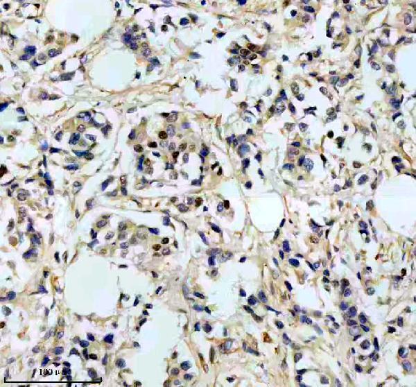
M00831 is a human-reactive anti-TIMELESS antibody with a paraffin-section IHC figure and listed IF/ICC applications; no IF figure is supplied (catalog: applications and reactivity; M00831 IHC caption).

Real IHC data IHC analysis of TIMELESS using anti-TIMELESS antibody (M00831). TIMELESS was detected in a paraffin-embedded section of human breast cancer tissue. Heat mediated antigen retrieval was performed in EDTA buffer (pH 8.0, epitope retrieval solution). The tissue section was blocked with 10% goat serum. The tissue section was then incubated with 1:50 rabbit anti-TIMELESS Antibody (M00831) overnight at 4°C. Peroxidase Conjugated Goat Anti-rabbit IgG was used as secondary antibody and incubated for 30 minutes at 37°C. The tissue section was developed using HRP Conjugated Rabbit IgG Super Vision Assay Kit (Catalog # SV0002) with DAB as the chromogen.
Anti-Timeless Rabbit Monoclonal Antibody
Cat # M00831

M00831 will render with an IHC figure from a paraffin-embedded human breast cancer section (M00831 IHC caption). IF/ICC is listed for M00831, but the payload supplies no IF image (catalog: applications and IF image alts).

Which to pick: Choose M00831 for human tissue IHC: it is a rabbit monoclonal, and its own caption documents paraffin-section staining at 1:50 after EDTA retrieval at pH 8.0 (catalog: host and clone; M00831 IHC caption). The fixative is unreported (M00831 IHC caption). For IF/ICC, M00831 is listed at 1:50, but has no IF figure; no cross-species reactivity is listed beyond human (catalog: applications, IF dilution, IF image alts and reactivity).

Each figure is that product's own IHC / IF validation image from its datasheet.

References

  1. UniProt Consortium. UniProt entry Q9UNS1 (TIM_HUMAN, Protein timeless homolog).
  2. Human Protein Atlas. TIMELESS tissue IHC expression (reliability: Enhanced).
  3. Human Protein Atlas. TIMELESS subcellular location (ICC-IF): Localized to the nucleoplasm..
  4. Human Protein Atlas. TIMELESS antibody validation summary (1 antibodies).
  5. An integrative evaluation of circadian gene TIMELESS as a pan-cancer immunological and predictive biomarker. European journal of medical research 2023 — PMC10696727.
  6. Activation of the clock gene TIMELESS by H3k27 acetylation promotes colorectal cancer tumorigenesis by binding to Myosin-9. Journal of experimental & clinical cancer research : CR 2021 — PMC8108341.
  7. Association of the Timeless Gene with Prognosis and Clinical Characteristics of Human Lung Cancer. Diagnostics (Basel, Switzerland) 2022 — PMC9688960.
  8. TIMELESS upregulates PD-L1 expression and exerts an immunosuppressive role in breast cancer. Journal of translational medicine 2023 — PMC10280842.
  9. PubMed PMID:9891984 — UniProt-cited evidence.
  10. PubMed PMID:9856465 — UniProt-cited evidence.
  11. PubMed PMID:16541075 — UniProt-cited evidence.