TIMM8A / Mitochondrial import inner membrane translocase subunit Tim8 A · IHC design guide

Design Immunohistochemistry for TIMM8A

Plan TIMM8A staining in paraffin sections using the catalog antibody’s IHC protocol. Compare granular cytoplasmic staining with HPA tissue IHC findings, including high staining in cardiomyocytes, distal tubules and hepatocytes (HPA tissue IHC).

Evidence assembled Oct 2026 · For research use; verify linked source records and product datasheet before use
Immunohistochemistry protocol sheet for TIMM8A (IHC for TIMM8A): expected localisation Granular cytoplasm (HPA tissue IHC); mitochondrial inner membrane (UniProt), antibody A07659-1, validated IHC image, and IHC protocol steps
Printable TIMM8A IHC protocol sheet — expected localisation Granular cytoplasm (HPA tissue IHC); mitochondrial inner membrane (UniProt), antibody A07659-1, controls and protocol steps. Open the full TIMM8A IHC guide →

TIMM8A Immunohistochemistry Experimental Design Guide

Expected localisation, validated protocols, controls and antibodies — the at-a-glance facts below, then the full design guide.

Must know before staining
Expected localisation Granular cytoplasm (HPA tissue IHC); mitochondrial inner membrane (UniProt)
Staining pattern Granular cytoplasmic staining in several tissues (HPA tissue IHC)
Antigen retrieval EDTA pH 8.0 HIER, heat-mediated (datasheet A07659-1)
Positive control ⓘ Cerebellum+4 more · see all
Negative control ⓘ Adipose tissue+4 more · see all
Important caveats
Reasons your staining may differ from the expected pattern.
Fixation Keep fixation consistent across sections (standard IHC practice; not target-specific)
Caveat Staining and RNA show medium consistency (HPA tissue IHC)
Regulation No specific expression regulator annotated (UniProt)
Isoform / epitope 0 isoforms annotated; one 1–97 chain (UniProt)
Section 1

Recommended TIMM8A IHC & IF Protocols

The catalog antibody protocol and these two published IHC protocols provide starting conditions for TIMM8A staining in paraffin sections (datasheet A07659-1; PMC13513568; PMC12408856).

Recommended immunohistochemistry (IHC-P) protocol parameters
SampleParaffin-embedded human melanoma tissue; fixative not specified (datasheet A07659-1)
FixationImage fixative and duration unreported (datasheet A07659-1); verify before use.
Sectioning4–5 µm sections on charged slides (standard)
DeparaffinisationXylene, graded ethanol series to water (standard)
Antigen retrievalHeat retrieval: EDTA pH 8.0 (datasheet A07659-1); 20 min, 95–100 °C (standard)
Peroxidase block3% H2O2, 10 min, room temperature (standard)
Blocking10% goat serum (datasheet A07659-1)
Primary antibodyRabbit anti-TIMM8A, 2-5μg/ml (datasheet A07659-1)
Primary incubationOvernight at 4 °C (datasheet A07659-1)
DetectionStreptavidin-biotin complex (SABC), DAB chromogen (datasheet A07659-1)
CounterstainHematoxylin, blue, dehydrate and mount (standard)
Expected resultTIMM8A-positive staining in synaptic glomeruli - core of cerebellum (HPA tissue IHC: High). HPA tissue profile: Granular cytoplasmic expression in several tissues. No signal in the no-primary control.
💡Decision noteStart with heat-mediated EDTA pH 8.0 retrieval for the catalog antibody (datasheet A07659-1); citrate pH 6.0 is a published alternative with a different antibody (PMC12408856).
Section 2

What Is the Expected TIMM8A Staining Pattern?

TIMM8A is associated with the mitochondrial inner membrane and has no transmembrane segment (UniProt O60220 topology). In paraffin section IHC, expect granular cytoplasmic staining in selected cells, including cardiomyocytes, kidney distal tubules and hepatocytes (HPA: High in each). HPA describes the tissue staining as Supported, with medium consistency between antibody staining and RNA expression; interpret individual positive cells in that context (HPA: reliability Supported).

What am I looking at on my slide?
Discrete cytoplasmic granules in cardiomyocytes, kidney distal tubules or hepatocytes, with little nuclear staining.This fits the reported granular cytoplasmic tissue pattern and the High staining in those cells (HPA: tissue IHC). Mitochondrial localization supports a granular cytoplasmic interpretation, although chromogenic IHC alone does not prove that each granule is a mitochondrion (UniProt O60220; HPA: ICC-IF mitochondria).
Strong, predominantly nuclear, cell-surface or extracellular staining replaces the expected cytoplasmic granules.Treat the compartment mismatch as a possible artifact or off-target signal and review controls before scoring it as TIMM8A. The expected compartment is mitochondrial, with no annotated transmembrane segment (UniProt O60220 topology); HPA reports granular cytoplasmic tissue staining (HPA: tissue IHC).
Strong staining dominates a cell population HPA reports as Not detected, such as adipocytes or lung alveolar cells.Cross-reactivity or endogenous detection activity is possible; compare a no-primary control and examine whether staining follows cell boundaries or tissue-wide deposits (standard IHC practice). HPA's Not detected calls are cell-type observations, so staining elsewhere in the same tissue needs separate interpretation (HPA: tissue IHC).
A uniform haze covers positive and negative cells, obscuring any discrete cytoplasmic pattern.Score the pattern as uninterpretable until background is resolved. Check the no-primary control, blocking and detection conditions (standard IHC practice). A diffuse field-wide signal does not match the reported granular cytoplasmic pattern (HPA: tissue IHC).
No signal appears in cardiomyocytes, kidney distal tubules or hepatocytes.First check section quality, primary-antibody and detection controls, and whether the relevant cells are present (standard IHC practice). These cells are High in HPA tissue IHC, so their absence of staining weakens confidence in a negative result elsewhere (HPA: High in each).
💡Expected TIMM8A appearanceCall a convincing positive when selected cells show clear granular cytoplasmic staining, especially HPA High cardiomyocytes, distal tubules or hepatocytes; dominant nuclear staining or uniform haze is suspect (HPA: tissue IHC; UniProt O60220 localization).
How each factor affects the staining
Compartment and topology (UniProt O60220)TIMM8A is associated with the mitochondrial inner membrane but has no transmembrane segment (UniProt O60220 topology). Read a granular cytoplasmic IHC pattern as consistent with localization; do not infer membrane insertion or exact organelle boundaries from chromogen alone (HPA: granular cytoplasmic tissue IHC).
Cell-specific tissue contrast (HPA: tissue IHC)HPA reports High staining in kidney distal tubules and Not detected staining in adipocytes and lung alveolar cells (HPA: tissue IHC). Use the named cell population when comparing sections; a whole-organ positive or negative label loses that distinction.
Expression evidence versus tissue staining (UniProt O60220; HPA: tissue IHC)UniProt lists expression in lung and skeletal muscle, while HPA reports Not detected IHC staining in lung alveolar cells and skeletal-muscle myocytes (UniProt O60220 tissue specificity; HPA: tissue IHC). Expression across a tissue does not guarantee visible staining in every sampled cell type.
Antibody evidence (HPA: HPA003628; HPA: tissue IHC)The listed rabbit polyclonal antibody has Supported IHC and ICC status (HPA: HPA003628). The tissue profile has medium consistency with RNA expression (HPA: reliability description); use matched controls and treat a mismatch as a finding to investigate, rather than overriding morphology.
IF/ICC Q&A: What localization should fluorescence show? (HPA: ICC-IF)A mitochondrial pattern is expected; HPA marks mitochondria as supported and lists ICC-IF images from A-431, U-251MG and U2OS (HPA: subcellular). That localization helps interpret IF/ICC images, while this guide's primary readout is chromogenic paraffin-section IHC.
Why is my staining missing, weak or wrong?
SituationLikely causeNext action
High-background section with no separable positive cells.Nonspecific primary binding or endogenous detection activity may contribute (standard IHC practice).Compare a no-primary control, review blocking and wash conditions, and reassess only after discrete granules can be distinguished from the field-wide haze (standard IHC practice; HPA: granular cytoplasmic tissue IHC).
Known HPA High cell types appear negative.A failed staining run, unsuitable retrieval or detection settings, or absent target cells is possible (standard IHC practice).Confirm the named cells are present, inspect run controls, and review the catalog antibody's validated IHC-P directions before changing retrieval or dilution (standard IHC practice; HPA: High cardiomyocytes, distal tubules and hepatocytes).
Strong nuclear or extracellular staining is the principal result.The compartment differs from mitochondrial localization (UniProt O60220; HPA: ICC-IF mitochondria).Check the no-primary control and inspect a known HPA High cell population for granular cytoplasmic staining before assigning the unusual signal to TIMM8A (standard IHC practice; HPA: tissue IHC).
Adipocytes or lung alveolar cells show strong staining.Those specific cell types are Not detected in HPA tissue IHC; cross-reactivity or endogenous activity is possible (HPA: tissue IHC; standard IHC practice).Identify the stained cells precisely, compare no-primary and positive-section controls, and avoid extending HPA's cell-specific calls to every cell in adipose tissue or lung (standard IHC practice; HPA: tissue IHC).
An organ expected to express TIMM8A gives a mixed or weak IHC result.UniProt tissue expression and HPA cell-specific staining describe different evidence; HPA also rates RNA-to-staining consistency as medium (UniProt O60220 tissue specificity; HPA: reliability description).Score identifiable cell populations and their staining pattern, using HPA's named High, Medium, Low or Not detected cells as the comparison where available (HPA: tissue IHC).
An IF/ICC image looks diffuse despite a positive chromogenic IHC control.HPA supports mitochondrial ICC-IF localization, so diffuse fluorescence needs separate evaluation (HPA: subcellular).Assess the IF/ICC image against its own controls and mitochondrial pattern; keep its interpretation separate from the paraffin-section IHC result (standard IF practice; HPA: ICC-IF mitochondria).

Sample controls for TIMM8A IHC & IF

🧪Run liver first: hepatocytes should stain (HPA: High in hepatocytes). Use adipose tissue as the negative comparator (HPA: Not detected in adipocytes); any unstained neighboring cells on the liver slide should show counterstain without specific chromogen, but HPA does not identify a validated negative liver cell type (HPA: High in hepatocytes).
Positive control tissue: Cerebellum (Synaptic glomeruli - core, HPA High)
Negative control tissue: Adipose tissue (HPA Not detected)
ICC-IF cell lines (HPA subcellular resource): HPA ICC-IF images show TIMM8A in A-431, U-251MG, U2OS, with annotated localisation: Mitochondria (supported) (HPA subcellular).
Technical controls: Include no-primary (secondary-only), rabbit IgG isotype matched to the primary antibody’s clonality when known, and TIMM8A knockout material if available (caption: rabbit primary and biotinylated goat anti-rabbit secondary). Quench endogenous peroxidase and check for endogenous biotin background in liver when using avidin-biotin DAB detection (caption: SABC-DAB; standard IHC practice).
⚠️Feasibility: A target-specific fixation window or fixation effect is unreported, and the selected paraffin-section caption does not state a fixative (caption: fixative not stated). Heat retrieval in EDTA at pH 8.0 was used for the pictured melanoma section, but retrieval dependence is unreported (caption: EDTA heat retrieval in melanoma). The supplied evidence does not establish frozen sections or IF as easier; IF/ICC does show supported mitochondrial localization (HPA: Mitochondria supported), while liver endogenous biotin can complicate avidin-biotin detection (caption: SABC-DAB; standard IHC practice).

HPA tissue IHC evidence for TIMM8A

Comprehensive Human Protein Atlas IHC scoring per tissue (reliability: Supported — Medium consistency between antibody staining and RNA expression data.). Rows are taken directly from the HPA tissue chart — click any row's HPA link to view the source.

Positive expression · recommended positive controls

TissueCell typeLevelEvidenceSource
Cerebellum Synaptic glomeruli - core High Protein (IHC) HPA →
Heart muscle Cardiomyocytes High Protein (IHC) HPA →
Kidney Distal tubules High Protein (IHC) HPA →
Liver Hepatocytes High Protein (IHC) HPA →
Adrenal gland Glandular cells Medium Protein (IHC) HPA →

Undetected expression · recommended negative controls

TissueCell typeLevelEvidenceSource
Adipose tissue Adipocytes Not detected Protein (IHC) HPA →
Bone marrow Hematopoietic cells Not detected Protein (IHC) HPA →
Gallbladder Glandular cells Not detected Protein (IHC) HPA →
Lung Alveolar cells Not detected Protein (IHC) HPA →
Oral mucosa Squamous epithelial cells Not detected Protein (IHC) HPA →
Section 3

Advanced TIMM8A IHC Tips

Use the catalog antibody’s paraffin-section example as the starting point, then judge TIMM8A staining by cell type, mitochondrial pattern and matched controls.

Which retrieval condition should I start with for TIMM8A paraffin-section IHC?
Start with heat-mediated retrieval in EDTA at pH 8.0 (datasheet A07659-1). The selected paraffin-section example used that condition before overnight incubation with 2 µg/mL primary antibody at 4°C (datasheet A07659-1). Apply the same heating, cooling and washing schedule to every section in a comparison, and include a section processed without primary antibody (standard IHC practice). If staining is weak, adjust heating time in small steps while checking tissue integrity; the caption does not specify a retrieval duration (datasheet A07659-1). Judge the result by interpretable granular cytoplasmic staining in suitable cells, alongside low background in controls (HPA tissue IHC; standard IHC practice).
How should I troubleshoot fixation when TIMM8A staining varies between sections?
Target-specific fixation sensitivity is unknown: the selected paraffin-section caption does not state a fixative, and the supplied evidence gives no TIMM8A fixation comparison (datasheet A07659-1). Record the fixative and processing history for each specimen before attributing different staining intensities to biology (standard IHC practice). Compare sections processed together using EDTA retrieval at pH 8.0 and the same primary-antibody conditions, including the reported 2 µg/mL starting concentration (datasheet A07659-1). Inspect morphology and staining across each section for uneven processing or edge effects, then repeat suspect samples with matched controls (standard IHC practice). Neither mitochondrial location nor phosphorylation sites establish a preferred fixative (UniProt O60220 localisation and modified residues).
What staining pattern supports a mitochondrial TIMM8A signal in tissue?
Look for granular cytoplasmic staining consistent with mitochondria, while recognizing that chromogenic IHC alone cannot resolve the side of a mitochondrial membrane (HPA tissue IHC; HPA subcellular; standard IHC practice). UniProt places TIMM8A at the mitochondrial inner membrane and annotates no transmembrane segment (UniProt O60220 localisation and topology). HPA reports high staining in cardiomyocytes, distal tubules and hepatocytes, providing documented cell populations for comparison (HPA tissue IHC). Check whether granules stay within intact cell boundaries across the section and whether the no-primary control remains clear (standard IHC practice). Predominantly nuclear or diffuse extracellular DAB should prompt a review of controls and tissue preservation before being called TIMM8A (standard IHC practice).
Could an isoform or epitope-access problem explain weak TIMM8A IHC?
The supplied UniProt record lists 0 isoforms and a single 1–97 chain, so an annotated isoform switch is not an evidence-based explanation here (UniProt O60220 isoforms and processing). It also lists no transmembrane segment and phosphorylation at several residues, including 57 and 87, but gives no antibody epitope (UniProt O60220 topology and modified residues; datasheet A07659-1). Do not assign a phosphorylation-dependent staining effect without direct validation (standard IHC practice). First reproduce EDTA retrieval at pH 8.0 and the reported 2 µg/mL antibody concentration, then compare matched controls (datasheet A07659-1; standard IHC practice). If signal remains weak, test retrieval exposure systematically while monitoring morphology and background (standard IHC practice).
How can I follow up a TIMM8A IHC pattern with multiplex IF?
Treat IF/ICC as a separate assay: the supplied paraffin-section caption establishes a chromogenic IHC workflow, not IF fixation or antibody performance (datasheet A07659-1). Pair TIMM8A with a mitochondrial marker and a marker identifying the expected cell population, such as cardiomyocytes or distal-tubule cells in the documented tissues (HPA subcellular; HPA tissue IHC; standard IF practice). Choose fluorophores after inspecting unstained tissue autofluorescence, and assign the dimmer channel to a cleaner spectral range (standard IF practice). Optimise permeabilisation empirically for access to an intramitochondrial epitope; TIMM8A has no annotated transmembrane segment, but its antibody epitope is unspecified (UniProt O60220 topology; datasheet A07659-1). Include single-label and no-primary controls before interpreting colocalisation (standard IF practice).
What should I change when TIMM8A DAB staining is widespread or muddy?
Check a no-primary section first; DAB signal there points to detection-system background rather than antibody-dependent staining (standard IHC practice). The selected example used a 10% goat-serum block, 2 µg/mL rabbit primary antibody overnight at 4°C, and a biotin-based DAB detection system (datasheet A07659-1). For that detection chemistry, verify the usual endogenous peroxidase block and assess whether endogenous biotin contributes to signal (standard IHC practice). If controls are clean but tissue staining remains muddy, titrate primary antibody and detection exposure while keeping retrieval at EDTA pH 8.0 initially (datasheet A07659-1; standard IHC practice). Recheck whether the remaining signal has the documented granular cytoplasmic pattern (HPA tissue IHC).
How should I score TIMM8A staining across tissue sections? ⚠ ANSWER MARKED FOR VERIFICATION
Define the cell population and analysis area before scoring, then exclude folds, necrosis and damaged edges using the same rules for every section (standard IHC practice). For cell-rich regions, record the percentage of positive cells and an intensity-weighted H-score; for spatially discrete regions, report positive-cell density per mm² (standard IHC practice). Normalise counts to viable tissue area or the number of eligible cells, and keep imaging and DAB development conditions matched (standard IHC practice). Report scores by cell type because HPA describes high staining in cardiomyocytes, distal tubules and hepatocytes, but no detection in several other sampled cell populations (HPA tissue IHC). Use no-primary controls to set a consistent background threshold (standard IHC practice).
How do I distinguish genuine TIMM8A staining from tissue or detection artefact?
A credible result combines granular cytoplasmic signal with the expected cell context and a clean no-primary control (HPA tissue IHC; standard IHC practice). TIMM8A is mitochondrial, while HPA tissue IHC reports high staining in cardiomyocytes, distal tubules and hepatocytes; these observations support pattern-based checks without proving antibody specificity in a new specimen (UniProt O60220 localisation; HPA tissue IHC; standard IHC practice). Treat nuclear staining, necrotic deposits, section-edge enrichment and signal in the no-primary control as reasons to investigate artefact (standard IHC practice). In a biotin-based DAB workflow, assess endogenous peroxidase and biotin contributions when staining survives omission of primary antibody (datasheet A07659-1; standard IHC practice). Interpret weak or absent staining alongside morphology and matched controls (standard IHC practice).
Boster reagents

Best TIMM8A / Mitochondrial import inner membrane translocase subunit Tim8 A IHC Antibodies

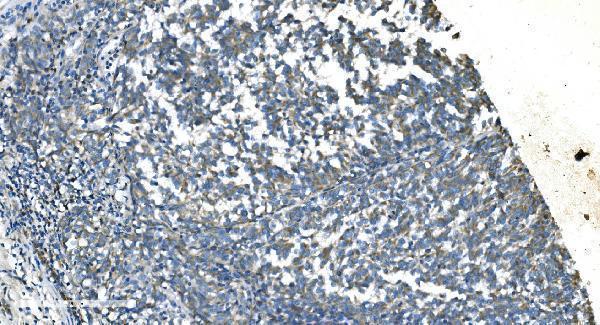
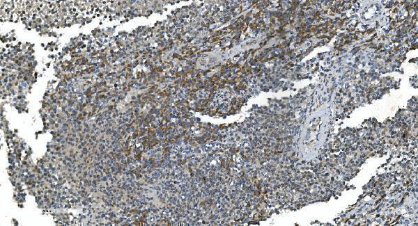
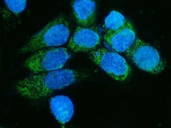
A07659-1 has IHC images from paraffin-embedded human melanoma, testicular cancer, lung cancer and tonsil cancer tissues, plus an IF image from A431 cells (catalog image captions).

Real IHC data IHC analysis of TIMM8A/DDP using anti-TIMM8A/DDP antibody (A07659-1). TIMM8A/DDP was detected in paraffin-embedded section of human melanoma tissue. Heat mediated antigen retrieval was performed in EDTA buffer (pH8.0, epitope retrieval solution). The tissue section was blocked with 10% goat serum. The tissue section was then incubated with 2μg/ml rabbit anti-TIMM8A/DDP Antibody (A07659-1) overnight at 4°C. Biotinylated goat anti-rabbit IgG was used as secondary antibody and incubated for 30 minutes at 37°C. The tissue section was developed using Strepavidin-Biotin-Complex (SABC) (Catalog # SA1022) with DAB as the chromogen.
Anti-TIMM8A/DDP Antibody ®
Cat # A07659-1

A07659-1 is listed for IHC and IF/ICC, with human paraffin-section IHC images and an A431-cell IF image (catalog applications and image captions). Its listed reactivity is human, mouse, rat and monkey; the application-specific listing names human and rat for IHC and human for IF/ICC (catalog dilution listing).

Which to pick: For tissue IHC, choose A07659-1: its own captions document paraffin sections with EDTA retrieval at pH 8.0 and primary antibody at 2 μg/ml; the fixative is unreported (A07659-1 IHC captions). For IF/ICC, the same rabbit antibody has an A431-cell IF image at 5 μg/ml (A07659-1 IF caption). For cross-species planning, A07659-1 lists human, mouse, rat and monkey reactivity, while its application-specific IHC listing names human and rat (catalog reactivity and dilution listing).

Each figure is that product's own IHC / IF validation image from its datasheet.

References

  1. UniProt Consortium. UniProt entry O60220 (TIM8A_HUMAN, Mitochondrial import inner membrane translocase subunit Tim8 A).
  2. Human Protein Atlas. TIMM8A tissue IHC expression (reliability: Supported).
  3. Human Protein Atlas. TIMM8A subcellular location (ICC-IF): Localized to the mitochondria..
  4. Human Protein Atlas. TIMM8A antibody validation summary (1 antibodies).
  5. Loss of TIMM8A Inhibits Breast Cancer Progression Through Promoting HSPA9 Ubiquitination and Degradation via Competitive Interaction with RNF4. International journal of molecular sciences 2026 — PMC13513568.
  6. ncRNAs-mediated high expression of TIMM8A correlates with poor prognosis and act as an oncogene in breast cancer. Cancer cell international 2022 — PMC9063222.
  7. TIMM8A-TIMM13 Complex Exerts Oncogenic Functions in Lung Cancer. Oncology research 2025 — PMC12408856.
  8. PubMed PMID:8841189 — UniProt-cited evidence.
  9. PubMed PMID:14702039 — UniProt-cited evidence.
  10. PubMed PMID:15772651 — UniProt-cited evidence.