TJP2 / Tight junction protein 2 · IHC design guide

Design Immunohistochemistry for TJP2

Plan TJP2 chromogenic IHC in paraffin sections using the observed cytoplasmic and membranous tissue pattern (HPA tissue IHC). Compare staining across tissues with the IHC-validated antibody, accounting for tissue-dependent isoform expression (UniProt).

Evidence assembled Oct 2026 · For research use; verify linked source records and product datasheet before use
Immunohistochemistry protocol sheet for TJP2 (IHC for TJP2): expected localisation Cytoplasmic and membranous staining in most tissues (HPA tissue IHC), antibody A02774-1, validated IHC image, and IHC protocol steps
Printable TJP2 IHC protocol sheet — expected localisation Cytoplasmic and membranous staining in most tissues (HPA tissue IHC), antibody A02774-1, controls and protocol steps. Open the full TJP2 IHC guide →

TJP2 Immunohistochemistry Experimental Design Guide

Expected localisation, validated protocols, controls and antibodies — the at-a-glance facts below, then the full design guide.

Must know before staining
Expected localisation Cytoplasmic and membranous staining in most tissues (HPA tissue IHC)
Staining pattern Glandular cells show cytoplasmic and membranous staining (HPA tissue IHC)
Antigen retrieval EDTA pH 8.0 HIER, heat-mediated (datasheet A02774-1)
Positive control ⓘ Adrenal gland+4 more · see all
Negative control ⓘ Adipose tissue+3 more · see all
Important caveats
Reasons your staining may differ from the expected pattern.
Fixation Keep paraffin-section fixation consistent between samples (standard IHC practice; not target-specific)
Caveat HPA staining may detect targets from more than one gene (HPA tissue IHC)
Regulation Isoform expression varies by tissue (UniProt)
Isoform / epitope Seven isoforms; confirm which isoforms the epitope covers (UniProt)
Section 1

Recommended TJP2 IHC & IF Protocols

The catalog antibody’s IHC protocol is followed by published TJP2 staining details from two articles (PMC4061468; PMC10039855).

Recommended immunohistochemistry (IHC-P) protocol parameters
SampleParaffin-embedded human ovarian serous adenocarcinoma tissue; fixative not specified (datasheet A02774-1)
FixationImage fixative and duration unreported (datasheet A02774-1); verify before use.
Sectioning4–5 µm sections on charged slides (standard)
DeparaffinisationXylene, graded ethanol series to water (standard)
Antigen retrievalHeat retrieval: EDTA pH 8.0 (datasheet A02774-1); 20 min, 95–100 °C (standard)
Peroxidase block3% H2O2, 10 min, room temperature (standard)
Blocking10% goat serum (datasheet A02774-1)
Primary antibodyRabbit anti-TJP2, 2μg/ml (datasheet A02774-1)
Primary incubationOvernight at 4 °C (datasheet A02774-1)
DetectionStreptavidin-biotin complex (SABC), DAB chromogen (datasheet A02774-1)
CounterstainHematoxylin, blue, dehydrate and mount (standard)
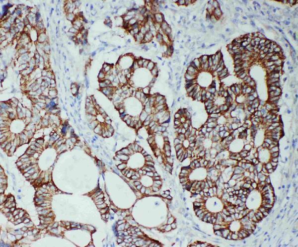
Expected resultTJP2-positive staining in glandular cells of adrenal gland (HPA tissue IHC: Medium). HPA tissue profile: Cytoplasmic and membranous expression in most tissues. No signal in the no-primary control.
💡Decision noteStart with heat-mediated EDTA retrieval at pH 8.0 for the catalog antibody (datasheet A02774-1). The cited articles do not specify retrieval conditions (PMC4061468; PMC10039855).
Section 2

What Is the Expected TJP2 Staining Pattern?

TJP2 is associated with tight and adherens junctions and has no transmembrane segment (UniProt Q9UDY2). In paraffin tissue sections, expect staining along epithelial cell borders, with cytoplasmic and membranous staining also reported across many tissues (HPA: tissue IHC). HPA rates its tissue pattern Supported, with medium agreement between staining and RNA data, and cautions that the antibody signal may include protein from more than one gene (HPA: tissue IHC).

What am I looking at on my slide?
Glandular or respiratory epithelial cells show membranous staining, especially at cell contacts, with some cytoplasmic signal.This fits the junction-associated location of TJP2 (UniProt Q9UDY2) and HPA's cytoplasmic and membranous tissue profile (HPA: tissue IHC). HPA reports Medium staining in glandular cells of colon, duodenum, breast and endometrium, and in bronchial respiratory epithelium (HPA: tissue IHC).
The strongest signal is uniformly nuclear, with little staining at epithelial borders.Treat this as a compartment mismatch for routine tissue interpretation (HPA: tissue IHC). Nuclear TJP2 remains possible under environmental stress and in migratory endothelial cells or subconfluent epithelial cultures (UniProt Q9UDY2). HPA also reports nucleoplasmic ICC-IF signal (HPA: subcellular). Nuclear staining alone therefore needs context and antibody controls before it is scored as specific (general IHC practice).
Strong staining appears in adipocytes or soft-tissue fibroblasts while nearby epithelium is weak.These cell types were Not detected in HPA tissue IHC (HPA: adipose tissue; soft tissue). Consider antibody cross-reactivity or endogenous detection activity, especially because HPA cautions that the tissue antibody signal may include protein from more than one gene (HPA: tissue IHC). A negative HPA observation is a comparison, not proof that every specimen must be negative.
Color covers stroma and empty spaces without recognizable cell borders.This is diffuse background rather than the expected cell-associated pattern (HPA: tissue IHC; UniProt Q9UDY2). Uneven blocking, residual detection reagent or excessive chromogen development are general IHC possibilities (general IHC practice). Judge whether the signal follows cells before assigning it to TJP2.
No staining is visible in glandular cells of a colon or duodenum section.HPA reports Medium staining in those cells, so the section is a useful positive comparison (HPA: tissue IHC). An absent signal can reflect assay failure or specimen variation; it does not establish TJP2 absence from one negative slide (general IHC practice). Review controls and the assay steps before drawing a biological conclusion.
💡Expected TJP2 appearanceCall a result compatible with TJP2 when epithelial cell borders show interpretable membranous staining, with possible cytoplasmic signal and approximately Medium staining in HPA-listed glandular or bronchial cells (UniProt Q9UDY2; HPA: tissue IHC); isolated stain in empty spaces or unexpected cell types is a potential false positive requiring controls (general IHC practice).
How each factor affects the staining
Junction association and topologyTJP2 has tight-junction and adherens-junction locations but no transmembrane segment (UniProt Q9UDY2). Score the pattern at cell contacts; a diffuse outline of every structure has less support from the reported tissue pattern (HPA: tissue IHC). Topology alone does not predict an antigen-retrieval condition.
Tissue and cell-type contextHPA describes cytoplasmic and membranous expression in most tissues, with Low staining in lung alveolar and oral squamous epithelial cells and Not detected staining in adipocytes (HPA: tissue IHC). Compare the named cell population rather than treating every tissue as uniformly positive.
Antibody evidenceHPA001813 and CAB009228 have Supported IHC status; HPA070714 has no listed IHC status (HPA: antibodies). HPA's overall tissue reliability is Supported and includes a caution about signal from more than one gene (HPA: tissue IHC). These ratings support cautious pattern comparison, not antibody-specific identity in an individual stained cell.
Isoforms and tissue distributionUniProt lists seven TJP2 isoforms and reports different tissue distributions for A1 and C1 (UniProt Q9UDY2). Because the supplied evidence does not map an IHC antibody epitope to an isoform, a tissue difference cannot be assigned to a particular isoform from staining alone.
Additional nuclear locationUniProt describes nuclear localization in specific stress or cell-state contexts, while HPA calls nucleoplasm an additional ICC-IF location (UniProt Q9UDY2; HPA: subcellular). Use this as context for an unexpected nuclear signal, without making nuclear staining the default positive pattern for paraffin tissue sections (HPA: tissue IHC).
Why is my staining missing, weak or wrong?
SituationLikely causeNext action
Known-positive glandular epithelium has no visible stain.HPA reports Medium glandular staining in colon and duodenum, so a fully blank result merits an assay check (HPA: tissue IHC).Check the positive control, primary-antibody step and chromogenic detection; compare any antigen-retrieval adjustment against the antibody's IHC-P instructions (general IHC practice). The supplied sources give no TJP2-specific retrieval condition.
Borders are hard to distinguish from broad cytoplasmic color.Some cytoplasmic staining is expected in tissue, but the pattern should remain interpretable by cell type (HPA: tissue IHC).Review adjacent cells and the counterstain, then compare the border pattern in a known-positive epithelial area (general IHC practice; HPA: tissue IHC). Avoid scoring diffuse color as junctional staining solely because the tissue is expected to express TJP2.
Adipocytes or fibroblasts stain strongly.Both are listed as Not detected in the specified HPA tissue samples; cross-reactivity or endogenous detection activity is possible (HPA: tissue IHC; general IHC practice).Inspect a no-primary control and reassess blocking and detection chemistry (general IHC practice). Compare staining with the expected epithelial pattern, while retaining HPA's caution that the tissue antibody may detect protein from more than one gene (HPA: tissue IHC).
Background is widespread across cells and spaces.A pattern that ignores tissue structures is inconsistent with HPA's cellular staining description (HPA: tissue IHC); assay background is possible (general IHC practice).Check the no-primary control, wash and blocking steps, and chromogen development (general IHC practice). Interpret a revised section only when cellular boundaries can be distinguished.
Nuclear staining dominates the IHC section.Nuclear TJP2 can occur in the contexts UniProt describes, but HPA's tissue profile emphasizes cytoplasmic and membranous staining (UniProt Q9UDY2; HPA: tissue IHC).Record the cell type and specimen context, compare epithelial borders, and assess controls before accepting a nuclear call (general IHC practice). Do not infer environmental stress from nuclear color alone.
Q: Should an IF/ICC image show the same pattern as this IHC-P section?A: HPA reports enhanced plasma-membrane and cell-junction locations plus approved nucleoplasmic localization in ICC-IF (HPA: subcellular). UniProt also allows nuclear localization in specific contexts (UniProt Q9UDY2).Use the IF/ICC guide for that application. Here, compare the IHC-P section with HPA's tissue cell types and cytoplasmic or membranous profile (HPA: tissue IHC); do not treat an ICC-IF nuclear image as an IHC-P positive control.

Sample controls for TJP2 IHC & IF

🧪Run breast first: its glandular cells should stain (HPA: Medium in breast glandular cells). Run adipose tissue as the negative tissue (HPA: Not detected in adipocytes); adipocytes within the breast section, where present, should show background-level staining (HPA: Not detected in adipocytes).
Positive control tissue: Adrenal gland (Glandular cells, HPA Medium)
Negative control tissue: Adipose tissue (HPA Not detected)
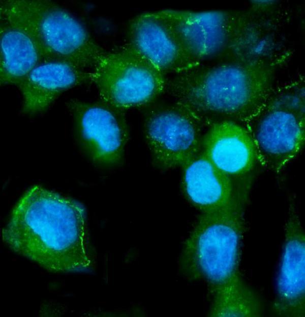
ICC-IF cell lines (HPA subcellular resource): HPA ICC-IF images show TJP2 in A-431, U-251MG, U2OS, HaCaT, with annotated localisation: Plasma membrane (enhanced), Cell Junctions (enhanced) (HPA subcellular).
Technical controls: Include no-primary (secondary-only), a rabbit IgG isotype control matched to the primary antibody’s clonality, and TJP2-knockout tissue as a biological negative (caption: rabbit anti-TJP2 primary; standard IHC controls). For the caption’s biotin-based DAB detection, block endogenous peroxidase and assess endogenous biotin background on the breast section (caption: biotinylated secondary, SABC and DAB; standard IHC practice).
⚠️Feasibility: A target-specific fixation window or fixation effect is unreported, and the selected A02774-1 tissue-IHC caption does not report a fixative (caption: fixative not stated). The caption uses heat retrieval in EDTA at pH 8.0, but does not establish whether retrieval is required; the supplied evidence does not establish that frozen sections or IF are easier (caption: heat-mediated EDTA retrieval; HPA: ICC-IF images). No breast-specific artefact is reported in the supplied evidence (HPA: breast glandular-cell staining; caption: ovarian tissue-IHC example).

HPA tissue IHC evidence for TJP2

Comprehensive Human Protein Atlas IHC scoring per tissue (reliability: Supported — Medium consistency between antibody staining and RNA expression data. Caution, targets protein from more than one gene.). Rows are taken directly from the HPA tissue chart — click any row's HPA link to view the source.

Positive expression · recommended positive controls

TissueCell typeLevelEvidenceSource
Adrenal gland Glandular cells Medium Protein (IHC) HPA →
Appendix Glandular cells Medium Protein (IHC) HPA →
Breast Glandular cells Medium Protein (IHC) HPA →
Bronchus Respiratory epithelial cells Medium Protein (IHC) HPA →
Cervix Glandular cells Medium Protein (IHC) HPA →

Undetected expression · recommended negative controls

TissueCell typeLevelEvidenceSource
Adipose tissue Adipocytes Not detected Protein (IHC) HPA →
Cerebellum Cells in granular layer Not detected Protein (IHC) HPA →
Soft tissue Fibroblasts Not detected Protein (IHC) HPA →
Spleen Cells in red pulp Not detected Protein (IHC) HPA →
Section 3

Advanced TJP2 IHC Tips

Troubleshoot TJP2 chromogenic staining in paraffin sections by checking retrieval, compartment, controls and scoring before interpreting changes in expression.

Which retrieval conditions should I start with for TJP2 in paraffin sections?
Start with heat-mediated retrieval in EDTA at pH 8.0 (datasheet A02774-1). The catalog antibody detected TJP2 in a paraffin section after that treatment, followed by 2 μg/mL primary antibody overnight at 4°C (datasheet A02774-1). If staining is weak, compare a modest change in heating time on adjacent sections while holding antibody concentration and detection constant (standard IHC practice). Check that epithelial junctions gain a coherent signal rather than diffuse cytoplasmic staining, because TJP2 localizes to cell junctions and can also occupy other compartments (UniProt Q9UDY2; HPA subcellular). Record retrieval conditions for every comparison so changes in intensity remain interpretable (standard IHC practice).
How should I troubleshoot variable staining when fixation history is uncertain?
Target-specific fixation sensitivity is unknown: the selected paraffin-section caption does not state a fixative (datasheet A02774-1). Document the actual fixative and fixation duration for each specimen, and compare sections with matched processing whenever possible (standard IHC practice). If staining varies, run adjacent sections through the same EDTA pH 8.0 retrieval and detection sequence before attributing the difference to biology (datasheet A02774-1; standard IHC practice). Include a consistently processed epithelial control and inspect tissue preservation alongside staining intensity (UniProt Q9UDY2 tissue specificity; standard IHC practice). Neither the reported tissue pattern nor TJP2 topology establishes how this antibody responds to fixation (HPA tissue IHC; UniProt Q9UDY2 topology).
Where should convincing TJP2 staining appear in chromogenic IHC?
Prioritize coherent staining along epithelial cell borders, especially junction-rich interfaces, because TJP2 is found in epithelial junctions (UniProt Q9UDY2). Cytoplasmic and membranous tissue staining has also been reported, so a broad DAB signal needs compartment-level review rather than automatic rejection (HPA tissue IHC). Nuclear staining can be biologically plausible under environmental stress, although that context cannot be established from an isolated section (UniProt Q9UDY2 subcellular). Compare the same cell types across sections and use a counterstain that leaves cell borders readable (standard IHC practice). Treat a diffuse signal that ignores epithelial architecture as a specificity question, particularly because the HPA tissue antibody may recognize proteins from more than one gene (HPA tissue IHC).
Can this antibody distinguish TJP2 isoforms or reveal a masked epitope?
Do not assign an IHC pattern to one isoform without epitope-mapping evidence for the catalog antibody (standard IHC interpretation). TJP2 has 7 listed isoforms, and A1 and C1 have different reported tissue distributions (UniProt Q9UDY2 isoforms and tissue specificity). Its PDZ, SH3 and guanylate kinase-like regions offer possible sequence contexts, but the supplied caption does not identify the antibody epitope (UniProt Q9UDY2 domains; datasheet A02774-1). The record also lists phosphorylation sites; staining changes alone cannot establish a modification-dependent effect (UniProt Q9UDY2 modified residues; standard IHC interpretation). If an isoform claim matters, compare mapped reagents or orthogonal isoform-specific evidence with matched section processing (standard IHC practice).
How can IF help resolve an ambiguous chromogenic TJP2 pattern?
Use IF on a separately optimized specimen to test whether TJP2 signal follows cell borders and overlaps an epithelial marker such as cytokeratin (UniProt Q9UDY2 tissue specificity; standard IF practice). Choose spectrally separated fluorophores, favoring a far-red channel for the weaker target where tissue autofluorescence is prominent (standard IF practice). TJP2 lacks a transmembrane segment, so test mild permeabilisation to expose intracellular junction-associated epitopes while checking that borders remain intact (UniProt Q9UDY2 topology; standard IF practice). Include single-stain and no-primary controls to assess bleed-through and autofluorescence (standard IF practice). HPA reports junctional and plasma-membrane enrichment with additional nucleoplasmic localization in ICC/IF, which provides a compartment reference rather than validation of this catalog antibody for IF (HPA subcellular).
What should I adjust when DAB obscures junctional TJP2 staining?
First inspect a no-primary section for detection-system background and endogenous enzyme activity before changing the primary antibody (standard chromogenic IHC practice). Apply a peroxidase block when using DAB detection, then maintain consistent wash and development conditions across sections (standard chromogenic IHC practice). The selected example used 10% goat serum blocking, 2 μg/mL primary overnight at 4°C, and a biotinylated secondary with DAB development (datasheet A02774-1). If background persists, titrate primary concentration and shorten DAB development while retaining a positive control (standard IHC practice). Evaluate whether remaining color follows epithelial borders rather than tissue edges or acellular material (UniProt Q9UDY2 subcellular; standard IHC practice).
How should I score TJP2 staining across paraffin sections? ⚠ ANSWER MARKED FOR VERIFICATION
Define the epithelial region and score comparable, viable cells using percent positive cells or an H-score with fixed intensity categories (UniProt Q9UDY2 tissue specificity; standard IHC quantification). If junction continuity is the endpoint, measure stained border length or junctional signal density per mm² of evaluable epithelium instead of pooling all DAB-positive pixels (UniProt Q9UDY2 subcellular; standard image analysis practice). Normalize to epithelial area or cell count and keep retrieval, exposure, staining batch and threshold consistent (standard IHC quantification). Score nuclear and cytoplasmic staining separately from border staining because TJP2 can occupy those compartments (UniProt Q9UDY2 subcellular; HPA tissue IHC). Exclude folds, necrosis and section edges before comparison (standard IHC quantification).
How do I distinguish true TJP2 signal from artefact?
A persuasive positive pattern follows intact epithelial cell interfaces and is reproducible in similarly processed sections (UniProt Q9UDY2 tissue specificity and subcellular; standard IHC interpretation). Nuclear signal warrants contextual review because TJP2 can localize there under environmental stress, while isolated nuclear DAB does not prove that state (UniProt Q9UDY2 subcellular; standard IHC interpretation). Flag staining concentrated at cut edges, folds or necrotic areas, and investigate no-primary staining or incomplete peroxidase block before calling it positive (standard chromogenic IHC practice). Compare cell identity and compartment with controls; HPA reports cytoplasmic and membranous tissue staining but cautions that its tissue antibody may recognize more than one gene (HPA tissue IHC). Interpret intensity changes alongside morphology and processing records (standard IHC interpretation).
Boster reagents

Best TJP2 / Tight junction protein 2 IHC Antibodies

Both antibodies have human paraffin-section IHC images and A431 cell IF images; listed species reactivity spans human, monkey, mouse and rat (catalog images; catalog reactivity).

Real IHC data IHC analysis of TJP2/ZO2 using anti-TJP2/ZO2 antibody (A02774-1). TJP2/ZO2 was detected in paraffin-embedded section of human ovarian serous adenocarcinoma tissue. Heat mediated antigen retrieval was performed in EDTA buffer (pH8.0, epitope retrieval solution). The tissue section was blocked with 10% goat serum. The tissue section was then incubated with 2μg/ml rabbit anti-TJP2/ZO2 Antibody (A02774-1) overnight at 4°C. Biotinylated goat anti-rabbit IgG was used as secondary antibody and incubated for 30 minutes at 37°C. The tissue section was developed using Strepavidin-Biotin-Complex (SABC) (Catalog # SA1022) with DAB as the chromogen.
Anti-TJP2/ZO2 Antibody ®
Cat # A02774-1
Real IHC data IHC analysis of TJP2 using anti-TJP2 antibody (PA1957). TJP2 was detected in a paraffin-embedded section of Human Intestinal Cancer tissue. Heat mediated antigen retrieval was performed in EDTA buffer (pH 8.0, epitope retrieval solution). The tissue section was blocked with 10% goat serum. The tissue section was then incubated with 1 μg/ml rabbit anti-TJP2 Antibody (PA1957) overnight at 4°C. Peroxidase Conjugated Goat Anti-rabbit IgG was used as secondary antibody and incubated for 30 minutes at 37°C. The tissue section was developed using HRP Conjugated Rabbit IgG Super Vision Assay Kit (Catalog # SV0002) with DAB as the chromogen.
Anti-Tight junction protein ZO-2 TJP2 Antibody ®
Cat # PA1957

A02774-1 has IHC images from human ovarian serous adenocarcinoma, rectal cancer and ovarian cancer paraffin sections, plus an A431 cell IF image (A02774-1 image captions). PA1957 has a human intestinal cancer paraffin-section IHC image and A431 and U2OS cell IF images (PA1957 image captions).

Which to pick: For chromogenic tissue IHC, PA1957 offers a listed 0.5–1 μg/ml paraffin-section range (PA1957 catalog dilution); its human intestinal cancer image used EDTA pH 8.0 retrieval and DAB, with the fixative unreported (PA1957 IHC caption). For IF/ICC, PA1957 has images from two cell lines, A431 and U2OS (PA1957 IF captions), while A02774-1 has an A431 image (A02774-1 IF caption); both are rabbit antibodies with clonality unreported (catalog). For broader listed species reactivity, choose A02774-1, which includes monkey alongside human, mouse and rat (A02774-1 catalog reactivity); its IHC images show human paraffin sections processed with EDTA pH 8.0 retrieval and DAB, with the fixative unreported (A02774-1 IHC captions).

Each figure is that product's own IHC / IF validation image from its datasheet.

References

  1. UniProt Consortium. UniProt entry Q9UDY2 (ZO2_HUMAN, Tight junction protein 2).
  2. Human Protein Atlas. TJP2 tissue IHC expression (reliability: Supported).
  3. Human Protein Atlas. TJP2 subcellular location (ICC-IF): Mainly localized to the plasma membrane and cell junctions. In addition localized to the nucleoplasm..
  4. Human Protein Atlas. TJP2 antibody validation summary (3 antibodies).
  5. Mutations in TJP2 cause progressive cholestatic liver disease. Nature genetics 2014 — PMC4061468.
  6. Diagnostic markers of urothelial cancer based on DNA methylation analysis. BMC cancer 2013 — PMC3691617.
  7. ZNF582 overexpression restrains the progression of clear cell renal cell carcinoma by enhancing the binding of TJP2 and ERK2 and inhibiting ERK2 phosphorylation. Cell death & disease 2023 — PMC10039855.
  8. PubMed PMID:7951235 — UniProt-cited evidence.
  9. PubMed PMID:11018256 — UniProt-cited evidence.
  10. PubMed PMID:14702039 — UniProt-cited evidence.