TMBIM6 / Bax inhibitor 1 · IHC design guide

Design Immunohistochemistry for TMBIM6

Plan TMBIM6 IHC in paraffin sections around the cytoplasmic staining reported in most tissues (HPA tissue IHC). The catalog antibody’s human IHC example uses 2 µg/mL primary antibody (datasheet A05462); compare stained cells with appropriate controls (standard IHC practice).

Evidence assembled Oct 2026 · For research use; verify linked source records and product datasheet before use
Immunohistochemistry protocol sheet for TMBIM6 (IHC for TMBIM6): expected localisation Cytoplasmic tissue staining (HPA tissue IHC); ER membrane location (UniProt), antibody A05462, validated IHC image, and IHC protocol steps
Printable TMBIM6 IHC protocol sheet — expected localisation Cytoplasmic tissue staining (HPA tissue IHC); ER membrane location (UniProt), antibody A05462, controls and protocol steps. Open the full TMBIM6 IHC guide →

TMBIM6 Immunohistochemistry Experimental Design Guide

Expected localisation, validated protocols, controls and antibodies — the at-a-glance facts below, then the full design guide.

Must know before staining
Expected localisation Cytoplasmic tissue staining (HPA tissue IHC); ER membrane location (UniProt)
Staining pattern Cytoplasmic staining in most tissues (HPA tissue IHC)
Antigen retrieval EDTA pH 8.0 HIER, heat-mediated (datasheet A05462)
Positive control ⓘ Bone marrow+4 more · see all
Negative control ⓘ Adipose tissue+4 more · see all
Important caveats
Reasons your staining may differ from the expected pattern.
Fixation Keep fixation consistent across sections. (standard IHC practice; not target-specific)
Caveat IHC staining has medium consistency with RNA data (HPA tissue IHC)
Regulation No staining-linked induction specified (UniProt)
Isoform / epitope 2 isoforms; map cytoplasmic versus ER-lumenal epitopes (UniProt)
Section 1

Recommended TMBIM6 IHC & IF Protocols

Use the catalog antibody’s IHC-P protocol (datasheet A05462) alongside the three published TMBIM6 IHC protocols below (PMC12219401; PMC8548939; PMC9837939).

Recommended immunohistochemistry (IHC-P) protocol parameters
SampleParaffin-embedded human endometrial adenocarcinoma tissue; fixative not specified (datasheet A05462)
FixationImage fixative and duration unreported (datasheet A05462); verify before use.
Sectioning4–5 µm sections on charged slides (standard)
DeparaffinisationXylene, graded ethanol series to water (standard)
Antigen retrievalHeat retrieval: EDTA pH 8.0 (datasheet A05462); 20 min, 95–100 °C (standard)
Peroxidase block3% H2O2, 10 min, room temperature (standard)
Blocking10% goat serum (datasheet A05462)
Primary antibodyRabbit anti-TMBIM6, 2 μg/ml (datasheet A05462)
Primary incubationOvernight at 4 °C (datasheet A05462)
DetectionHRP-conjugated secondary, DAB chromogen (datasheet A05462)
CounterstainHematoxylin, blue, dehydrate and mount (standard)
Expected resultTMBIM6-positive staining in hematopoietic cells of bone marrow (HPA tissue IHC: High). HPA tissue profile: Cytoplasmic expression in most tissues. No signal in the no-primary control.
💡Decision noteStart with heat-mediated EDTA retrieval at pH 8.0 (datasheet A05462); a glioma TMA study used heat-induced retrieval at pH 9 (PMC12219401).
Section 2

What Is the Expected TMBIM6 Staining Pattern?

TMBIM6 is a six-pass endoplasmic reticulum membrane protein (UniProt P55061 topology). In paraffin-section IHC, expect cytoplasmic staining in many tissues, including high staining in bronchial respiratory epithelial cells and cortical neurons (HPA: tissue IHC). Treat this as a working pattern: the HPA IHC profile is Approved, with medium consistency between staining and RNA expression and external verification pending (HPA: tissue IHC).

What am I looking at on my slide?
Cytoplasmic chromogen in bronchial respiratory epithelial cells or cortical neurons, with nuclei relatively clear.This fits the reported high staining in those cells (HPA: tissue IHC). Interpret the cytoplasmic pattern alongside, rather than as direct proof of, endoplasmic reticulum membrane localisation (UniProt P55061 subcellular location).
Strong, predominantly nuclear or sharply cell-surface staining in an otherwise well-preserved positive tissue.That differs from HPA's cytoplasmic tissue profile (HPA: tissue IHC) and the annotated endoplasmic reticulum membrane location (UniProt P55061). Check morphology and detection controls before assigning such staining to TMBIM6.
Prominent chromogen in adipocytes or liver cholangiocytes, especially when neighboring expected-positive cells behave normally.Those specific cell populations are reported as not detected (HPA: adipose tissue; HPA: liver). Consider cross-reactivity or endogenous detection activity; do not treat either tissue as uniformly TMBIM6-negative.
Uniform haze over cells and extracellular spaces, obscuring cell borders and the counterstain.A field-wide deposit is difficult to score as cell-specific IHC (general IHC practice). Compare reagent-omission controls and tissue morphology; a true expected result should resolve to identifiable cytoplasmic cells (HPA: tissue IHC).
No visible chromogen in bronchial respiratory epithelial cells or cortical neurons.Both are high-staining reference populations (HPA: bronchus; HPA: cerebral cortex). A blank result calls for a run-level check before interpreting the study tissue as negative; HPA's Approved profile still awaits external verification (HPA: tissue IHC).
💡Expected TMBIM6 appearanceCall positive when identifiable bronchial respiratory epithelial cells or cortical neurons show strong cytoplasmic staining (HPA: bronchus; HPA: cerebral cortex); isolated nuclear signal or field-wide haze does not match the reported tissue pattern (HPA: tissue IHC; general IHC practice).
How each factor affects the staining
Compartment and topologySix membrane-spanning segments support an endoplasmic reticulum membrane assignment (UniProt P55061 topology; subcellular location). Chromogenic IHC may show a cytoplasmic distribution at tissue-section resolution; it cannot by itself establish membrane topology (general IHC practice).
Choice of reference cellsUse a named high-staining cell population, such as bronchial respiratory epithelial cells, to judge whether the run detected signal (HPA: bronchus). Compare named not-detected populations separately, such as adipocytes (HPA: adipose tissue); a whole-organ label can hide cell-level differences.
Antibody-validation limitThe listed rabbit polyclonal antibody HPA017378 is Approved for IHC (HPA: antibody validation). The tissue profile has medium staining–RNA consistency and awaits external verification (HPA: tissue IHC), so an unexpected compartment or cell population merits independent checking.
Isoforms and epitope informationTwo isoforms are listed (UniProt P55061 isoforms). The supplied record does not map an IHC epitope to either one; do not infer isoform-specific staining or attribute a negative section to isoform loss without separate evidence.
IF/ICC: what localisation is supported?UniProt places TMBIM6 at the endoplasmic reticulum membrane (UniProt P55061 subcellular location). HPA gives only a broad membrane summary, with no main location or ICC-IF image cell line supplied (HPA: subcellular; antibody validation); this does not verify a specific IF pattern.
Why is my staining missing, weak or wrong?
SituationLikely causeNext action
Known high-staining reference tissue is blank.A failed staining run or weak detection is possible (general IHC practice); HPA reports high staining in bronchial respiratory epithelial cells (HPA: bronchus).Review the reference section, reagent order, detection chemistry and counterstain, then compare a parallel positive control (general IHC practice). Avoid scoring the study tissue negative until the reference responds.
Expected cells stain weakly or inconsistently across sections.Section handling, reagent conditions or antigen retrieval may vary (general IHC practice). The supplied sources do not report TMBIM6-specific fixation or retrieval sensitivity.Compare matched sections while changing one general IHC parameter at a time, and retain the same positive reference cell population (general IHC practice; HPA: bronchus). Do not presume a TMBIM6-specific retrieval requirement.
Most nuclei appear positive while cytoplasm is faint.Predominantly nuclear staining conflicts with the reported cytoplasmic tissue profile (HPA: tissue IHC) and endoplasmic reticulum membrane annotation (UniProt P55061).Check tissue morphology, counterstain and reagent-omission control; repeat with an independent detection condition if available (general IHC practice). Record the nuclear pattern as unresolved.
Adipocytes or cholangiocytes show convincing-looking chromogen.Those populations are listed as not detected (HPA: adipose tissue; HPA: liver); cross-reactivity or endogenous detection activity is possible (general IHC practice).Inspect cell identity and omission controls, then compare staining with a named high-staining population (general IHC practice; HPA: bronchus). Avoid extrapolating the negative call to every cell in either tissue.
Diffuse brown deposit obscures boundaries throughout the section.Non-specific reagent binding or endogenous detection activity can produce background in chromogenic IHC (general IHC practice). HPA's expected tissue profile is cellular and cytoplasmic (HPA: tissue IHC).Check reagent-omission controls, blocking and washes; adjust detection conditions against a positive reference (general IHC practice). Score only distinguishable cells after background is controlled.
An IF/ICC image seems inconsistent with the IHC slide.The supplied HPA subcellular entry lacks a main-location call and ICC-IF image cell lines (HPA: subcellular); the available antibody validation lists IHC as Approved and gives no ICC status (HPA: antibody validation).Keep the IHC interpretation anchored to named tissue cells and cytoplasmic staining (HPA: tissue IHC). Treat a proposed IF compartment as unverified here; assess it within the separate IF/ICC guide.

Sample controls for TMBIM6 IHC & IF

🧪Run bronchus first and look for staining in respiratory epithelial cells (HPA: High in bronchus respiratory epithelial cells). Run adipose tissue as the negative comparator, focusing on adipocytes (HPA: Not detected in adipocytes); on the bronchus slide, adjacent stromal cells should show little or no background staining, although their TMBIM6-negative status is unverified (standard IHC practice).
Positive control tissue: Bone marrow (Hematopoietic cells, HPA High)
Negative control tissue: Adipose tissue (HPA Not detected)
ICC-IF cell lines (HPA subcellular resource): HPA carries no ICC-IF cell line for TMBIM6; derive a cell-line control from the positive tissue's cell type (Hematopoietic cells) and confirm it by RNA or western blot first.
Technical controls: Include no-primary (secondary-only) and host- and clonality-matched rabbit IgG controls, plus a TMBIM6 knockout specimen if available (selected IHC caption: rabbit primary; standard IHC practice). Block endogenous peroxidase and check blood and inflammatory cells in bronchus for nonspecific chromogen signal (standard chromogenic IHC practice).
⚠️Feasibility: The selected A05462 paraffin-section caption does not report a fixative, and no target-specific fixation window or effect is reported (selected IHC caption). Heat retrieval in EDTA, pH 8.0 was used, but retrieval dependence was not established (selected IHC caption). The supplied evidence demonstrates chromogenic paraffin-section IHC; it does not establish whether frozen sections or IF are easier (selected IHC caption).

HPA tissue IHC evidence for TMBIM6

Comprehensive Human Protein Atlas IHC scoring per tissue (reliability: Approved — Medium consistency between antibody staining and RNA expression data. Pending external verification.). Rows are taken directly from the HPA tissue chart — click any row's HPA link to view the source.

Positive expression · recommended positive controls

TissueCell typeLevelEvidenceSource
Bone marrow Hematopoietic cells High Protein (IHC) HPA →
Bronchus Respiratory epithelial cells High Protein (IHC) HPA →
Caudate Neuronal cells High Protein (IHC) HPA →
Cerebral cortex Neuronal cells High Protein (IHC) HPA →
Cervix Glandular cells High Protein (IHC) HPA →

Undetected expression · recommended negative controls

TissueCell typeLevelEvidenceSource
Adipose tissue Adipocytes Not detected Protein (IHC) HPA →
Liver Cholangiocytes Not detected Protein (IHC) HPA →
Lymph node Germinal center cells Not detected Protein (IHC) HPA →
Ovary Ovarian stroma cells Not detected Protein (IHC) HPA →
Parathyroid gland Glandular cells Not detected Protein (IHC) HPA →
Section 3

Advanced TMBIM6 IHC Tips

Troubleshoot TMBIM6 staining in paraffin sections by checking retrieval, cellular distribution, controls, and scoring against the reported ER localisation (UniProt P55061).

Which retrieval condition should I start with for TMBIM6 paraffin sections?
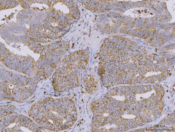
Use heat-mediated EDTA retrieval at pH 8.0 for paraffin sections stained with A05462 (datasheet A05462). The selected image used that condition before overnight incubation with 2 µg/ml antibody at 4°C (datasheet A05462). If staining is weak, check heating consistency and section adhesion before comparing a second retrieval condition on matched sections (standard IHC practice). Score the same cell population across conditions, since endometrial glandular cells provide a reported high-expression reference (HPA: High in endometrial glandular cells). Watch for tissue damage and diffuse DAB when adjusting heat exposure, and retain the condition that preserves morphology and specific staining (standard IHC practice).
Can fixation explain weak or uneven TMBIM6 staining?
The A05462 tissue caption describes a paraffin section but does not state its fixative, so target-specific fixation sensitivity remains unknown (datasheet A05462). Record the fixative, fixation duration, section age, and processing history for each specimen before comparing staining intensity (standard IHC practice). If staining varies across a block, examine morphology and compare similarly processed sections using the same EDTA retrieval at pH 8.0 (datasheet A05462; standard IHC practice). Include a positive reference such as endometrial glandular cells where available, and apply identical detection conditions to that reference (HPA: High in endometrial glandular cells; standard IHC practice). Do not assign weak staining to a TMBIM6-specific fixation effect without a controlled fixation comparison (datasheet A05462: fixative unreported).
Where should convincing TMBIM6 staining appear in a paraffin section?
Expect predominantly cytoplasmic, potentially reticular staining: TMBIM6 is assigned to the ER membrane, while tissue IHC reports cytoplasmic expression in most tissues (UniProt P55061 subcellular location; HPA: tissue profile). Its 6 transmembrane segments support a membrane-associated pattern, although chromogenic light microscopy cannot resolve individual ER membranes (UniProt P55061 topology; standard IHC practice). Interpret signal within identifiable cells rather than treating extracellular deposits or section-wide haze as localisation (standard IHC practice). Endometrial glandular cells offer a reported high-staining comparison for the A05462 endometrial adenocarcinoma image (HPA: High in endometrial glandular cells; datasheet A05462). A dominant nuclear-only pattern warrants review of controls and antibody specificity before biological interpretation (UniProt P55061 subcellular location; standard IHC practice).
Could isoform choice or epitope accessibility alter the staining pattern?
TMBIM6 has 2 annotated isoforms, but the supplied A05462 caption does not identify its epitope or establish isoform selectivity (UniProt P55061 isoforms; datasheet A05462). The protein has 6 transmembrane segments, with annotated cytoplasmic and lumenal loops that could differ in antibody accessibility after processing (UniProt P55061 topology; standard IHC practice). Request the immunogen or epitope position before attributing staining differences to a particular isoform or membrane face (standard IHC practice). Compare matched sections under the documented EDTA pH 8.0 retrieval condition and include a positive tissue reference (datasheet A05462; HPA: High in endometrial glandular cells). A changed pattern alone cannot establish isoform-specific detection without independent validation (standard IHC practice).
How should I adapt the tissue result for multiplex TMBIM6 immunofluorescence?
Treat IF as a separate assay: the supplied A05462 image documents chromogenic paraffin-section IHC, not IF performance (datasheet A05462). Pair TMBIM6 with a marker identifying the expected cell population, such as a glandular-cell marker when examining endometrium, and verify that signals occupy the same cells (HPA: High in endometrial glandular cells; standard IF practice). Select spectrally separated fluorophores and favour a far-red channel if tissue autofluorescence obscures shorter-wavelength signal (standard IF practice). Set permeabilisation according to the confirmed antibody epitope: TMBIM6 has both cytoplasmic and lumenal regions, while this antibody’s epitope is unspecified (UniProt P55061 topology; datasheet A05462). Include single-colour and no-primary controls before interpreting colocalisation or apparent ER-like signal (standard IF practice).
How can I reduce diffuse or nonspecific brown staining?
Begin with the documented 10% goat-serum block and 2 µg/ml primary incubation overnight at 4°C as reference conditions for A05462 (datasheet A05462). Check no-primary sections for secondary-reagent binding and DAB deposits before changing the primary concentration (standard IHC practice). Quench endogenous peroxidase before HRP detection, and control DAB development time consistently across samples (standard IHC practice). If background persists, optimise washing and titrate primary antibody on matched sections while preserving a reported positive cell population (standard IHC practice; HPA: High in endometrial glandular cells). Diffuse staining across unrelated structures should be assessed against controls before being called TMBIM6 expression (UniProt P55061 subcellular location; standard IHC practice).
How should I quantify TMBIM6 staining across tissue samples? ⚠ ANSWER MARKED FOR VERIFICATION
Define the scored cell population and compartment before counting; TMBIM6 is assigned to the ER membrane and tissue staining is described as cytoplasmic (UniProt P55061 subcellular location; HPA: tissue profile). For cellular staining, report the percentage of positive cells and an H-score from 0–300, calculated from intensity grades 0–3 weighted by their cell percentages (standard IHC practice). For spatial counts, report positive-cell density per mm² of viable tissue and document excluded necrotic or damaged regions (standard IHC practice). Normalise comparisons to the same identified cell type, viable area, staining batch, and scoring threshold (standard IHC practice). Keep glandular and stromal measurements separate when analysing endometrial sections (HPA: High in endometrial glandular cells; standard IHC practice).
Which findings distinguish TMBIM6 expression from staining artefact?
A plausible positive result is cellular cytoplasmic staining consistent with an ER membrane protein, especially in a population reported to express TMBIM6 (UniProt P55061 subcellular location; HPA: tissue profile). Endometrial glandular cells are reported High, while cholangiocytes are reported Not detected; compare equivalent, well-preserved cell populations rather than whole-tissue colour alone (HPA: endometrium and liver tissue IHC; standard IHC practice). Edge-restricted colour, necrotic regions, and staining in no-primary controls favour processing or detection artefact (standard IHC practice). Endogenous peroxidase can contribute to DAB signal, so review the peroxidase-block control when staining is unexpectedly broad (standard IHC practice). HPA rates its tissue staining Approved with medium RNA agreement and pending external verification, so resolve discordant patterns with independent controls (HPA: reliability description).
Boster reagents

Best TMBIM6 / Bax inhibitor 1 IHC Antibodies

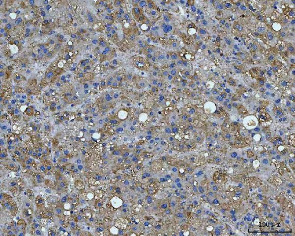
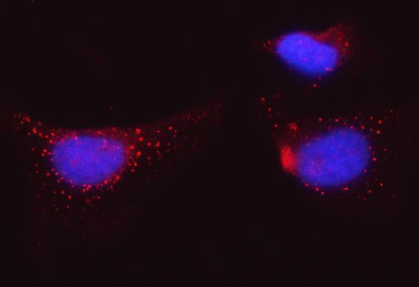
A05462 has IHC images from human paraffin-embedded tumor sections and IF images from HeLa cells and human paraffin-embedded liver cancer tissue (catalog image captions).

Real IHC data IHC analysis of BI-1/TMBIM6 using anti-BI-1/TMBIM6 antibody (A05462). BI-1/TMBIM6 was detected in a paraffin-embedded section of human endometrial adenocarcinoma tissue. Heat mediated antigen retrieval was performed in EDTA buffer (pH 8.0, epitope retrieval solution). The tissue section was blocked with 10% goat serum. The tissue section was then incubated with 2 μg/ml rabbit anti-BI-1/TMBIM6 Antibody (A05462) overnight at 4°C. Peroxidase Conjugated Goat Anti-rabbit IgG was used as secondary antibody and incubated for 30 minutes at 37°C. The tissue section was developed using HRP Conjugated Rabbit IgG Super Vision Assay Kit (Catalog # SV0002) with DAB as the chromogen.
Anti-BI-1/TMBIM6 Antibody ®
Cat # A05462

A05462 lists human reactivity and IHC, IF, and ICC applications (catalog applications and reactivity). Its IHC captions show human endometrial adenocarcinoma, liver cancer, lung cancer, and prostate cancer paraffin sections; its IF captions show HeLa cells and human liver cancer paraffin sections (A05462 image captions).

Which to pick: For tissue IHC, choose A05462: its own IHC caption shows a human endometrial adenocarcinoma paraffin section, EDTA retrieval at pH 8.0, and 2 μg/mL primary antibody; the fixative is unreported (A05462 IHC caption). For IF/ICC, A05462 lists both applications and has IF images from HeLa cells and human liver cancer paraffin sections; its host is rabbit and clonality is unreported (catalog applications and host; A05462 IF captions; catalog clone field). No cross-species choice is supported because A05462 lists Human reactivity only (catalog reactivity).

Each figure is that product's own IHC / IF validation image from its datasheet.

References

  1. UniProt Consortium. UniProt entry P55061 (BI1_HUMAN, Bax inhibitor 1).
  2. Human Protein Atlas. TMBIM6 tissue IHC expression (reliability: Approved).
  3. Human Protein Atlas. TMBIM6 subcellular location (ICC-IF): Membrane.
  4. Human Protein Atlas. TMBIM6 antibody validation summary (1 antibodies).
  5. TMBIM6 promotes glioma progression according to integrated bioinformatics and experimental evidence. Scientific reports 2025 — PMC12219401.
  6. TMBIM6 regulates redox-associated posttranslational modifications of IRE1α and ER stress response failure in aging mice and humans. Redox biology 2021 — PMC8476450.
  7. miR‑302d‑3p regulates the viability, migration and apoptosis of breast cancer cells through regulating the TMBIM6‑mediated ERK signaling pathway. Molecular medicine reports 2021 — PMC8548939.
  8. STRIP2 motivates non-small cell lung cancer progression by modulating the TMBIM6 stability through IGF2BP3 dependent. Journal of experimental & clinical cancer research : CR 2023 — PMC9837939.
  9. PubMed PMID:8530040 — UniProt-cited evidence.
  10. PubMed PMID:14702039 — UniProt-cited evidence.
  11. PubMed PMID:16541075 — UniProt-cited evidence.