This website uses cookies to ensure you get the best experience on our website.


- Table of Contents
Plan chromogenic IHC on paraffin sections using 2–5 μg/ml of the IHC-validated antibody (datasheet A14820-1). Compare staining with the reported cytoplasmic and membranous tissue pattern, while accounting for its uncertain reliability (HPA tissue IHC).

Expected localisation, validated protocols, controls and antibodies — the at-a-glance facts below, then the full design guide.
| Expected localisation | Cytoplasmic and membranous tissue staining (HPA tissue IHC) | |
| Staining pattern | Glandular cells: cytoplasmic/membranous staining (HPA tissue IHC) | |
| Antigen retrieval | EDTA pH 8.0 HIER, heat-mediated (datasheet A14820-1) | |
| Positive control | Bone marrow+4 more · see all | |
| Negative control | Adipose tissue+3 more · see all |
| Fixation | Keep fixation consistent across paraffin sections (standard IHC practice; not target-specific); Matched tissue-IHC evidence does not establish the fixation claim. Validate the specimen-specific method before use. (selected-SKU IHC image A14820-1) | |
| Caveat | Staining has low concordance with RNA expression (HPA tissue IHC) | |
| Regulation | No expression regulator annotated (UniProt) | |
| Isoform / epitope | 2 isoforms; map the epitope to extracellular or cytoplasmic regions (UniProt) |
The catalog antibody’s IHC-P protocol is paired with a published pancreatic tissue microarray example (PMC6412825).
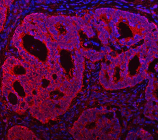
| Sample | Paraffin-embedded human colorectal adenocarcinoma tissue; fixative not specified (datasheet A14820-1) |
| Fixation | Image fixative and duration unreported (datasheet A14820-1); verify before use. |
| Sectioning | 4–5 µm sections on charged slides (standard) |
| Deparaffinisation | Xylene, graded ethanol series to water (standard) |
| Antigen retrieval | Heat retrieval: EDTA pH 8.0 (datasheet A14820-1); 20 min, 95–100 °C (standard) |
| Peroxidase block | 3% H2O2, 10 min, room temperature (standard) |
| Blocking | 10% goat serum (datasheet A14820-1) |
| Primary antibody | Rabbit anti-TMC7, 2-5 μg/ml (datasheet A14820-1) |
| Primary incubation | Overnight at 4 °C (datasheet A14820-1) |
| Detection | HRP-conjugated secondary, DAB chromogen (datasheet A14820-1) |
| Counterstain | Hematoxylin, blue, dehydrate and mount (standard) |
| Expected result | TMC7-positive staining in hematopoietic cells of bone marrow (HPA tissue IHC: High). HPA tissue profile: General cytoplasmic and membranous expression. No signal in the no-primary control. |
TMC7 is a nine-pass membrane protein (UniProt Q7Z402 topology). In tissue IHC, HPA reports general cytoplasmic and membranous staining, including High staining in breast and prostate glandular cells, bronchial respiratory epithelial cells, and kidney glomerular cells (HPA tissue IHC). Treat these as provisional reference patterns: HPA rates tissue staining Uncertain because antibody staining and RNA expression have low consistency (HPA tissue IHC reliability).
| Membranous and cytoplasmic staining appears in breast or prostate glandular cells, bronchial respiratory epithelium, or kidney glomerular cells. | This resembles the reported High cell-specific IHC pattern (HPA tissue IHC). Score the relevant cells and compartments separately; agreement with HPA supports interpretation but does not independently establish specificity because tissue IHC reliability is Uncertain (HPA tissue IHC reliability). |
| The dominant signal is nuclear, with little membranous or cytoplasmic staining. | A nuclear-only pattern does not match the HPA tissue profile or UniProt membrane annotation (HPA tissue IHC; UniProt Q7Z402 subcellular location). Inspect counterstain overlap and detection controls before assigning the nuclear signal to TMC7 (general IHC practice). |
| Strong signal appears chiefly in adipocytes or skeletal myocytes rather than the expected cell population. | HPA reports TMC7 Not detected in those cell types (HPA tissue IHC). Consider antibody cross-reactivity or endogenous chromogen-producing activity; compare a no-primary control and a reported positive cell population on the same run (general IHC practice; HPA tissue IHC). |
| Chromogen covers many tissue compartments without clear cell boundaries or contrast with surrounding tissue. | This does not resolve the cytoplasmic and membranous profile reported by HPA (HPA tissue IHC). Diffuse background can reflect nonspecific binding, inadequate washing, or detection-system background; inspect no-primary and detection controls before scoring cells (general IHC practice). |
| No convincing signal is seen in a section containing breast glandular or bronchial respiratory epithelial cells. | Those cells are reported High by HPA, so absent staining warrants an assay check (HPA tissue IHC). Confirm the cells are present and assess retrieval, antibody conditions, and detection with run controls; a negative result alone cannot settle expression given HPA's Uncertain tissue reliability (general IHC practice; HPA tissue IHC reliability). |
| Membrane topology and antibody access (UniProt Q7Z402 topology) | TMC7 has 9 transmembrane segments and both extracellular and cytoplasmic regions (UniProt Q7Z402 topology). Epitope position is not supplied, so topology alone cannot choose a retrieval condition or predict which membrane face the IHC antibody recognizes (UniProt Q7Z402 topology; evidence limit). |
| Tissue evidence and validation (HPA tissue IHC; HPA antibody HPA029465) | HPA lists Uncertain tissue IHC reliability and Uncertain IHC validation for HPA029465; its ICC validation is Approved (HPA tissue IHC; HPA antibody HPA029465). Use the tissue examples as comparisons, not a fixed pass/fail threshold (HPA tissue IHC reliability). |
| Isoforms and modifications (UniProt Q7Z402) | UniProt lists 2 isoforms and five glycosylation sites, but supplies no epitope or isoform-specific IHC pattern here (UniProt Q7Z402 isoforms and glycosylation; evidence limit). Do not infer that an absent or altered stain identifies a particular isoform or glycosylation state. |
| IF/ICC Q&A: where might TMC7 appear? (HPA subcellular ICC-IF) | HPA reports Approved vesicle localization in ICC-IF, with images from A-431, U-251MG, and U2OS (HPA subcellular ICC-IF). This complements, but does not replace, the cytoplasmic and membranous tissue IHC profile (HPA tissue IHC); IF/ICC assay conditions belong in its separate guide. |
| Situation | Likely cause | Next action |
|---|---|---|
| Reported positive cells are blank. | The cells may be absent from the section, or the IHC run may have failed; HPA's High examples are provisional (HPA tissue IHC reliability). | Verify cell identity on the counterstained section, then review run controls, retrieval, antibody dilution, and detection settings (general IHC practice). |
| Signal is mainly nuclear. | That compartment conflicts with the reported tissue profile and membrane annotation (HPA tissue IHC; UniProt Q7Z402 subcellular location). | Check the no-primary section and chromogen against the nuclear counterstain; only score staining that can be assigned to the reported compartments (general IHC practice; HPA tissue IHC). |
| Brown deposit appears in many cell types, including reported negative cells. | Nonspecific binding or endogenous detection activity may obscure the reported cell pattern (general IHC practice; HPA tissue IHC). | Compare no-primary and reported negative cell populations; review blocking, washes, and endogenous-peroxidase control for chromogenic IHC (general IHC practice). |
| Patchy tissue staining is hard to score. | A mixed section may contain reported positive and negative cell populations (HPA tissue IHC); section quality can also complicate scoring (general IHC practice). | Score identifiable cell types separately and compare intact tissue regions with the counterstain; exclude damaged regions from interpretation (general IHC practice). |
| A reported Not detected population stains as strongly as a reported High population. | The pattern disagrees with HPA's cell-specific observations, whose tissue reliability is Uncertain (HPA tissue IHC). | Repeat comparison with appropriate run and no-primary controls, and report the discordance rather than assigning it to TMC7 without further validation (general IHC practice; HPA tissue IHC reliability). |
| Vesicular ICC-IF signal seems inconsistent with diffuse tissue IHC staining. | HPA reports vesicles in ICC-IF and general cytoplasmic and membranous tissue IHC staining; these are observations from different applications (HPA subcellular ICC-IF; HPA tissue IHC). | Interpret each image against its own application and cell context; consult the separate IF/ICC guide for that assay (HPA subcellular ICC-IF; HPA tissue IHC). |


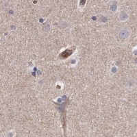

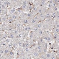
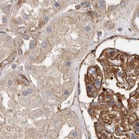


Comprehensive Human Protein Atlas IHC scoring per tissue (reliability: Uncertain — Low consistency between antibody staining and RNA expression data. Pending external verification.). Rows are taken directly from the HPA tissue chart — click any row's HPA link to view the source.
| Tissue | Cell type | Level | Evidence | Source |
|---|---|---|---|---|
| Bone marrow | Hematopoietic cells | High | Protein (IHC) | HPA → |
| Breast | Glandular cells | High | Protein (IHC) | HPA → |
| Bronchus | Respiratory epithelial cells | High | Protein (IHC) | HPA → |
| Colon | Endothelial cells | High | Protein (IHC) | HPA → |
| Kidney | Cells in glomeruli | High | Protein (IHC) | HPA → |
| Tissue | Cell type | Level | Evidence | Source |
|---|---|---|---|---|
| Adipose tissue | Adipocytes | Not detected | Protein (IHC) | HPA → |
| Cerebellum | Cells in granular layer | Not detected | Protein (IHC) | HPA → |
| Hippocampus | Glial cells | Not detected | Protein (IHC) | HPA → |
| Skeletal muscle | Myocytes | Not detected | Protein (IHC) | HPA → |
Troubleshoot TMC7 chromogenic IHC in paraffin sections using the catalog antibody’s tissue image and the stated limits of its localisation evidence.
A14820-1 has IHC images from human colorectal adenocarcinoma and liver cancer paraffin sections, plus an IF image from a human intestinal cancer paraffin section (catalog image captions).
A14820-1 will render with its human colorectal adenocarcinoma paraffin-section IHC image; a second IHC caption describes human liver cancer (catalog IHC image captions). Its application list includes IHC and IF, its IF caption shows human intestinal cancer paraffin tissue, and its listed reactivity is human, mouse, and rat (catalog applications, IF image caption, reactivity).
Which to pick: Choose A14820-1 for human paraffin-section chromogenic IHC: its IHC captions document EDTA pH 8.0 retrieval and DAB detection, but do not report the fixative (catalog IHC image captions). For IF, A14820-1 has a human paraffin-section image; ICC validation is not documented in the supplied payload (catalog IF image caption, applications). For mouse or rat work, A14820-1 lists reactivity with both species, while the supplied IHC and IF images show human tissue only (catalog reactivity, image captions).