This website uses cookies to ensure you get the best experience on our website.


- Table of Contents
Plan TMEM192 IHC-P around granular cytoplasmic staining in most cells (HPA tissue IHC). The catalog antibody A11938 has an IHC starting concentration of 5 μg/mL (datasheet); lung macrophages provide a high-staining reference (HPA tissue IHC).

Expected localisation, validated protocols, controls and antibodies — the at-a-glance facts below, then the full design guide.
| Expected localisation | Granular cytoplasm (HPA tissue IHC); lysosome membrane and late endosome (UniProt) | |
| Staining pattern | Granular cytoplasmic staining in most cells (HPA tissue IHC) | |
| Antigen retrieval | Citrate pH 6.0 HIER, 95–98 °C, 20 min (rule: cytoplasmic / membrane antigen) | |
| Positive control | Adrenal gland+4 more · see all | |
| Negative control | None in HPA (detected in all 45 tissues); use no-primary + isotype controls |
| Fixation | Keep fixation conditions consistent across sections. (standard IHC practice; not target-specific) | |
| Caveat | High staining in lung macrophages can affect tissue-level scoring (HPA tissue IHC) | |
| Regulation | No expression regulator annotated (UniProt) | |
| Isoform / epitope | 2 isoforms; verify epitope coverage across cytoplasmic and lumenal regions (UniProt) |
The catalog antibody’s IHC-P protocol is accompanied by a published mouse brain cryosection IHC method (PMC5546430).
| Sample | Tissue sections; selected-image fixative not specified (standard IHC workflow) |
| Fixation | Image fixative and duration unreported (datasheet A11938); verify before use. |
| Sectioning | 4–5 µm sections on charged slides (standard) |
| Deparaffinisation | Xylene, graded ethanol series to water (standard) |
| Antigen retrieval | Heat-induced epitope retrieval in citrate buffer, pH 6.0, 20 min at 95–98 °C (standard rule: cytoplasmic / membrane antigen) |
| Peroxidase block | 3% H2O2, 10 min, room temperature (standard) |
| Blocking | 10% normal serum of the secondary host, 30 min, room temperature (standard) |
| Primary antibody | Rabbit anti-TMEM192, 5 μg/mL (datasheet A11938) |
| Primary incubation | Overnight at 4 °C (standard) |
| Detection | HRP-polymer secondary, DAB chromogen 5–10 min (standard) |
| Counterstain | Hematoxylin, blue, dehydrate and mount (standard) |
| Expected result | TMEM192-positive staining in glandular cells of adrenal gland (HPA tissue IHC: High). HPA tissue profile: Granular cytoplasmic expression in most cells. No signal in the no-primary control. |
TMEM192 is a four-pass lysosomal membrane protein also found in late endosomes (UniProt Q8IY95 topology and subcellular location). In paraffin-section IHC, expect granular cytoplasmic staining, including in lung macrophages, hippocampal neurons and several glandular cell populations (HPA tissue IHC: High; granular cytoplasmic expression in most cells). HPA rates tissue staining Enhanced, with medium consistency against RNA data and external verification pending (HPA tissue IHC reliability).
| Discrete cytoplasmic granules in lung macrophages, with stronger staining than nearby cells. | This fits a plausible positive IHC result: HPA reports High staining in lung macrophages and granular cytoplasmic expression in most cells (HPA tissue IHC). Score the macrophages separately from surrounding lung cells; the HPA level does not specify an intensity threshold for every slide. |
| Predominantly smooth plasma-membrane, diffuse cytoplasmic or dominant nuclear staining in a paraffin section. | Recheck specificity and morphology: the expected IHC profile is granular cytoplasm (HPA tissue IHC), consistent with lysosome and late-endosome localisation (UniProt Q8IY95). Nuclear signal needs context: HPA also reports an additional nucleoplasmic location in ICC-IF (HPA subcellular), so its presence alone does not prove artefact. |
| Strong staining concentrated in vaginal squamous epithelium, ovarian stromal cells or soft-tissue fibroblasts. | HPA lists these cell populations as Low (HPA tissue IHC). Unexpectedly strong staining warrants a specificity check, including antibody cross-reactivity and endogenous chromogenic detection activity as general IHC possibilities. Compare cell identity and staining distribution before calling the whole tissue positive. |
| Broad, even colour across tissue, stroma or blank areas, obscuring individual cytoplasmic granules. | This does not match the reported granular cytoplasmic profile (HPA tissue IHC). In general chromogenic IHC practice, incomplete blocking, excess antibody or detection reagent, or residual endogenous enzyme activity can produce background; inspect a detection-only control before interpreting weak cell staining. |
| No convincing granules in an adequately represented lung-macrophage or other HPA High cell population. | Check the run before assigning biological absence: HPA reports High staining in lung macrophages and selected neuronal, glandular, placental and Leydig cells (HPA tissue IHC). HPA tissue reliability is Enhanced but has only medium RNA–staining consistency and awaits external verification, so a single negative section is not decisive. |
| Cell population and tissue context | HPA records High staining in lung macrophages; hippocampal neuronal cells; cerebellar molecular-layer cells; adrenal, parathyroid and thyroid glandular cells; placental cytotrophoblasts; and testicular Leydig cells (HPA tissue IHC). Its Low entries for selected other cells are comparison points, not validated negative controls. |
| Organelle location and topology | TMEM192 has four membrane-spanning segments and cytoplasmic and lumenal regions (UniProt Q8IY95 topology). Lysosome-membrane and late-endosome localisation supports a punctate intracellular interpretation (UniProt Q8IY95; HPA tissue IHC). Antibody epitope location is not supplied, so topology cannot determine a retrieval or permeabilisation setting. |
| Isoforms and processing | Two isoforms are listed, with no signal peptide or propeptide and a chain annotated from residues 1–271 (UniProt Q8IY95). The payload does not identify antibody epitopes or isoform coverage. An altered staining pattern therefore cannot be assigned to an isoform or processing event from these records. |
| Strength of tissue-pattern evidence | HPA calls tissue IHC reliability Enhanced but describes only medium agreement between staining and RNA expression, pending external verification (HPA tissue IHC). HPA014717 and HPA024110 each have Enhanced IHC status (HPA antibodies); those ratings do not independently validate an unspecified catalog antibody or every observed compartment. |
| Paraffin-section detection conditions | No TMEM192-specific fixation, retrieval, dilution or endogenous-enzyme result is supplied. For chromogenic IHC, assess retrieval, blocking and detection controls using general IHC practice and the chosen antibody's validated IHC-P instructions; do not infer a TMEM192 fixation effect from topology or HPA staining levels. |
| Situation | Likely cause | Next action |
|---|---|---|
| A known HPA High cell population is blank. | Possible run failure, weak detection or poorly represented target cells; HPA's High classification is a population observation, not a guarantee for each section (HPA tissue IHC). | Confirm the cell population on the counterstain, check that the positive-control section stained, and review the chosen antibody's IHC-P instructions for retrieval and detection. Repeat the run before reporting absence. |
| Signal is uniformly diffuse instead of granular. | Diffuse colour conflicts with HPA's granular cytoplasmic profile (HPA tissue IHC); nonspecific reagent deposition or incomplete background control are general IHC possibilities. | Compare with a detection-only control, inspect unstained spaces, and adjust blocking, washing or reagent concentration within the chosen IHC-P workflow. Score discrete cell-associated granules only after background is controlled. |
| Unexpectedly strong colour appears in an HPA Low cell population. | HPA lists vaginal squamous cells, ovarian stromal cells and soft-tissue fibroblasts as Low (HPA tissue IHC). Cell misidentification, cross-reactivity or endogenous chromogenic activity may explain a discordant result. | Verify cell identity and compartment on the counterstain; compare a detection-only control and an HPA High cell population processed in the same run. Treat the discordance as unresolved until those checks support the stain. |
| Staining is dominated by a cell-surface rim or nuclei. | A rim conflicts with lysosome-membrane and late-endosome localisation (UniProt Q8IY95). Dominant nuclear IHC colour differs from HPA's granular tissue profile, although nucleoplasm is an additional ICC-IF location (HPA tissue IHC; HPA subcellular). | Recheck morphology and background controls, then compare independent staining evidence if available. Describe nuclear IHC signal separately; do not discard it solely because the main expected compartment is cytoplasmic. |
| Two plausible positive tissues give different staining strengths. | HPA reports cell-specific High entries and an overall granular profile, while its tissue reliability description notes medium consistency with RNA data (HPA tissue IHC). Section composition and assay variation are general IHC considerations. | Compare the same named cell populations, counterstain quality and controls across sections. Record intensity and the fraction of stained target cells separately, without converting HPA's High category into a universal numeric cutoff. |
| IF/ICC Q&A: what localisation should an IF result show? | HPA reports supported endosomal and lysosomal locations, plus an approved additional nucleoplasmic location in ICC-IF (HPA subcellular). These ICC-IF observations do not define a paraffin-IHC protocol. | For IF interpretation, look for intracellular endosomal or lysosomal puncta and document any nucleoplasmic signal separately (HPA subcellular). Use the dedicated IF/ICC guide for experimental settings; this section supplies no IF protocol option. |
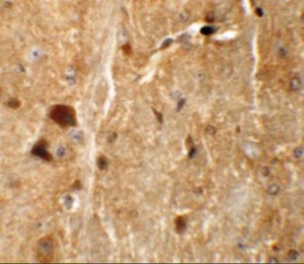
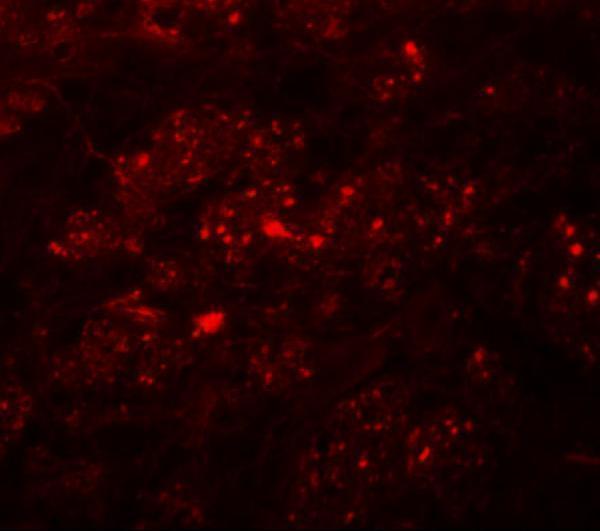


Comprehensive Human Protein Atlas IHC scoring per tissue (reliability: Enhanced — Medium consistency between antibody staining and RNA expression data. Pending external verification.). Rows are taken directly from the HPA tissue chart — click any row's HPA link to view the source.
| Tissue | Cell type | Level | Evidence | Source |
|---|---|---|---|---|
| Adrenal gland | Glandular cells | High | Protein (IHC) | HPA → |
| Cerebellum | Cells in molecular layer | High | Protein (IHC) | HPA → |
| Hippocampus | Neuronal cells | High | Protein (IHC) | HPA → |
| Lung | Macrophages | High | Protein (IHC) | HPA → |
| Parathyroid gland | Glandular cells | High | Protein (IHC) | HPA → |
| Tissue | Cell type | Level | Evidence | Source |
|---|---|---|---|---|
| None in HPA: TMEM192 is detected in all 45 scored tissues. Use a no-primary (secondary-only) and an isotype control instead. | ||||
Troubleshoot TMEM192 staining in paraffin sections by checking retrieval, compartmental pattern, controls, and scoring before comparing specimens.
A11938 has mouse brain IHC and IF images (catalog image captions) and lists Human, Mouse, and Rat reactivity (catalog reactivity).
A11938 is the figure-backed card: mouse brain IHC at 5 μg/mL and IF at 20 μg/mL (A11938 image captions). M11938 lists IF/ICC but has no IHC or IF image in the payload, so it has no figure-backed card here (M11938 applications and image alts).
Which to pick: Choose A11938 for paraffin-section IHC: IHC-P is listed, and its own caption documents mouse brain tissue staining at 5 μg/mL (A11938 applications and IHC image caption); the fixative is unreported (A11938 IHC image caption). For IF/ICC, A11938 has a mouse brain IF image, while rabbit monoclonal M11938 lists IF and ICC without a supplied image (A11938 IF image caption; M11938 host, clone, applications and image alts). Both list Human, Mouse, and Rat reactivity, but the supplied staining images document mouse tissue only (catalog reactivity; A11938 image captions).