This website uses cookies to ensure you get the best experience on our website.


- Table of Contents
Plan chromogenic TMX1 IHC in paraffin sections with the IHC-validated antibody at 1:50–1:200 (datasheet: A07462). Assess cytoplasmic and membranous staining in positive cell populations, including lung macrophages (HPA tissue IHC).

Expected localisation, validated protocols, controls and antibodies — the at-a-glance facts below, then the full design guide.
| Expected localisation | Cytoplasmic and membranous in tissue IHC (HPA tissue IHC) | |
| Staining pattern | Glandular cells and lung macrophages; cytoplasmic/membranous (HPA tissue IHC) | |
| Antigen retrieval | Citrate pH 6.0 HIER, 95–98 °C, 20 min (rule: cytoplasmic / membrane antigen) | |
| Positive control | Appendix+4 more · see all | |
| Negative control | Adipose tissue+4 more · see all |
| Fixation | Keep paraffin-section fixation consistent across samples. (standard IHC practice; not target-specific) | |
| Caveat | Antibody staining and RNA show medium consistency (HPA tissue IHC) | |
| Regulation | Thrombin stimulates secretion (UniProt) | |
| Isoform / epitope | No isoforms; map epitope to residues 27–180 or 204–280 (UniProt) |
The catalog antibody protocol is accompanied by one published TMX1 IHC protocol using paraffin sections of human skin and melanoma (PMC6669928).
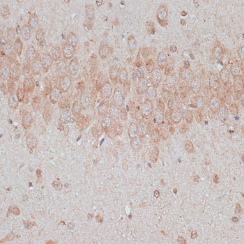
| Sample | Paraffin-embedded rat brain tissue; fixative not specified (datasheet A07462) |
| Fixation | Image fixative and duration unreported (datasheet A07462); verify before use. |
| Sectioning | 4–5 µm sections on charged slides (standard) |
| Deparaffinisation | Xylene, graded ethanol series to water (standard) |
| Antigen retrieval | Heat-induced epitope retrieval in citrate buffer, pH 6.0, 20 min at 95–98 °C (standard rule: cytoplasmic / membrane antigen) |
| Peroxidase block | 3% H2O2, 10 min, room temperature (standard) |
| Blocking | 10% normal serum of the secondary host, 30 min, room temperature (standard) |
| Primary antibody | Rabbit anti-TMX1, 1:50-1:200 (datasheet A07462) |
| Primary incubation | Overnight at 4 °C (standard) |
| Detection | HRP-polymer secondary, DAB chromogen 5–10 min (standard) |
| Counterstain | Hematoxylin, blue, dehydrate and mount (standard) |
| Expected result | TMX1-positive staining in glandular cells of appendix (HPA tissue IHC: Medium). HPA tissue profile: Cytoplasmic and membranous expression in several tissues. No signal in the no-primary control. |
TMX1 is predominantly an endoplasmic reticulum membrane protein with one transmembrane segment at residues 181–203 (UniProt Q9H3N1 topology and localization). In paraffin IHC, expect cytoplasmic and membranous staining in selected cells, including appendix glandular cells and lung macrophages, both reported at medium intensity (HPA tissue IHC). The HPA tissue antibody is Approved, with medium consistency between staining and RNA data (HPA tissue IHC).
| Cytoplasmic and membranous staining in appendix or duodenal glandular cells. | This fits the reported medium staining in those cells (HPA tissue IHC) and TMX1’s predominant ER membrane localization (UniProt Q9H3N1 localization). Chromogenic IHC need not resolve the ER as a distinct network (general IHC practice). |
| Strong staining confined to nuclei, with little cytoplasmic signal. | This does not match the reported tissue IHC profile (HPA tissue IHC). Check specificity and section background before scoring it as TMX1. Nucleolar localization is separately supported in ICC-IF, so an IF nucleolar signal alone is not grounds to reject that assay (HPA subcellular ICC-IF). |
| Prominent staining in kidney glomerular cells or liver cholangiocytes. | Neither cell population had detectable staining in the cited HPA images (HPA tissue IHC). Investigate nonspecific binding or endogenous detection activity (general IHC practice). These cell-specific observations do not establish that an entire kidney or liver sample lacks TMX1 (HPA tissue IHC; UniProt Q9H3N1 tissue specificity). |
| Widespread, hazy chromogen that obscures cell boundaries. | A haze cannot be assigned confidently to TMX1’s reported cytoplasmic and membranous pattern (HPA tissue IHC). Review negative controls, blocking, washing and chromogen development for assay background (general IHC practice). |
| No staining in appendix glandular cells or lung macrophages. | Those cell populations showed medium staining in HPA tissue IHC (HPA tissue IHC). A negative result warrants checking tissue preservation, retrieval and detection with appropriate controls (general IHC practice); HPA provides no TMX1-specific fixation-sensitivity finding. |
| Compartment and topology | TMX1 is predominantly on the ER membrane and spans the membrane at residues 181–203 (UniProt Q9H3N1 topology and localization). Score a cellular cytoplasmic or membranous pattern in IHC rather than requiring resolution of an ER network (HPA tissue IHC; general IHC practice). |
| Choice of positive and comparator cells | Appendix glandular cells and lung macrophages are medium-staining examples; adipocytes and esophageal squamous cells were not detected (HPA tissue IHC). Use the specified cell populations as comparators, since the HPA calls are cell-specific (HPA tissue IHC). |
| Strength of validation | The tissue IHC profile is Approved and has medium consistency with RNA expression data (HPA tissue IHC). HPA003085 is Approved for IHC, while the supplied antibody record does not designate it Enhanced (HPA antibodies). Treat an unexpected pattern as a result to verify, not as established TMX1 distribution. |
| Expression versus sampled cells | UniProt describes TMX1 as ubiquitous and highly expressed in kidney and liver (UniProt Q9H3N1 tissue specificity). HPA reports no detection specifically in glomerular cells and cholangiocytes (HPA tissue IHC). Those statements concern different levels of sampling; neither predicts staining in every cell. |
| Secretion context | UniProt reports extracellular secretion following thrombin stimulation (UniProt Q9H3N1 localization). That context does not establish extracellular staining in routine paraffin tissue sections; interpret extracellular chromogen against controls and the reported tissue IHC pattern (HPA tissue IHC; general IHC practice). |
| IF/ICC: what pattern should I expect? | HPA supports mainly ER localization and additional nucleolar localization in ICC-IF (HPA subcellular ICC-IF). This is a separate imaging observation; the IHC decision here uses the cytoplasmic and membranous tissue profile (HPA tissue IHC). |
| Situation | Likely cause | Next action |
|---|---|---|
| Expected positive cells show no chromogen. | The run may have failed, or the selected cells may be poorly preserved (general IHC practice). HPA reports medium staining in appendix glandular cells and lung macrophages (HPA tissue IHC); it does not establish TMX1-specific fixation sensitivity. | Confirm the intended cells are present, check a same-run positive control, and review retrieval and detection against the chosen IHC procedure (general IHC practice). Do not infer TMX1 loss from a failed control. |
| Only nuclei stain strongly in tissue IHC. | Nuclear-only tissue staining diverges from HPA’s cytoplasmic and membranous profile (HPA tissue IHC). Nucleoli are an additional location in ICC-IF, a different assay (HPA subcellular ICC-IF). | Compare with a negative reagent control and an HPA-positive tissue cell population; review primary-antibody specificity and counterstain interpretation (HPA tissue IHC; general IHC practice). |
| Unexpected cell populations stain prominently. | Cross-reactivity or endogenous detection activity may contribute (general IHC practice). Glomerular cells and cholangiocytes were not detected in HPA’s sampled tissue images (HPA tissue IHC). | Identify the stained cell type before comparing it with HPA; examine reagent controls and, for enzyme detection, the relevant endogenous-activity block (general IHC practice). Avoid a whole-organ negative call from one cell type (HPA tissue IHC). |
| Diffuse haze obscures the expected cells. | Nonspecific reagent binding or excess chromogen development can obscure compartment assignment (general IHC practice). The HPA tissue profile is cytoplasmic and membranous (HPA tissue IHC). | Inspect negative controls, then review blocking, washes and development time within the established IHC workflow (general IHC practice). Score TMX1 only where cellular staining can be distinguished from background (HPA tissue IHC). |
| A kidney or liver section seems inconsistent with published expression. | UniProt reports high expression in those organs, while HPA reports no detection in kidney glomerular cells and liver cholangiocytes specifically (UniProt Q9H3N1 tissue specificity; HPA tissue IHC). | Annotate the actual compartment and cell population before comparing results. Use a documented HPA-positive cell population and same-run controls to assess assay performance (HPA tissue IHC; general IHC practice). |
| Extracellular staining appears alongside weak cellular signal. | Thrombin-associated secretion is reported by UniProt, but the supplied tissue IHC profile describes cytoplasmic and membranous expression (UniProt Q9H3N1 localization; HPA tissue IHC). The extracellular signal has no established expectation for these sections. | Check whether extracellular color persists in negative controls, then report cellular and extracellular observations separately (general IHC practice). Do not assign extracellular color to TMX1 solely from the secretion annotation (UniProt Q9H3N1 localization). |
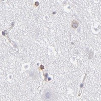
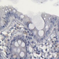
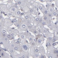
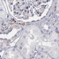


Comprehensive Human Protein Atlas IHC scoring per tissue (reliability: Approved — Medium consistency between antibody staining and RNA expression data.). Rows are taken directly from the HPA tissue chart — click any row's HPA link to view the source.
| Tissue | Cell type | Level | Evidence | Source |
|---|---|---|---|---|
| Appendix | Glandular cells | Medium | Protein (IHC) | HPA → |
| Bone marrow | Hematopoietic cells | Medium | Protein (IHC) | HPA → |
| Breast | Glandular cells | Medium | Protein (IHC) | HPA → |
| Caudate | Glial cells | Medium | Protein (IHC) | HPA → |
| Duodenum | Glandular cells | Medium | Protein (IHC) | HPA → |
| Tissue | Cell type | Level | Evidence | Source |
|---|---|---|---|---|
| Adipose tissue | Adipocytes | Not detected | Protein (IHC) | HPA → |
| Esophagus | Squamous epithelial cells | Not detected | Protein (IHC) | HPA → |
| Gallbladder | Glandular cells | Not detected | Protein (IHC) | HPA → |
| Heart muscle | Cardiomyocytes | Not detected | Protein (IHC) | HPA → |
| Kidney | Cells in glomeruli | Not detected | Protein (IHC) | HPA → |
Troubleshoot TMX1 chromogenic IHC in paraffin sections using the tissue staining evidence, protein topology, and controls appropriate to each specimen.
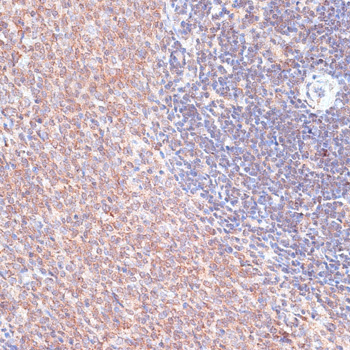
The catalog antibody has real IHC images from paraffin-embedded rat brain and spleen (catalog IHC captions); Human, Mouse and Rat reactivity is listed (catalog reactivity). No IF data are supplied (catalog IF images).
A07462 is the sole card and is listed for IHC (catalog applications). Its IHC images show paraffin-embedded rat brain and spleen at 1:100 with microwave retrieval in 10 mM PBS, pH 7.2 (A07462 IHC captions).
Which to pick: Choose A07462 for tissue IHC in paraffin sections: its rat brain and spleen captions document that processing, while the fixative is unreported (A07462 IHC captions). It is listed as a rabbit antibody reactive with Human, Mouse and Rat, though the supplied IHC images show Rat only; clonality is unreported (catalog host, reactivity and clone; A07462 IHC captions). No SKU is listed for IF/ICC because A07462 has no IF application or IF image in the payload (catalog applications and IF images).