This website uses cookies to ensure you get the best experience on our website.
- Table of Contents
2 Citations
Facts about Tumor necrosis factor receptor superfamily member 10A.
The consequent death-inducing signaling complex (DISC) performs caspase-8 proteolytic activation which initiates the subsequent cascade of caspases (aspartate-specific cysteine proteases) mediating apoptosis. Promotes the activation of NF-kappa-B.
Human | |
---|---|
Gene Name: | TNFRSF10A |
Uniprot: | O00220 |
Entrez: | 8797 |
Belongs to: |
---|
No superfamily |
APO2; CD261 antigen; CD261; cytotoxic TRAIL receptor; Death receptor 4; DR4 TRAIL receptor 1; DR4; TNF-related apoptosis inducing ligand receptor 1; TNF-related apoptosis-inducing ligand receptor 1; TNFRSF10A; TRAIL R1; TRAILR1; TRAIL-R1; TRAILR-1; TRAILR1MGC9365; tumor necrosis factor receptor superfamily member 10A; tumor necrosis factor receptor superfamily, member 10a
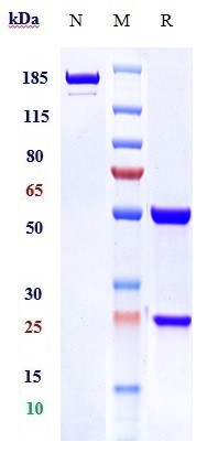
Mass (kDA):
50.089 kDA
Human | |
---|---|
Location: | 8p21.3 |
Sequence: | 8; NC_000008.11 (23190452..23225102, complement) |
Membrane; Single-pass type I membrane protein.
Among the hottest topics in biotechnology right now is TNFRSF10A. This marker is a critical part of many cell and tissue culture studies. Its tunable reactivity is critical for the detection of a variety of cancers. While TNFRSF10A is commonly used in cancer research, it can be used in many other settings, including irradiation, immunohistochemistry, and more.
The TNFRSF10A gene encodes a protein that is a member of the TNF receptor superfamily. These proteins are involved in immune responses to inflammatory conditions and are responsible for the development of a wide variety of therapeutic agents. Boster offers a variety of high-affinity primary antibodies that are highly cited in the scientific literature. These antibodies have been validated for use in ELISA, Immunohistochemistry, and Western Blotting.
The TNFRSF10A marker, also called TRAIL R1, is found on the surface of human cells. The cDNA for this protein codes for a 468 amino acid precursor protein. The protein contains extracellular cysteine-rich domains, transmembrane domains, and cytoplasmic death domains. The two proteins share 55% amino acid sequence identity. The binding of trimeric TRAIL to TRAIL R1 induces apoptosis in cells. The binding is likely facilitated by oligomerization. The chimera can neutralize the apoptosis-inducing effects of TRAIL in humans.
GSLCs for the TNFR-SF10A marker were performed in 21 DMD patients from VaC1 and 46 patients from DiCs. These results validate the SOLiD method. The oligonucleotides used in the polymerase chain reaction (PCR) were listed in Supplementary Table S2A. Genomic DNA was amplified by polymerase chain reaction with 30 cycles. Each PCR was performed in a 25-ml volume containing 100 ng of genomic DNA, 1.5 mmol MgCl2, 0.2 mmol dNTPs, and 0.4 ml of each primer.
The expression of DR4 in GSLC and non-GSLC cells was determined using a western blot. Expression of DR4 was significantly decreased in U87-sph cells. We also tested DR5 expression in GSLC using western blot. All three GSLC were expressed in DR5, whereas DR4 was not. This study highlights the utility of GSLCs for the TNFRSF10A marker in the detection of glioma.
The DR5 pathway is activated in GSLCs after irradiation or TRAIL treatment. DR5 is a protein implicated in the resistance to irradiation and TRAIL treatment. When GSLCs are irradiated, they produce more DR5 and fewer activated caspases. Hence, they are more resistant to radiation and TRAIL.
TNFRSF10A SNPs may play an important role in the association between age at onset of LoA and the presence of CS treatment. These SNPs may help predict the response to VBP15, a new anti-inflammatory that improves muscle dystrophy without adverse CS side effects. If this is the case, these results are further strengthened. They suggest that GSLCs for the TNFRSF10A marker may influence the outcome of CS therapy in patients with DMD.
The TNFRSF10A gene is expressed in a variety of cancer cells. Its expression can be determined using flow cytometry. The cells were seeded into six-well plates and divided into four groups. One group was non-treated; the other received TRAIL 50 ng/ml and irradiation (8 Gy) alone. Cells were harvested 48 h after treatment. The dose of TRAIL was increased over time.
Irradiation of the TNFR-SF10A gene marker in Boster Bio was also tested in the GSLCs. The treatment increased expression of apoptosis-related genes. The most notable increase was observed for TNFRSF10B (DR5). These findings suggest that irradiation of the TNFRSF10A gene promotes cell death.
For the RT-PCR analysis, we used primary antibodies from Proteintech 50599-2-ig, 10442-1-AP, and boster BM0627. The real-time PCR data was analysed using a CFX Connect Real-Time System. The RNA expression levels were normalized using an endogenous control gene, GAPDH. The relative levels of mRNA expression were calculated using the 2-DDCt method.
After the cell culture, we determined the cell proliferation rate. For this, we used the cell counting kit-8. The cells were seeded at 5 x 10³/well in a PLL-coated 96-well plate. Then, we used the Boster Bio reagent to treat the cells with 50 ng/ml TRAIL. After the treatment, cells were cultured for 14 days at 37 degC. Then, we fixed the cells with methanol and stained them with 0.5% crystal violet. Then, we determined the efficiency of plating the cells and calculating the survival fraction by counting the number of colonies that consisted of 50+.
The TNFRSF10A marker is part of the B7 family of TNF receptors. It is involved in the delivery of both costimulation and co-suppression signals to lymphocytes. Targeting this gene can enhance immune response by offsetting inhibition signals. The TNFRSF10A marker can be used in a wide variety of applications, including immunoassays.
Antibodies that recognize the TNFRSF10A marker include the full-length antibody. Each antibody contains at least two complete heavy chains and a light chain. The antigen-binding portion of the antibody is made up of the variable regions of the light and heavy chains. The Fd fragment is one such variable region. The full-length antibody may include the variable regions of the light and heavy chains.
TNF receptors are a group of proteins that activate the immune system. When activated, they promote the production of immune system cells. TNF receptors are part of the TNF superfamily. These molecules are released by immune cells to fight infections and cancer. Those cells that are activated by TNF receptors respond to these signals. The TNF receptor superfamily is the most common immune system receptors, so antibodies targeting this gene are an effective tool for detecting this cytokine.
PMID: 9082980 by Pan G., et al. The receptor for the cytotoxic ligand TRAIL.
PMID: 9430227 by Chaudhary P.M., et al. Death receptor 5, a new member of the TNFR family, and DR4 induce FADD-dependent apoptosis and activate the NF-kappaB pathway.
*More publications can be found for each product on its corresponding product page