This website uses cookies to ensure you get the best experience on our website.


- Table of Contents
Plan TNFRSF21 paraffin-section IHC around cytoplasmic staining in CNS tissue (HPA tissue IHC). Use the catalog antibody’s IHC workflow (datasheet A04348-2) and assess staining by cell type and compartment.

Expected localisation, validated protocols, controls and antibodies — the at-a-glance facts below, then the full design guide.
| Expected localisation | Cytoplasmic tissue staining (HPA tissue IHC) | |
| Staining pattern | Cytoplasmic staining in cortical and hippocampal neurons (HPA tissue IHC) | |
| Antigen retrieval | EDTA pH 8.0 HIER, heat-mediated (datasheet A04348-2) | |
| Positive control | Cerebellum+4 more · see all | |
| Negative control | Bone marrow |
| Fixation | Keep fixation consistent (standard IHC practice; not target-specific); Matched tissue-IHC evidence does not establish the fixation claim. Validate the specimen-specific method before use. (selected-SKU IHC image A04348-2) | |
| Caveat | ROS-triggered endocytosis may shift surface signal inward (UniProt) | |
| Regulation | Higher in Alzheimer disease brain (UniProt) | |
| Isoform / epitope | No isoforms; extracellular vs cytoplasmic epitope matters (UniProt) |
Compare the catalog antibody’s IHC-P protocol (datasheet A04348-2) with two published TNFRSF21 IHC protocols (PMC12027272; PMC9962795).
| Sample | Paraffin-embedded human gastric cancer tissue; fixative not specified (datasheet A04348-2) |
| Fixation | Image fixative and duration unreported (datasheet A04348-2); verify before use. |
| Sectioning | 4–5 µm sections on charged slides (standard) |
| Deparaffinisation | Xylene, graded ethanol series to water (standard) |
| Antigen retrieval | Heat retrieval: EDTA pH 8.0 (datasheet A04348-2); 20 min, 95–100 °C (standard) |
| Peroxidase block | 3% H2O2, 10 min, room temperature (standard) |
| Blocking | 10% goat serum (datasheet A04348-2) |
| Primary antibody | Rabbit anti-TNFRSF21, 2μg/ml (datasheet A04348-2) |
| Primary incubation | Overnight at 4 °C (datasheet A04348-2) |
| Detection | Streptavidin-biotin complex (SABC), DAB chromogen (datasheet A04348-2) |
| Counterstain | Hematoxylin, blue, dehydrate and mount (standard) |
| Expected result | TNFRSF21-positive staining in cells in molecular layer of cerebellum (HPA tissue IHC: High). HPA tissue profile: Cytoplasmic expression in several tissues, most abundant in CNS. No signal in the no-primary control. |
TNFRSF21 is a cell membrane receptor with an extracellular region at residues 42–349 and a cytoplasmic region at 371–655 (UniProt O75509 topology). In paraffin tissue IHC, expect predominantly cytoplasmic staining, strongest in selected CNS cells, including cortical and hippocampal neurons (HPA tissue IHC). HPA rates its tissue staining “Approved,” with medium consistency between staining and RNA expression (HPA tissue IHC reliability).
| Strong staining in cortical or hippocampal neurons, or cells of the cerebellar molecular layer (HPA tissue IHC). | This matches HPA’s High staining in these cell populations; a predominantly cytoplasmic appearance is consistent with its tissue profile (HPA tissue IHC). Compare the cell population and compartment, since intensity alone is insufficient to judge a slide (general IHC practice). |
| A cell-edge signal accompanies cytoplasmic staining in an otherwise plausible cell population. | A membrane component is biologically plausible because TNFRSF21 spans the membrane (UniProt O75509 topology); HPA also reports plasma membrane and cytosol localisation by ICC-IF (HPA subcellular). The tissue IHC profile is predominantly cytoplasmic, so membrane-only staining needs comparison with tissue controls (HPA tissue IHC; general IHC practice). |
| Nuclear staining dominates, with little staining at the cell edge or in the cytoplasm. | This does not match UniProt’s membrane topology or HPA’s plasma membrane and cytosol localisation (UniProt O75509 topology; HPA subcellular). Treat it as an unconfirmed pattern and check the counterstain, detection controls and antibody conditions before assigning it to TNFRSF21 (general IHC practice). |
| Strong staining appears throughout bone marrow hematopoietic cells. | HPA reports these cells as Not detected (HPA tissue IHC). Widespread strong signal there raises concern about cross-reactivity or endogenous detection activity, though this comparison alone cannot identify the cause (HPA tissue IHC; general IHC practice). |
| Diffuse color covers cells and surrounding tissue, obscuring cell boundaries. | The slide cannot support a compartment or cell-type call while background masks morphology (general IHC practice). Compare a no-primary control and review blocking, washing and detection conditions before interpreting the apparent signal (general IHC practice). |
| Membrane topology and unknown antibody epitope | TNFRSF21 has extracellular residues 42–349, a transmembrane segment at 350–370 and cytoplasmic residues 371–655 (UniProt O75509 topology). The supplied antibody information gives no epitope, so topology cannot predict which compartment this IHC antibody will emphasize. |
| Oxidation-associated internalisation | UniProt reports endocytosis following oxidation in response to reactive oxygen species (UniProt O75509 subcellular). Intracellular staining is therefore biologically plausible, but a tissue stain alone cannot establish that oxidation or endocytosis occurred in that specimen. |
| Protein processing and glycosylation | UniProt lists a 1–41 signal peptide, a 42–655 chain and six glycosylation sites (UniProt O75509 processing; glycosylation). The payload does not identify the antibody epitope or show that these features alter staining under a particular retrieval condition. |
| Tissue reference and validation limit | HPA reports High staining in selected CNS cells, Medium staining in several other listed cell populations, and no detection in bone marrow hematopoietic cells (HPA tissue IHC). Its “Approved” tissue assessment has medium staining-to-RNA consistency; these observations guide comparison but do not guarantee a result in every section (HPA tissue IHC reliability). |
| Situation | Likely cause | Next action |
|---|---|---|
| No stain in cortical or hippocampal neurons. | These are High reference populations, so a blank slide may reflect assay failure or unsuitable conditions; the image alone cannot establish which (HPA tissue IHC; general IHC practice). | Confirm the control section contains the relevant neurons, then check primary antibody application, retrieval, detection reagents and counterstain visibility (general IHC practice). The supplied sources give no TNFRSF21-specific retrieval setting. |
| Only faint staining in an otherwise intact section. | Some listed populations are Low, including pancreatic exocrine cells and cardiomyocytes, so faint staining there can fit the reference pattern (HPA tissue IHC). | Identify the cell population before changing conditions; compare it with a CNS positive control processed in the same run (HPA tissue IHC; general IHC practice). |
| Strong signal persists in a no-primary control. | That pattern points toward endogenous detection activity or background from detection reagents rather than primary antibody binding (general IHC practice). | Review the detection system’s appropriate endogenous-activity block and the blocking and washing steps, then repeat the control (general IHC practice). |
| Staining is widespread but tissue detail is still visible. | Excessive primary or detection signal, insufficient blocking, or inadequate washing can reduce cell-type contrast (general IHC practice). | Compare no-primary and known-positive controls, then adjust antibody dilution, blocking or washes one variable at a time (general IHC practice). No dilution is supplied for this section. |
| A sharply nuclear pattern is the main result. | Nuclear dominance conflicts with the supplied membrane, cytosol and cytoplasmic localisation evidence (UniProt O75509 topology; HPA subcellular; HPA tissue IHC). | Recheck the counterstain and no-primary control, and compare the same antibody in a CNS positive section before scoring nuclear staining as TNFRSF21 (general IHC practice; HPA tissue IHC). |
| Can ICC-IF show both membrane and cytosolic signal? | Yes. HPA reports supported plasma membrane and approved cytosol localisation, with ICC-IF images in U-251MG and U2OS (HPA subcellular). | Interpret those images as ICC-IF localisation evidence; use the separate IF/ICC guide for its workflow, while using HPA tissue IHC to judge paraffin tissue staining (HPA subcellular; HPA tissue IHC). |
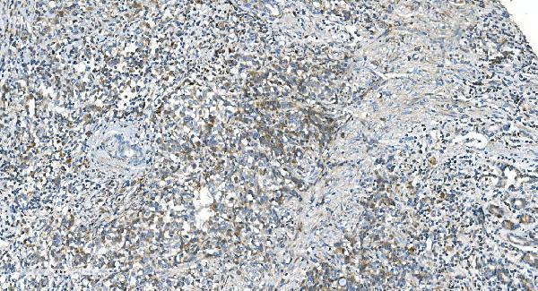
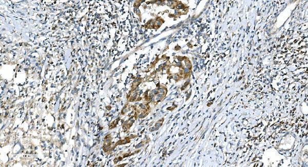
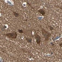


Comprehensive Human Protein Atlas IHC scoring per tissue (reliability: Approved — Medium consistency between antibody staining and RNA expression data.). Rows are taken directly from the HPA tissue chart — click any row's HPA link to view the source.
| Tissue | Cell type | Level | Evidence | Source |
|---|---|---|---|---|
| Cerebellum | Cells in molecular layer | High | Protein (IHC) | HPA → |
| Cerebral cortex | Neuronal cells | High | Protein (IHC) | HPA → |
| Hippocampus | Neuronal cells | High | Protein (IHC) | HPA → |
| Adipose tissue | Adipocytes | Medium | Protein (IHC) | HPA → |
| Adrenal gland | Glandular cells | Medium | Protein (IHC) | HPA → |
| Tissue | Cell type | Level | Evidence | Source |
|---|---|---|---|---|
| Bone marrow | Hematopoietic cells | Not detected | Protein (IHC) | HPA → |
Troubleshoot TNFRSF21 staining in paraffin section IHC by checking retrieval, compartment, cell type and controls before interpreting chromogenic signal.
A04348-2 has IHC images from paraffin-embedded human gastric cancer, bladder cancer, and renal carcinoma sections (catalog image captions). Human, mouse, and rat reactivity is listed (catalog reactivity).
A04348-2 is listed for IHC and has images from paraffin-embedded human gastric cancer, bladder cancer, and renal carcinoma sections (catalog applications; image captions). It is listed as reactive with human, mouse, and rat, but the supplied IHC images show human tissue only (catalog reactivity; image captions).
Which to pick: Choose A04348-2 for paraffin-section IHC; its human tissue captions report EDTA retrieval at pH 8.0 and 2 μg/ml primary antibody (catalog image captions). No IF/ICC application or image is supplied, so an IF/ICC choice cannot be supported here (catalog applications; image captions). Human, mouse, and rat reactivity is listed for A04348-2, but the supplied IHC images establish only human tissue use; the fixative is unreported (catalog reactivity; image captions).