TNNC1 / Troponin C, slow skeletal and cardiac muscles · IHC design guide

Design Immunohistochemistry for TNNC1

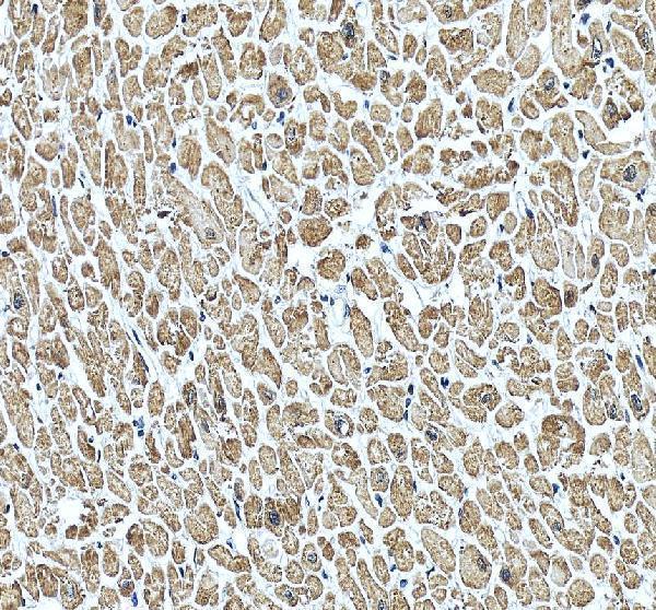
Plan paraffin-section TNNC1 IHC using heart or skeletal muscle as a positive control (HPA tissue IHC). Assess cytoplasmic staining in myocytes and compare it with an appropriate negative control (HPA tissue IHC; standard IHC practice).

Evidence assembled Oct 2026 · For research use; verify linked source records and product datasheet before use
Immunohistochemistry protocol sheet for TNNC1 (IHC for TNNC1): expected localisation Cytoplasmic in myocytes (HPA tissue IHC), antibody PA1779, validated IHC image, and IHC protocol steps
Printable TNNC1 IHC protocol sheet — expected localisation Cytoplasmic in myocytes (HPA tissue IHC), antibody PA1779, controls and protocol steps. Open the full TNNC1 IHC guide →

TNNC1 Immunohistochemistry Experimental Design Guide

Expected localisation, validated protocols, controls and antibodies — the at-a-glance facts below, then the full design guide.

Must know before staining
Expected localisation Cytoplasmic in myocytes (HPA tissue IHC)
Staining pattern Selective cytoplasmic staining in myocytes (HPA tissue IHC)
Antigen retrieval EDTA pH 8.0 HIER, heat-mediated (datasheet PA1779)
Positive control ⓘ Heart muscle+1 more · see all
Negative control ⓘ Adipose tissue+4 more · see all
Important caveats
Reasons your staining may differ from the expected pattern.
Fixation Keep fixation consistent across sections. (standard IHC practice; not target-specific)
Caveat Non-myocytes may lack staining in positive tissue (HPA tissue IHC)
Regulation High in heart and skeletal myocytes (HPA tissue IHC)
Isoform / epitope No annotated isoforms; one chain spans residues 1–161 (UniProt)
Section 1

Recommended TNNC1 IHC & IF Protocols

The catalog antibody's IHC-P protocol (datasheet: PA1779) is followed by 4 published TNNC1 IHC protocols (PMC5356821; PMC11529595; PMC4185407; PMC12142794).

Recommended immunohistochemistry (IHC-P) protocol parameters
SampleTissue sections; selected-image fixative not specified (standard IHC workflow)
FixationImage fixative and duration unreported (datasheet PA1779); verify before use.
Sectioning4–5 µm sections on charged slides (standard)
DeparaffinisationXylene, graded ethanol series to water (standard)
Antigen retrievalHeat retrieval: EDTA pH 8.0 (datasheet PA1779); 20 min, 95–100 °C (standard)
Peroxidase block3% H2O2, 10 min, room temperature (standard)
Blocking10% goat serum (datasheet PA1779)
Primary antibodyRabbit anti-TNNC1, 0.5-1μg/ml (datasheet PA1779)
Primary incubationOvernight at 4 °C (datasheet PA1779)
DetectionHRP-conjugated secondary, DAB chromogen (datasheet PA1779)
CounterstainHematoxylin, blue, dehydrate and mount (standard)
Expected resultTNNC1-positive staining in cardiomyocytes of heart muscle (HPA tissue IHC: High). HPA tissue profile: Selective cytoplasmic expression in myocytes. No signal in the no-primary control.
💡Decision noteStart with heat-mediated EDTA retrieval at pH 8.0 for the catalog antibody (datasheet: PA1779). Two published protocols also specify EDTA retrieval (PMC11529595; PMC12142794).
Section 2

What Is the Expected TNNC1 Staining Pattern?

In paraffin-section IHC, expect predominantly cytoplasmic TNNC1 staining in cardiomyocytes and skeletal myocytes (HPA: selective cytoplasmic expression in myocytes; High in both tissues). HPA rates the tissue pattern Enhanced, reflecting high consistency between antibody staining and RNA expression (HPA: tissue IHC). TNNC1 has no transmembrane segment, so a membrane-restricted pattern would need separate support (UniProt P63316: topology).

What am I looking at on my slide?
Strong cytoplasmic staining follows cardiomyocytes in heart muscle or myocytes in skeletal muscle.This matches the reported positive cells, compartment and High staining level (HPA: tissue IHC). Interpret intensity alongside cell morphology; the HPA rating supports the overall pattern, rather than guaranteeing identical intensity in every section (HPA: Enhanced; general IHC practice).
Staining is confined to nuclei or cell borders, with little cytoplasmic staining in myocytes.That distribution conflicts with the selective cytoplasmic tissue-IHC profile (HPA: tissue IHC). Check morphology, counterstain and staining controls before calling it TNNC1. Nuclear signal in ICC-IF has separate HPA support and does not establish a nuclear-only paraffin-IHC pattern (HPA: subcellular ICC-IF).
Strong signal appears in adipocytes or adrenal glandular cells instead of the expected myocytes.Those cell types were Not detected in the reported tissue-IHC samples (HPA: adipose tissue; adrenal gland). Assess possible antibody cross-reactivity or endogenous chromogen activity using appropriate controls; staining alone cannot distinguish those causes (general IHC practice).
Chromogen spreads across the section without clear cell boundaries or a myocyte-linked pattern.Diffuse background makes compartment and cell-type scoring unreliable (general IHC practice). Compare a no-primary control and inspect the section for uneven reagent coverage or inadequate washing before interpreting faint color as target staining (general IHC practice).
No convincing staining appears in identifiable heart cardiomyocytes or skeletal myocytes.This is discordant with the reported High staining in those cells (HPA: tissue IHC). First establish that the intended cells are present, then review run controls and the validated staining conditions; a single blank section does not establish biological absence (general IHC practice).
💡Expected TNNC1 appearanceCall a result positive when High, predominantly cytoplasmic chromogen follows cardiomyocytes or skeletal myocytes (HPA: tissue IHC); isolated nuclear, membrane-rim or nonmyocyte staining is discordant with that tissue pattern and warrants control review (HPA: tissue IHC; general IHC practice).
How each factor affects the staining
Tissue and cell selectionHeart cardiomyocytes and skeletal myocytes are reported High, while the listed adipocytes and adrenal glandular cells are Not detected (HPA: tissue IHC). HPA reports tongue RNA group enrichment, but this payload gives no tongue protein-staining level; do not assign one (HPA: RNA specificity).
Subcellular evidence by applicationTissue IHC reports selective cytoplasmic expression in myocytes (HPA: tissue IHC). ICC-IF separately approves nucleoplasm, actin-filament and mitochondrial locations (HPA: subcellular ICC-IF). Keep those application-specific observations distinct when scoring paraffin sections.
Antibody validationThe three listed antibodies have Enhanced IHC status; only HPA044848 also has Approved ICC status in this payload (HPA: antibodies). This supports the reported IHC pattern but does not supply a catalog-antibody dilution, retrieval condition or IF protocol.
Topology and processingTNNC1 is a 161-residue chain with no signal peptide, propeptide or transmembrane segment (UniProt P63316: processing and topology). These annotations do not establish an epitope location or target-specific fixation sensitivity; neither can be inferred for this staining run.
Why is my staining missing, weak or wrong?
SituationLikely causeNext action
Known-positive heart or skeletal muscle has no myocyte signal.The absent signal conflicts with the reported High level in those cells (HPA: tissue IHC); the run, tissue preservation or antibody conditions may need review (general IHC practice).Confirm myocytes are present, inspect a run-matched positive control, and check the IHC-validated antibody's documented dilution, retrieval and detection steps before changing conditions (general IHC practice).
Adipocytes or adrenal glandular cells stain strongly.Those sampled cell types are reported Not detected (HPA: tissue IHC). Cross-reactivity or endogenous chromogen activity are possible explanations, not diagnoses from appearance alone (general IHC practice).Compare a no-primary control, review endogenous-enzyme blocking appropriate to the chromogen system, and confirm cell identity on the counterstain (general IHC practice).
Only nuclei or sharp cell-border rims stain in a positive tissue.A nuclear-only or rim-only IHC pattern differs from HPA's selective cytoplasmic myocyte profile (HPA: tissue IHC); morphology or background may complicate compartment assignment (general IHC practice).Recheck focus, counterstain and controls; score the result against the cytoplasmic myocyte pattern before interpreting isolated compartment signal as specific (HPA: tissue IHC; general IHC practice).
Weak color covers most structures and obscures myocytes.Diffuse background prevents reliable localization or intensity scoring (general IHC practice). The supplied TNNC1 sources do not identify a target-specific cause for this appearance.Inspect no-primary staining and reagent coverage; review blocking, washes, antibody concentration and chromogen development against the validated IHC procedure (general IHC practice).
Myocyte staining varies across one section.The HPA High designation summarizes observed cell staining, not a requirement for uniform intensity across every field (HPA: tissue IHC). Section quality or staining distribution may also contribute (general IHC practice).Compare well-preserved fields, cell morphology and run controls; record the extent and intensity of cytoplasmic myocyte staining instead of treating one bright field as the whole section (general IHC practice).
Does ICC-IF nuclear or mitochondrial signal change the expected paraffin-IHC call?HPA approves nucleoplasm, mitochondria and actin filaments in ICC-IF, while its tissue-IHC summary describes selective cytoplasmic myocyte expression (HPA: subcellular ICC-IF; tissue IHC).Use the cytoplasmic myocyte pattern to interpret this IHC section (HPA: tissue IHC). Consult the separate IF/ICC guide for that application's localization and controls; no IF protocol is specified here.

Sample controls for TNNC1 IHC & IF

🧪Run heart muscle first; cardiomyocytes should stain (HPA: High in cardiomyocytes). Use adipose tissue as the negative tissue (HPA: Not detected in adipocytes); on the heart slide, interstitial spaces should lack the cardiomyocyte staining pattern, while nonmyocyte cells should be assessed by morphology rather than assumed negative (HPA: High in cardiomyocytes).
Positive control tissue: Heart muscle (Cardiomyocytes, HPA High)
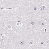
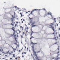
Negative control tissue: Adipose tissue (HPA Not detected)
ICC-IF cell lines (HPA subcellular resource): HPA ICC-IF images show TNNC1 in HeLa, Hep-G2, U2OS, with annotated localisation: Nucleoplasm (approved), Actin filaments (approved), Mitochondria (approved) (HPA subcellular).
Technical controls: Include a no-primary, secondary-only control and a primary-host-matched nonimmune IgG control for a polyclonal antibody, or an isotype-matched control for a monoclonal antibody (standard IHC practice). A TNNC1-knockout sample, if available, provides a biological negative; quench endogenous peroxidase and assess cardiac pigment as potential chromogenic background (standard IHC practice).
⚠️Feasibility: A target-specific fixation window or fixation effect is unreported, and the fixative is unreported in the selected PA1779 rat cardiac muscle IHC(P) caption (PA1779 caption: fixative not stated). Retrieval dependency is unreported, so assess retrieval conditions using the positive heart section (standard IHC practice). The supplied evidence does not establish whether frozen sections or IF are easier; cardiac muscle autofluorescence can complicate IF assessment (standard IF practice).

HPA tissue IHC evidence for TNNC1

Comprehensive Human Protein Atlas IHC scoring per tissue (reliability: Enhanced — High consistency between antibody staining and RNA expression data.). Rows are taken directly from the HPA tissue chart — click any row's HPA link to view the source.

Positive expression · recommended positive controls

TissueCell typeLevelEvidenceSource
Heart muscle Cardiomyocytes High Protein (IHC) HPA →
Skeletal muscle Myocytes High Protein (IHC) HPA →

Undetected expression · recommended negative controls

TissueCell typeLevelEvidenceSource
Adipose tissue Adipocytes Not detected Protein (IHC) HPA →
Adrenal gland Glandular cells Not detected Protein (IHC) HPA →
Appendix Glandular cells Not detected Protein (IHC) HPA →
Bone marrow Hematopoietic cells Not detected Protein (IHC) HPA →
Breast Adipocytes Not detected Protein (IHC) HPA →
Section 3

Advanced TNNC1 IHC Tips

Troubleshoot TNNC1 staining in paraffin section IHC using the catalog antibody’s retrieval guidance and tissue expression as controls; assess IF separately.

What retrieval should I try when TNNC1 staining is weak?
Start with heat-mediated EDTA pH 8.0 antigen retrieval for paraffin section IHC (datasheet PA1779). Keep heating, cooling and detection conditions identical across a small retrieval time series, then compare signal in cardiomyocytes with staining outside myocytes (standard IHC practice; HPA: High in cardiomyocytes). If staining remains weak, test an alternative retrieval condition only as a documented fallback, using adjacent sections and the same detection system (standard IHC practice). Excessive retrieval can damage morphology or increase background, so choose the shortest condition that gives interpretable cellular staining (standard IHC practice). Record the final buffer, pH and heating time with every image (standard IHC practice).
Could fixation explain weak or uneven TNNC1 staining?
Target-specific fixation sensitivity is unknown for this antibody; the rat cardiac muscle IHC(P) caption does not state a fixative (catalog caption: PA1779). Compare sections with documented fixation and processing histories before changing retrieval or antibody concentration, because those variables can alter epitope accessibility in paraffin sections (standard IHC practice). Inspect whether weak staining tracks specimen edges, thick areas or poorly preserved morphology rather than myocyte distribution (standard IHC practice; HPA: selective cytoplasmic expression in myocytes). Run an adequately preserved positive section alongside each processing condition, and keep detection settings constant (standard IHC practice; HPA: High in cardiomyocytes). Do not assign a TNNC1-specific fixation effect without a controlled comparison.
Where should a convincing TNNC1 IHC signal appear?
In tissue IHC, look first for selective cytoplasmic staining of myocytes, with strong cardiomyocyte and skeletal myocyte signals as reference patterns (HPA tissue IHC: selective cytoplasmic expression; High in both cell types). TNNC1 has no annotated transmembrane segment, so a crisp membrane-only outline needs additional verification (UniProt P63316: topology). Cell-based IF reports nucleoplasm, mitochondria and actin filaments, but those observations do not establish the dominant pattern in paraffin tissue IHC (HPA subcellular; HPA tissue IHC). Examine matched morphology and adjacent negative cell populations before interpreting fine intracellular patterns (standard IHC practice). Compare compartmental staining under identical counterstain and exposure conditions (standard IHC practice).
How can epitope uncertainty affect TNNC1 IHC interpretation?
The supplied TNNC1 record lists 0 alternative isoforms and four EF-hand regions across the 161-residue chain (UniProt P63316 record). It also lists N-acetylmethionine at residue 1 and phosphoserine at residue 98, but gives no PA1779 epitope map (UniProt P63316 record; supplied catalog evidence). Therefore, altered staining cannot be assigned to one EF-hand, modification or isoform from IHC alone (supplied record; standard IHC interpretation). If specificity is in doubt, compare the IHC-validated antibody with an independently validated reagent recognizing a documented distinct epitope, using matched sections and controls (standard IHC practice). Report the actual reagent and validation evidence used (standard IHC practice).
How should I adapt the TNNC1 question to multiplex IF?
For a separate IF experiment, pair TNNC1 with a validated marker of the expected myocyte population and check cellular overlap, using cardiomyocytes or skeletal myocytes as tissue context (HPA tissue IHC: High in both). Choose spectrally separated fluorophores and place the weaker signal in a channel with low tissue autofluorescence, verified using unstained controls (standard IF practice). Because the PA1779 epitope is unspecified, optimize mild permeabilisation only if intracellular access is needed, while checking whether morphology and marker signal are preserved (supplied catalog evidence; standard IF practice). Include single-stain and secondary-only controls to assess bleed-through and nonspecific signal (standard IF practice). Do not infer IF validation from the PA1779 IHC(P) caption (catalog caption: PA1779).
How do I reduce diffuse brown staining without losing TNNC1 signal?
Use a no-primary control to separate detection-system background from antibody-dependent staining, and compare it with a known myocyte-positive section in the same run (standard IHC practice; HPA: High in cardiomyocytes). Quench endogenous peroxidase before chromogenic detection and optimize protein blocking and washes as general IHC workflow steps (standard IHC practice). Titrate the catalog antibody across documented concentrations, keeping retrieval, section thickness and DAB development time constant so changes are interpretable (standard IHC practice). Diffuse staining in cell populations reported as negative should trigger review of antibody concentration, detection chemistry and tissue handling (HPA: Not detected in adipocytes and several other listed cell populations; standard IHC practice).
What is a defensible way to score TNNC1 IHC? ⚠ ANSWER MARKED FOR VERIFICATION
Define cardiomyocytes or skeletal myocytes as the scored population before measuring, because TNNC1 tissue staining is selective for myocytes (HPA tissue IHC: selective cytoplasmic expression). For intensity comparisons, use an H-score based on the percentage of cells at each staining grade, with a fixed grading rubric (standard IHC practice). Alternatively, report % positive myocytes using one prespecified threshold; use positive-cell density per mm² only when tissue area is the relevant denominator (standard IHC practice). Normalize to viable myocyte number or viable myocyte area, exclude damaged regions by a written rule, and keep retrieval and DAB development matched (standard IHC practice).
How can I distinguish true TNNC1 staining from artefact?
A plausible tissue result is cytoplasmic staining in myocytes, supported by strong cardiomyocyte and skeletal myocyte signals (HPA tissue IHC: selective cytoplasmic expression; High in both). Treat isolated staining in an unexpected cell type or an exclusively membrane pattern as a finding requiring independent validation (HPA tissue IHC; UniProt P63316: no transmembrane segment). Compare suspicious signal with morphology, section edges and necrotic areas, where staining artefacts can occur (standard IHC practice). A no-primary control helps reveal endogenous enzyme or detection-system colour, while a matched positive section checks the staining run (standard IHC practice). Interpret unusual nuclear staining cautiously because HPA cell-based IF reports nucleoplasmic localization (HPA subcellular).
Boster reagents

Best TNNC1 / Troponin C, slow skeletal and cardiac muscles IHC Antibodies

PA1779 has IHC images from rat cardiac muscle and human heart paraffin sections (PA1779 IHC image captions). Listed reactivity covers Human, Mouse, and Rat (PA1779 catalog reactivity).

Real IHC data Anti-cardiac Troponin C antibody, PA1779, IHC(P) IHC(P): Rat Cardiac Muscle Tissue
Anti-cardiac Troponin C/TNNC1 Antibody ®
Cat # PA1779

PA1779 is the sole rendering SKU and is listed for IHC (catalog applications). Its IHC captions show rat cardiac muscle and a paraffin-embedded human heart section (PA1779 IHC image captions).

Which to pick: For tissue IHC, choose PA1779: its own captions document paraffin sections, but the fixative is unreported (PA1779 IHC image captions). No listed SKU has IF/ICC validation or an IF image (catalog applications and image alts). For cross-species IHC, PA1779 lists Human, Mouse, and Rat reactivity; pictured tissue data cover Human and Rat (PA1779 catalog reactivity; PA1779 IHC image captions).

Each figure is that product's own IHC / IF validation image from its datasheet.

References

  1. UniProt Consortium. UniProt entry P63316 (TNNC1_HUMAN, Troponin C, slow skeletal and cardiac muscles).
  2. Human Protein Atlas. TNNC1 tissue IHC expression (reliability: Enhanced).
  3. Human Protein Atlas. TNNC1 subcellular location (ICC-IF): Localized to the nucleoplasm, mitochondria and actin filaments..
  4. Human Protein Atlas. TNNC1 antibody validation summary (3 antibodies).
  5. MFAP5 and TNNC1: Potential markers for predicting occult cervical lymphatic metastasis and prognosis in early stage tongue cancer. Oncotarget 2017 — PMC5356821.
  6. In-depth study of pyroptosis-related genes and immune infiltration in colon cancer. PeerJ 2024 — PMC11529595.
  7. Calcium-dependent FAK/CREB/TNNC1 signalling mediates the effect of stromal MFAP5 on ovarian cancer metastatic potential. Nature communications 2014 — PMC4185407.
  8. Screening Differentially Expressed Proteins in Areca Nut-Related Oral Squamous Cell Carcinoma Using Tandem Mass Tag Proteomics. International dental journal 2025 — PMC12142794.
  9. PubMed PMID:3951483 — UniProt-cited evidence.
  10. PubMed PMID:3166492 — UniProt-cited evidence.
  11. PubMed PMID:2250022 — UniProt-cited evidence.