This website uses cookies to ensure you get the best experience on our website.


- Table of Contents
Plan paraffin-section TNNI2 IHC around cytoplasmic staining in skeletal myocytes (HPA tissue IHC). Use the catalog antibody's 0.5–1 μg/mL IHC range (datasheet A07355-2) to guide chromogenic staining and scoring.

Expected localisation, validated protocols, controls and antibodies — the at-a-glance facts below, then the full design guide.
| Expected localisation | Cytoplasmic staining in skeletal muscle (HPA tissue IHC) | |
| Staining pattern | High cytoplasmic staining in skeletal myocytes (HPA tissue IHC) | |
| Antigen retrieval | Citrate pH 6 HIER, heat-mediated (datasheet A07355-2) | |
| Positive control | Skeletal muscle | |
| Negative control | Adipose tissue+4 more · see all |
| Fixation | Matched tissue-IHC evidence does not establish the fixation claim. Validate the specimen-specific method before use. (selected-SKU IHC image A07355-2) | |
| Caveat | Fast-skeletal identity may affect staining across fibers (UniProt) | |
| Regulation | No expression regulator specified (UniProt) | |
| Isoform / epitope | 2 isoforms; antibody epitope coverage is unknown (UniProt) |
The catalog antibody’s IHC-P protocol is paired with one published mouse skeletal-tissue IHC protocol (PMC4207604).
| Sample | Paraffin-embedded human skeletal muscle tissues; fixative not specified (datasheet A07355-2) |
| Fixation | Image fixative and duration unreported (datasheet A07355-2); verify before use. |
| Sectioning | 4–5 µm sections on charged slides (standard) |
| Deparaffinisation | Xylene, graded ethanol series to water (standard) |
| Antigen retrieval | Heat retrieval: Citrate pH 6, 20 min (datasheet A07355-2) |
| Peroxidase block | 3% H2O2, 10 min, room temperature (standard) |
| Blocking | 10% goat serum (datasheet A07355-2) |
| Primary antibody | Rabbit anti-TNNI2, 0.5-1μg/ml (datasheet A07355-2) |
| Primary incubation | Overnight at 4 °C (datasheet A07355-2) |
| Detection | Streptavidin-biotin complex (SABC), DAB chromogen (datasheet A07355-2) |
| Counterstain | Hematoxylin, blue, dehydrate and mount (standard) |
| Expected result | TNNI2-positive staining in myocytes of skeletal muscle (HPA tissue IHC: High). HPA tissue profile: Cytoplasmic expression in skeletal muscle. No signal in the no-primary control. |
In paraffin-section IHC, expect TNNI2 staining in the cytoplasm of skeletal muscle myocytes; HPA reports high staining with Enhanced reliability, reflecting consistency between antibody staining and RNA expression (HPA tissue IHC). TNNI2 is a thin-filament regulatory protein that binds actin and tropomyosin and has no annotated transmembrane segment (UniProt P48788 function, subunit, topology).
| Strong cytoplasmic chromogen in skeletal muscle myocytes, with a readable counterstain. | This matches the reported high myocyte staining and cytoplasmic tissue pattern (HPA tissue IHC). Score signal in identifiable myocytes rather than counting general tissue colour as positive. HPA's Enhanced rating supports the tissue pattern, but does not validate every antibody or preparation (HPA tissue IHC). |
| Predominantly nuclear, extracellular, or sharply surface-restricted staining in skeletal muscle. | Treat the compartment mismatch as a possible artefact until it is checked against cytoplasmic staining in a positive section (HPA tissue IHC; general IHC practice). UniProt lists no transmembrane segment and does not annotate a subcellular location, so topology alone cannot identify the cause (UniProt P48788 topology, subcellular record). |
| Clear staining in adipocytes or bone marrow hematopoietic cells. | These cells are reported as not detected in tissue IHC (HPA tissue IHC). Consider antibody cross-reactivity or endogenous detection activity, especially if the signal appears outside identifiable skeletal myocytes; test detection-only and negative-tissue controls before assigning TNNI2 expression (general IHC practice). |
| Diffuse colour across myocytes, stroma, and blank areas of the section. | A widespread, poorly localized deposit does not reproduce the cytoplasmic myocyte pattern (HPA tissue IHC). Examine background in a section processed without primary antibody, then review blocking, washing, detection chemistry, and antibody concentration as general IHC variables (general IHC practice). |
| No detectable staining in a skeletal muscle positive-control section. | That conflicts with HPA's high myocyte signal (HPA tissue IHC). Check section integrity, primary-antibody and detection steps, and counterstain visibility before interpreting test sections as negative (general IHC practice). No TNNI2-specific fixation or retrieval sensitivity is established by the supplied records. |
| Positive and negative tissue context | Skeletal muscle myocytes are the documented high-staining reference; adipocytes and bone marrow hematopoietic cells are documented as not detected (HPA tissue IHC). Use those contrasts to judge specificity within the sampled material, while recognizing that HPA observations do not guarantee every preparation's result. |
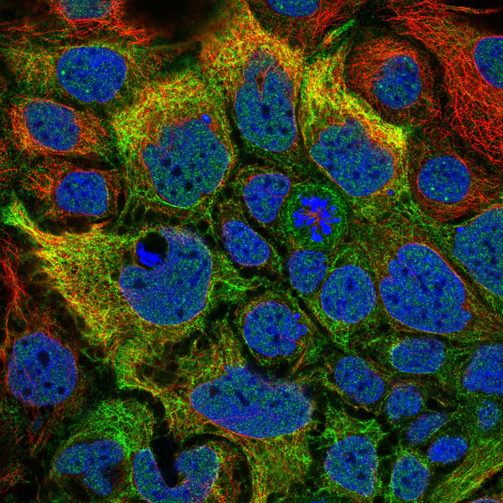
| Tissue IHC versus IF/ICC localisation | How should IF/ICC be read? HPA reports a main plasma-membrane location and an additional intermediate-filament location in ICC-IF, whereas tissue IHC reports cytoplasmic skeletal muscle expression (HPA subcellular ICC-IF; HPA tissue IHC). Interpret each assay in its own specimen and detection context. |
| Antibody evidence | HPA lists HPA055938 and CAB018697 as IHC Enhanced; HPA055938 is also ICC Approved (HPA antibodies). Those statuses support the reported applications and tissue-pattern assessment. They do not establish a dilution, retrieval setting, or validation status for a different catalog antibody. |
| Isoforms and protein features | UniProt lists two isoforms and modified residues, including acetylation and phosphorylation (UniProt P48788 isoforms, modified residues). Without an antibody epitope or isoform-specific validation in the supplied evidence, do not infer which form contributes to a positive section or whether a modification changes staining. |
| Paraffin-section workflow | Antigen retrieval and blocking are general IHC variables; compare documented conditions and include suitable controls when optimizing an assay (general IHC practice). The supplied UniProt and HPA records do not report a TNNI2-specific retrieval requirement or fixation effect. |
| Situation | Likely cause | Next action |
|---|---|---|
| Skeletal muscle positive control is blank. | A missed or ineffective primary-antibody, retrieval, or detection step can eliminate visible chromogen (general IHC practice). The result conflicts with high skeletal myocyte staining (HPA tissue IHC); the source records do not identify a TNNI2-specific failing step. | Confirm that tissue and counterstain are visible, then check each reagent and incubation against the antibody's documented IHC procedure. Compare an appropriately controlled retrieval condition if needed (general IHC practice). |
| Colour obscures cell boundaries throughout the section. | Excess antibody or detection background, inadequate blocking, or insufficient washing can reduce contrast (general IHC practice). Diffuse colour cannot be scored as the reported cytoplasmic myocyte pattern (HPA tissue IHC). | Review the antibody's documented working concentration, strengthen routine blocking and washing, and inspect a section without primary antibody (general IHC practice). Score only cell-associated signal that remains interpretable. |
| Adipocytes or marrow hematopoietic cells stain as strongly as myocytes. | Those cells are listed as not detected in the supplied tissue profile (HPA tissue IHC). Cross-reactivity or endogenous detection activity is possible, although the staining alone cannot distinguish them (general IHC practice). | Compare a detection-only control with the stained section, review endogenous-enzyme blocking if using enzyme-based detection, and reassess antibody specificity with documented controls (general IHC practice). |
| Signal appears mainly in nuclei or extracellular spaces. | This conflicts with the cytoplasmic tissue-IHC profile (HPA tissue IHC). Tissue deposits, detection background, or misidentified structures can produce misleading localisation (general IHC practice); UniProt's lack of a transmembrane segment does not diagnose the artefact (UniProt P48788 topology). | Inspect morphology alongside the counterstain, compare with the skeletal muscle positive control, and check a section processed without primary antibody before calling the signal TNNI2 (general IHC practice). |
| Signal is uneven across a section or between staining runs. | Uneven reagent coverage, section handling, or detection development can vary apparent intensity (general IHC practice). HPA reports high myocyte staining but does not establish uniform staining in every field or run (HPA tissue IHC). | Check section coverage and processing consistency, then compare runs using the same skeletal muscle control and scoring criteria (general IHC practice). Avoid treating technical variation alone as a change in TNNI2 expression. |


Comprehensive Human Protein Atlas IHC scoring per tissue (reliability: Enhanced — High consistency between antibody staining and RNA expression data.). Rows are taken directly from the HPA tissue chart — click any row's HPA link to view the source.
| Tissue | Cell type | Level | Evidence | Source |
|---|---|---|---|---|
| Skeletal muscle | Myocytes | High | Protein (IHC) | HPA → |
| Tissue | Cell type | Level | Evidence | Source |
|---|---|---|---|---|
| Adipose tissue | Adipocytes | Not detected | Protein (IHC) | HPA → |
| Adrenal gland | Glandular cells | Not detected | Protein (IHC) | HPA → |
| Appendix | Glandular cells | Not detected | Protein (IHC) | HPA → |
| Bone marrow | Hematopoietic cells | Not detected | Protein (IHC) | HPA → |
| Breast | Adipocytes | Not detected | Protein (IHC) | HPA → |
Troubleshoot TNNI2 staining in paraffin sections by checking retrieval, tissue pattern, controls, and scoring before interpreting signal.
The catalog antibody has real IHC data from human, mouse and rat paraffin skeletal muscle sections, plus IF data from mouse and rat paraffin skeletal muscle sections (catalog image captions).
A07355-2 will render with its human skeletal muscle paraffin-section IHC figure (IHC image caption). The same SKU is listed for human, mouse and rat IHC, with additional mouse and rat paraffin-section IF images (catalog applications, reactivity and image captions).
Which to pick: Choose A07355-2 for tissue IHC because its IHC captions show staining in human, mouse and rat skeletal muscle paraffin sections (IHC image captions). For IF, the same SKU has mouse and rat skeletal muscle paraffin-section images; ICC validation and clonality are unreported (IF image captions; catalog). The IHC captions do not report a fixative, so fixation conditions need separate validation (IHC image captions).